![]()
![]()
![]()
Use LEFT and RIGHT arrow keys to navigate between flashcards;
Use UP and DOWN arrow keys to flip the card;
H to show hint;
A reads text to speech;
28 Cards in this Set
- Front
- Back
|
What is the efferent division divided into? |
Somatic motor neurons (skeletal muscles) Autonomic neurons (smooth and cardiac muscle, many glands, and some adipose tissue) |
|
|
Autonomic nervous system |
-Not under voluntary control -Also known as the visceral nervous system -2 divisions: Sympathetic (fight or flight) and parasympathetic (rest and digest) |
|
|
What is the sympathetic nervous system mediated by? |
The hypothalamus. This system can create a total body response to a crisis. But activating one sympathetic pathway will not activate them all. ie. Blood flow to tissues is controlled by this system |
|
|
Are the sympathetic and the parasympathetic all or none (ie one is active and the other one is completely inactive) |
No! It is a seesaw between the two systems in order to maintain homeostasis within the body. |
|
|
What does the ANS work closely with to maintain homeostasis? |
The endocrine system and the behavioral state system. |
|
|
Spinal reflexes |
Autonomic reflexes that happen without input from the brain, but can be influenced by the brain sometimes |
|
|
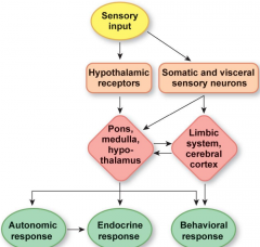
Integration of autonomic function |
|
|
|
What are the three autonomic control centers of the brain? |
-Hypothalamus: Water balance, temperature, and hunger -Pons: respiration -Medulla: Respiration, cardiac, vomiting, and swallowing |
|
|
Cannon's properties of homeostasis |
1) Preservation of fitness of the internal environment 2) Up-down regulation by tonic control 3) Antagonistic control 4) Chemical signals with different effects in different tissues |
|
|
What determines response in some pathways? |
The type of neurotransmitter receptor. ie. The catecholamines epinephrine and norepinephrine bind either with alpha (constriction of blood vessels) or beta (dilated blood vessels) receptors. |
|
|
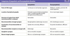
Sympathetic autonomic pathways |
-Thoracolumbar division. -Ganglia are found in chains along the bony vertebral columns, and some descending along the aorta -short preganglionic neurons -long post ganglionic neurons -Divergence is an important characteristic. 1 preganglionic neuron synapses with 8-9 postganglionic neurons (all the way up to 32). Allows the CNS to affect many things at once |
|
|
Parasympathetic autonomic pathways |
-Craniosacral division -Ganglia are located on or near the target organ -long preganglionic neurons -short postganglionic neurons |
|
|
Preganglionic neuron |
The first neuron. Originates from the CNS and projects to the 2nd neuron |
|
|
Autonomic ganglia |
Cluster of cell bodies outside the CNS. Located in different regions depending on whether it is sympathetic (along vertebral column/aorta) or parasympathetic (on or at target organ/tissue) |
|
|
Postganglionic neuron |
Where the pre and postganglionic neurons synapse (in ganglia). This neuron projects axon to target tissue. |
|
|
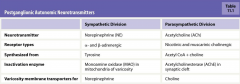
Rules for sympathetic and parasympathetic neurotransmitters and receptors: |
1) Both types of preganglionic neurons release ACh onto nicotinic cholinergic receptors (excitatory) on the postganglionic cell. 2) Most postganglionic sympathetic neurons secrete norepinephrine onto adrenergic receptors on the target cell 3) Most postganglionic parasympathetic neurons secrete ACh onto Mscantaric cholinergic receptors on target cells. |
|
|
Neuroeffector junction |
Synapse between postganglionic autonomic neuron and its target cell |
|
|
Varicosities |
Autonomic postganglionic axons contain these series of swollen areas at distal ends, and contain vesicles filled with neurotransmitters. The more neurotransmitter released in the synapse, the longer or stronger the response will be (also determined by how rapidly it is broken down) |
|
|
Norepinephrine (NE) release and removal at a sympathetic neuroeffector junction |
1)NE is synthesized from the amino acid tyrosine 2) An action potential arrives at a varicosity 3) Depolarization opens voltage gated calcium channels 4) Calcium entry triggers exocytosis of synaptic vessicles 5) NE binds to adrenergic receptor on the target 6) Receptor activation ceases when NE diffuses away from the synapse 7) NE is removed from the synapse by active transport 8) NE can be taken back into the synaptic vesicles for re-release 9) NE can be metabolized by monoamine oxidase (MAO) |
|
|
Differences between sympathetic and parasympathetic postsympathetic neurotransmitters |
|
|
|
Sympathetic receptor and subtypes |
-Sympathetic pathways secrete catecholamines (norepinephrine and epinephrine) that bind to adrenergic receptors (GPCR) on their target cells. -Alpha receptors: Most commone. Respond strongly to NE and weakly to E. Causes muscle contractions or exocytosis. -Beta receptors: Have 3 different subtypes all differing in their affinities for NE and E. All are cAMP and cause phosphorylation of intracellular proteins. - Results in modification of proteins or synthesis of new proteins, creating lasting metabolic effects. Each subtype has different second messenger pathways |
|
|
Parasympathetic receptor and subtypes |
-Release of ACh at target cells -Muscantaric cholinergic receptors (GPCR) to open potassium or calcium channels |
|
|
Adrenal medulla |
specialized neuroendocrine tissue associated with the sympathetic nervous system. Primarily secretes epinephrine. -core of adrenal glands. Often described as modified sympathetic ganglion. -large quantities of epinephrine is released during fight or flight |
|
|
Comparison of Sympathetic and parasympathetic branches |
|
|
|
Somatic motor division |
Controls skeletal muscles. A single neuron originates in the CNS and projects an axon to the target (skeletal muscle). Always excitatory. |
|
|
Somatic motor neurons |
Cell bodies located either in the ventral horn or the spinal cord or in the brain. Myelinated 1+ meters in length Branch close to targets. Cluster of enlarged axon terminals that lay on the surgace of skeletal muscle fiber. Branching allows control of multiple muscle fibers at once. |
|
|
Neuromuscular junction |
Synapse of a somatic motor neuron on a muscle fiber. Has 3 main components: 1) presynaptic terminal filled with synaptic vesicles and mitochondria 2) Synaptic cleft 3) Postsynaptic membrane of skeletal muscle fiber |
|
|
Motor end plate |
Postsynaptic muscle cell membrane modified into this a series of folds that look like shallow gutters. On the upper edge of each fold, nicotinic ACh receptor (always excitatory to make muscle contractions) channels cluster in an active zone -The synapse contains acetylcholinesterase - no dual innervation for antagonistic control. Stops when somatic motor neurons in the CNS are inhibited, preventing the release of ACh. |