![]()
![]()
![]()
Use LEFT and RIGHT arrow keys to navigate between flashcards;
Use UP and DOWN arrow keys to flip the card;
H to show hint;
A reads text to speech;
121 Cards in this Set
- Front
- Back
|
CNS and PNS |
Two branches of the Nervous system |
|
|
Brain and spinal cord |
Two parts of the CNS |
|
|
Autonomic NS and Somatic NS |
Two parts of the PNS |
|
|
Autonomic NS |
Controls self regulated action of internal organ and glands |
|
|
Parasympathic Nerves and Sympathetic Nerves |
Two parts of Autonomic NS |
|
|
Sympathetic Nerves |
Arousing |
|
|
Parasympathetic Nerves |
Calming |
|
|
Somatic NS |
Responsible for the sensory input & motor output (controls skeletal muscles) |
|
|
Autonomic NS |
consists of neurons that receive information from and send commands to the heart, intestines, and other organs. |
|
|
Sympathetic NS |
consists of chains of ganglia just to the left and right of the spinal cord's central regions (the thoracic and lumbar areas). |
|
|
Fight or flight |
These ganglia are connected by axons to the spinal cord. Sympathetic axons prepare the organs for____ increasing breathing and heart rate and decreasing digestive activity. |
|
|
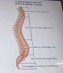
Spinal Column |
|
|
|
Parasympathetic NS |
Constricts pupil |
|
|
Parasympathetic NS |
Stimulates salivation |
|
|
Parasympathetic NS |
Constricts Bronchi, decreases heart rate |
|
|
Parasympathetic NS |
Stimulates digestive activity |
|
|
Parasympathetic NS |
Stimulates gallbladder |
|
|
Parasympathetic NS |
Inhibition of adrenaline production |
|
|
Parasympathetic NS |
Contracts bladder |
|
|
Parasympathetic NS |
Relaxes rectum |
|
|
Sympathetic NS |
Dilates pupil |
|
|
Sympathetic NS |
Inhibits salivation |
|
|
Sympathetic NS |
Relaxes bronchi, increases heart rate |
|
|
Sympathetic NS |
Inhibits digestive activity |
|
|
Sympathetic NS |
Stimulates glucose release by liver |
|
|
Sympathetic NS |
Stimulates epinephrine and norepinephrine release |
|
|
Sympathetic NS |
Relaxes bladder |
|
|
Sympathetic NS |
Orgasm, ejaculation, contracts rectum |
|
|
Spinal cord |
Most of PNS project from the |
|
|
Cranial nerves |
These nerves project from the brain |
|
|
diagnosis |
Functions of the various cranial nerves are commonly assessed by neurologists as a basis for |
|
|
I. Olfactory |
Smell(Identify odors with eyes closed) |
|
|
II. Optic Nerves |
Vision(Test peripheral vision with1 eye covered) |
|
|
III. Oculomotor Nerves |
Eye movement & pupillary reaction(Peripheral vision, eye chart, reaction to light) |
|
|
IV. Trochlear |
Eye movement (Test ability to depress & adduct eye) |
|
|
V. Trigeminal |
Face sensation & mastication ( Face sensation & clench teeth) |
|
|
VI. Abducens |
Eye movement (Test ability to abduct eye past midline) |
|
|
VII. Facial |
Facial muscles & taste (Close eyes & smile; detect various & tastetastes-sweet, sour, salty, bitter) |
|
|
VIII. Vestibulocochlear (Acoustic) |
Hearing & Balance (Hearing; feet together, eyes open/closed 5sec; test for past-pointing |
|
|
IX. Glossopharyngeal |
Swallow, voice, gag reflex (swallow and say "ahh" . Use tongue depressor to elicit gag reflex) |
|
|
X. Vagus |
Swallow, Voice, Gag Reflex (swallow and say "ahh" . Use tongue depressor to elicit gag reflex) |
|
|
XI. Spinal Accessory |
SCM & Trapezius (Rotate/SB neck; shrug shoulders) |
|
|
XII. Hypoglossal |
Tongue movement [Protrude tongue (watch for lateraldeviation)] |
|
|
Somatic NS |
Consists of a network of nerves that connect either to sensory receptors or to muscles that you can move voluntarily, such as muscles in your limbs, back, neck, and chest |
|
|
Somatic NS |
Consists of afferent and efferent neurons |
|
|
Meninges |
CNS encased In bone andcovered by three |
|
|
Dura mater |
Tough outer membrane |
|
|
Arachnoid membrane |
Web-like |
|
|
Pia Mater |
Adheres to CNS surface |
|
|
Cerebrospinal fluid (CSF) |
Fluid serves as cushion |
|
|
Blood brain barrier |
tightly-packed cells of blood vessel walls prevent entry of many molecules |
|
|
Blood brain barrier |
Chemical protection |
|
|
Physical protection |
Skull, meninges, CSF |
|
|
Nucleus |
Controls the entire neuron |
|
|
Axon |
Transfers signals toother cells and organs |
|
|
Myelin Sheath |
Increases the speed of the signal |
|
|
Axon Terminal |
Forms junctions with other cells |
|
|
Dendrites |
Receive signals from other cells |
|
|
Cell Body |
Organizes and keeps the cell functional |
|
|
Cell membrane |
Protects the cell |
|
|
Axon hillock |
Generates impulse in the neuron |
|
|
Node of Ranvier |
Allow diffusion of ions |
|
|
Schwann Cell |
Produces the myelin sheath |
|
|
Unipolar neuron, Bipolar neuron, Multipolar neuron, multipolar interneuron |
Classes of Neurons |
|
|
Glial cells |
Nourish and insulate neurons toprevent interference from otherelectrical signals |
|
|
Glial cells |
Direct the growth of neurons, andsupport mature neurons |
|
|
Glial cells |
Removes waste products that influence that growth of neurons |
|
|
Microglia |
They respond to injury ordisease by multiplyingengulfing cellular debris,and triggering inflammatoryresponses |
|
|
Astrocytes |
largest glial cells |
|
|
Astrocytes |
allowing the passage of somechemicals from the blood intoCNS neurons and in blockingother chemicals |
|
|
Astrocytes |
shown to send and receivesignals from neurons and otherglial cells, |
|
|
Astrocytes |
Modulate neural activity |
|
|
Astrocytes |
Control and maintain thesynapses |
|
|
Astrocytes |
Maintain function of axons |
|
|
Oligodendrocytes |
are glial cells with extensions that wrap around the axons of some neurons of the central nervous system. |
|
|
Forebrain (prosencephalon), Midbrain(Mesencephalon), Hindbrain(Rhombencephalon) |
Three Primary Brain Vesicles |
|
|
Telencephalon,Diencephalon, Mesencephalon, Metencephalon, Myelencephalon |
Five secondary brain vesicles |
|
|
Cerebrum |
In the Telencephalon is |
|
|
Eye cup, Thalamus, Hypothalamus, Epithalamus |
In the Diencephalon are |
|
|
Midbrain |
In the Mesencephalon is |
|
|
Pons, Cerebellum |
In the Metencephalon is |
|
|
Medulla Oblongata |
In the Myelencephalon is |
|
|
Telencephalon, Diencephalon |
Forebrain is broken down into |
|
|
Mesencephalon |
Midbrain is broken into |
|
|
Metencephalon, Myelencephalon |
Hindbrain is broken into |
|
|
Myelencephalon |
composed largely of tracts carryingsignals between the rest of thebrain and the body. |
|
|
Reticular Formation |
includes a group of cells that control vital reflexes, such as respiration, heart rate, and blood pressure. |
|
|
FOR YOUR INFORMATION |

|
|
|
Pons |
means bridge. A bridge to interconnect messages between the spinal cord and the brain. |
|
|
Pons |
makes chemicals involved in sleep |
|
|
Cerebellum |
Involved in coordinating motor movements but not in initiating, voluntary movements. |
|
|
Cerebellum |
Also involved in performing timed motor responses,such as those in playing games orsports, and in automatic reflexive learning, such as blinking the eye.More recently, it is related to cognitive functions ex. Decision making and language |
|
|
Mesencephalon |
reward pleasure center which is stimulated by food, sex, money,music, looking at attractive faces,and some drugs, has areas for visual and auditory reflexes, such as automatically turning your head toward a noise. |
|
|
Thalamus |
involved in receiving sensory information, doing some initial processing, and the relaying the sensory information to areas of the cortex |
|
|
Hypothalamus |
-lregulates many motivational behaviors, including,eating, drinking, sexual responses,emotional behaviors, such as arousing the body when fighting or fleeing. |
|
|
Hypothalamus |
It controls the autonomic nervous system |
|
|
Telencephalon |
responsible for an incredible number of complex functions. Learning and memory, speaking and language, emotional responses, experiencing sensations, initiating voluntary movements, planning and making decisions. |
|
|
Occipital Lobe |
Processing visual information, which includes seeing colors and perceiving and recognizing objects, animals, and people. |
|
|
Primary Visual Cortex |
which is located at the very back of the occipital lobe. |
|
|
Primary Visual Cortex |
receives electrical signals from receptors in the eyes and transforms these signals into meaningless basic visual sensations, such as lights, lines, shadows, colors, textures. |
|
|
Visual Association Area |
transforms basic sensations into complete meaningful visual perceptions such as persons, objects or animals |
|
|
Temporal Lobe |
Located directly below the parietal lobe |
|
|
Temporal Lobe |
Involves in hearing, speaking coherently, and understanding verbal and written material |
|
|
Broca's Area |
located at the left temporal lobe, necessary for combining sounds into words and arranging words into meaningful sentences (Speech production). |
|
|
Paul Broca |
Termed the Broca's area |
|
|
Wernicke's Area |
left temporal lobe. Necessary for speaking incoherent sentences and for understanding speech (language comprehension, interpretation) |
|
|
Primary Auditory Cortex |
receives electrical signals from receptors in the ears and transforms these signals into meaningless sound sensations, such as vowels and consonants (Individual sounds, noises, clicks) |
|
|
Auditory Association Area |
transforms basic sensory information such as noises or sounds, into recognizable auditory information such as words or music. |
|
|
Parietal Lobe |
Located behind the frontal lobe. |
|
|
Parietal Lobe |
Functions in processing sensory information from body parts, which includes touching, locating positions of limbs, and feeling temperature and pain, and attending to perceiving and analyzing objects in space. |
|
|
Motor Cortex |
located at the back edge of the frontal lobe. Involved in the initiation of all voluntary movements. Right controls left, left controls right. Damage: disruption of personality,emotional swings, attention, remembering decision making, planning and organizing |
|
|
Frontal Lobe |
Largest of the brain's four lobes. Located at the front part of the brain |
|
|
Frontal Lobe |
Functions: performing voluntary movements, interpreting and performing emotional behaviors behaving normally in social situations, maintaining a healthy personality, paying attention to things in the environment, making decisions, and executing plans |
|
|
Frontal Lobe |
Because the ____ is involved in making decisions, planning, reasoning, and carrying out behaviors, it is said to have executive functions, much like the duties of a company's executive officer. |
|
|
Phineas Gage |
The case of Phineas Gage |
|
|
Telecenphalon(Cerebrum) |
-Conscious thought processes, Intellectual functions -Memory storage and processing -Conscious and subconscious regulation of skeletal muscle contractions |
|
|
Diencephalon |
-THALAMUS(Relay and processing centers for sensory information) -HYPOTHALAMUS(Centers controlling emotions, autonomic functions, and hormone production) |
|
|
Mesencephalon(Midbrain) |
Processing of visual and auditory data, generation of reflexive somatic motor responses, Maintenance of consciousness |
|
|
Metencephalon (Cerebellum) |
Coordinates complex somatic motor patterns, adjusts output of other somatic motor centers in brain and spinal cord |
|
|
Metencephalon (Pons) |
Relays sensory information to cerebellum and thalamus, Subconscious somatic and visceral motor centers |
|
|
Myelencephalon (Medulla Oblongata) |
Relays sensory Information to thalamus, Autonomic centers for regulation of visceral functions suchas cardiovascular, respiratory, and digestive activities |