![]()
![]()
![]()
Use LEFT and RIGHT arrow keys to navigate between flashcards;
Use UP and DOWN arrow keys to flip the card;
H to show hint;
A reads text to speech;
93 Cards in this Set
- Front
- Back
|
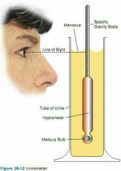
Urinometer |
|
|
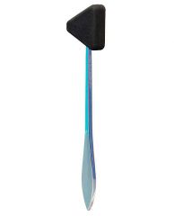
Mallet-sensory |
|
|
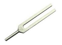
Tuning fork-sensory |
|
|
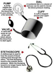
Blood pressure cuff and Stethoscope |
|

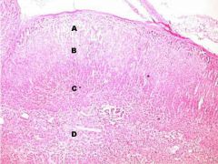
A |
Inspiratory Reserve Volume (IRV) Air forcibly taken in above TV ~3000 ml |
|

B |
Tidal volume (TV) Air volume moved per breath ~500ml |
|

C |
Expiratory reserve volume (ERV) Air forcibly moved out past TV ~1000 ml |
|
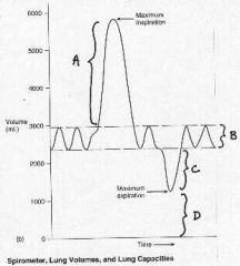
D |
Residual volume (RV) Air that remains in lungs ~500 ml |
|

ABC |
Vital Capacity |
|
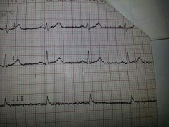
Electrical pathway |
SA node-AV node-Bundles of His-Purkinje fibers |
|

1 |
PR interval Depolarization from SA through AV |
|

2 |
PR segment |
|

6 |
QT interval Ventricular dramatization + repolarization |
|

8 |
T wave Ventricular repolarization |
|

9 |
U wave |
|

10 |
P wave Atrial depolarization |
|
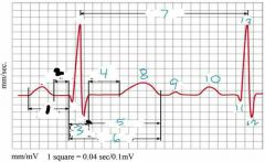
11 |
Q wave |
|

12 |
S wave |
|

13 |
R wave |
|

From beginning of 8 to end of 10 |
TP interval Ventricular diastole |
|

3 |
QRS interval Ventricle depolarization Atrial repolarization (cannot see) |
|
|
Forced Vital Capacity |
Vital capacity when forcibly exhale Healthy=FVC greater or equal to predicted Restrictive pulmonary disease=less than predicted |
|
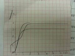
FEV1,2 &3 |
Forced Expiratory Volume Volume forcibly exhaled after 1,2 &3 seconds |
|
|
Pulmonary function equation |
FEV1/FVC×100 80%=healthy Less than 80%= obstructive pulmonary disease |
|
MV |
Minute Volume Volume of air per minute Respiratory rate × tidal volume |
|
|
MVV |
Maximum voluntary ventilation Volume of air maximally moved per minute RR×TV |
|
|
Vital Capacity |
Max air moved through lungs TV+IRV+ERV ~4500 ml |
|
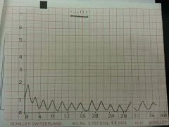
Calculate HR: 3-second method |
Count R peaks in 3 seconds Multiply by 20 (3×20=60sec) |
|
Calculate HR: 6 second method |
Count R peaks in 6 seconds Multiply by 10 |
|
Calculate HR: R-R (most accurate) |
Seconds from one R to another 1 beat/seconds=x beats/60 seconds (1÷seconds×60=BPM) |
|
|
5 Cardinal Rules: Interpretation of Dysrhythms |
1. Rhythm should be regular 2. HR should be 60-100 BPM 3. P-QRS ratio should be one to one 4. P-R interval= 0.12-0.20 seconds 5. QRS interval= 0.049-0.10 seconds |
|
|
Regular Rhythm means |
Time span does not vary between one R-R interval and others |
|
|
When to use 3 or 6 second methods vs R-R methods |
R-R only if regular rhythm 3 or 6 for irregular or regular 60-100 BPM |
|
|
P-QRS ration is one to one if |
For every P wave there is a QRS that follows |
|
|
What is the P-R interval |
Start of P to start of Q Conduction/depol. from SA node through AV node 0.12-0.20 seconds |
|
|
What is the QRS interval |
From start of Q to end of S Conduction from AV node through purkinje fibers Ventricular depolarization Atrial repolarization 0.04-0.10 seconds |
|
|
The PQRST wave represents |
Systole From start of P to end of T |
|
|
Where is diastole on an EKG |
End of T to start of P |
|
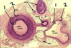
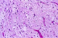
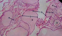
1 |
Tunica media Smooth muscle (w/elastin in arteries) Vasodilation & constriction |
|

2 |
Tunica externa Elastic & collagenous Elasticity of vessel |
|

1 |
Bundle of nerves |
|
2 |
Artery (Thick tunica media) |
|

3 |
Vein |
|
|
Tunica intima |
Endothelium Keeps things moving smoothly (Thinnest) |
|
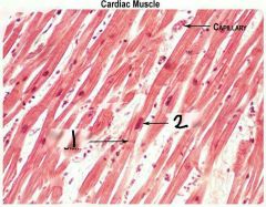
1 |
Intercalated disk (Perpendicular to nucleus) |
|

2 |
Nucleus |
|

1 |
Intercalated disc Connects individual heart muscle cells |
|
2 |
Sarcomere Contractile unit of a myofibril |
|
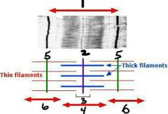
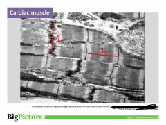
1 |
Sarcomere |
|

2 |
M line |
|

3 (center line & white on both sides) |
H zone |
|

4 |
A band Thick filaments, M line in middle |
|

5 |
Z disc |
|

6 |
I band Thin filaments, z disc in middle |
|
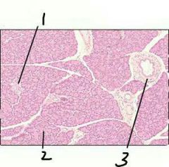
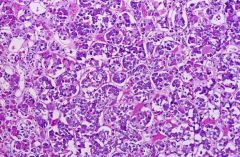
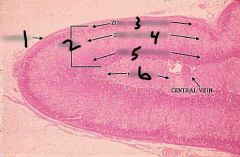
1 |
Islet of Langerhans (pancreas) Alpha cells》glucagon Beta cells》insulin |
|
|
Islets of Langerhans
High blood glucose-Beta cells produce insulin-cells uptake glucose, liver stores glycogen
Low blood glucose-Alpha cells produce glucagon-liver releases glucose |
|

1 |
Anterior pituitary Hypothalamus-portal vein Tropic/regulating hormones 6 (TSH, ACTH, FSH, LH, PRL, GH) |
|
2 |
Posterior Pituitary Hypothalamus-nerves ADH, Oxytocin |
|
|
Anterior pituitary Hypothalamus-portal vein Tropic/regulating hormones |
|
|
Posterior Pituitary Hypothalamus-nerves Oxytocin, ADH High blood osmolarity-osmoreceptor- hypothalamus/post.pituitary/ADH- Kidneys- Reabsorb H2O: less urine formed- Lower blood osmalarity |
|

1 |
Cuboid follicular cells Thyroid hormones T3 and T4 Basal metabolic rate |
|
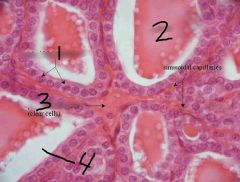
2 |
Colloid (thyroid) |
|

3 |
Parafollicular cells Calcitonin when high calcium (Thyroid) |
|

1 |
Cuboid follicular cell of thyroid Thyroid hormones T3, T4 Basal metabolic rate |
|

2 |
Colloid (thyroid) |
|
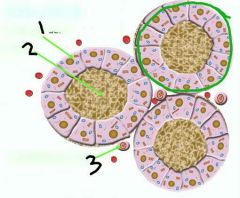
3 |
Parafollicular cells of Thyroid Calcitonin High blood calcium- Thyroid releases calcitonin- Osteoblasts store calcium in bone- Lower blood calcium |
|

Whole unit (circled) |
Thyroid follicle |
|
Which organ is this and what does it help regulate? |
Thyroid Metabolism & blood calcium (lowers Ca) |
|

1 |
Adrenal capsule |
|
2 |
Adrenal cortex Glucocortisoids, mineralcorticoids, androgens |
|

4 |
Zona fasciculata Cortisol-stress |
|

3 |
Zona glomerulosa Aldosterone-blood volume
|
|

5 |
Zona reticularis Sex hormones |
|

6 |
Adrenal medulla Epinephrine & Norepinephrine |
|

A: name gland, zone, hormone and function |
Adrenal gland Zona glomerulosa Aldosterone Low blood volume》inceases sodium absorption》water follows》 Also promotes potassium excretion |
|

B: gland, zone, hormone, purpose? |
Adrenal gland Zona fasciculata Cortisol Up blood blood glucose & liver glycogen, lower inflammation & immune response |
|
C: gland, zone, hormones, purpose? |
Adrenal gland Zona reticularis Androgens Sex hormones |
|

D: gland, zone, hormones, purpose? |
Adrenal gland Adrenal medulla Epinephrine & Norepinephrine Stimulates glucose & glycogen use, release of lipids & adipocytes Increases HR, BP Vasoconstricts BV |
|
Gland and hormones? |
Ovary Estrogen, testosterone and progesterone progesterone |
|
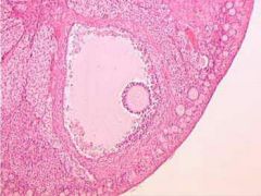
Gland, hormones, and what's that big opening? |
Ovary Estrogen, testosterone, progesterone Graafian follicle (contains fluid) |
|
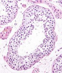
Organ, hormone and structure? |
Testis Testosterone (leydic cells) Seminiferous tubule |
|
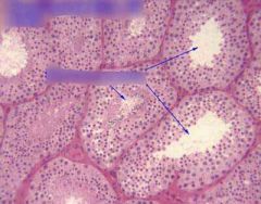
Gland & hormones? |
Testis Testosterone (leydic cells) |
|

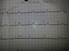
1 |
0.5 mV or 5 mm |
|

4 |
5 mV or 5 mm |
|

2 |
0.04 seconds |
|

3 |
0.2 seconds |
|
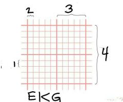
How many large squares equal 1 second? (horizontal) |
5 |
|
|
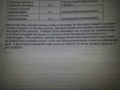
I feel like since this has * on the sheet, we should know it |
|

CNII |
Optic Cranial Nerve-sensory/afferent Carries info into brain Right CNII damaged= Light in Right eye: neither pupils constrict & Light in Left eye: both pupils constrict |
|
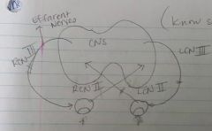
CNIII |
Oculormotor Cranial Nerve-motor/efferent Right CNIII damaged= Light in Right Eye: left eye constricts Light in Left Eye: left eye constricts |
|
|
Rinne Test |
Rinne test: Tuning fork base at mastoid process, when no sound present move fork to front of ear. Then reverse order on ear Conduction deafness: sound by mastoid after air or no sound in air after mastoid |
|
|
Weber Test |
Tuning fork on vertex of skull Conduction deafness: Sounds better in poor ear-simulated by putting cotton in one ear |
|
|
Plantar (Babinski) Reflex |
Test for Upper Motor Neuron lesion, Corticospinal damage Normal Adult: Plantar flexion (downward movement) Normal Infant: Plantar Extension (Big toe upward, toes may fan) |