![]()
![]()
![]()
Use LEFT and RIGHT arrow keys to navigate between flashcards;
Use UP and DOWN arrow keys to flip the card;
H to show hint;
A reads text to speech;
11 Cards in this Set
- Front
- Back
|
What is the Etiology of Pericardial Effusion? |
1) Tuberculosis. |
|
|
What is the pathophysiology of pericardial effusion?
|
1) Pericarditis leads to fluid into the pericardial space. |
|
|
What are the physical signs of Pericardial Effusion?
|
1) Chase pain. C
2) Dyspnea. D 3) Distended neck veins. DNV 4) Friction Rub. FR 5) Pulsus Paradoxus. PP |
|
|
What are the Echo-findings of Pericardial Effusion?
|
1) The pericardium fluid is seen between the epicardium & pericardium.
2)Decrease echo gain to identify pericardium effusion (brightness). |
|
|
What are the Echo-findings of Pericardial Effusion?
|
3) Isolated anterior space may be a pericardial fat path. |
|
|
How many types of Aneurysm are there?
|
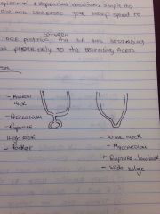
Pseudoaneurysm. True aneurysm |
|
|
What is the etiology of Constrictive Pericarditis?
|
1) Idiopathic. |
|
|
What is the pathophysiology of Constrictive Pericarditis?
|
1) Fibrosis or Calcification of pericardium results in restriction of ventricular filling. |
|
|
What are the physical signs of Constrictive Pericarditis?
|
1) Dyspnea. D
2) Edema. E 3) Distended Neck Veins. DNV 4) Pericardial Nock during Diastole. PNDD |
|
|
What are the echo-findings in Constrictive Pericarditis?
|
1) Normal LV diastolic function. |
|
|
What are the Doppler findings in Pericardial Effusion?
|
1) TR & MR are usually present.
2) Respiration Variation will increase. |