![]()
![]()
![]()
Use LEFT and RIGHT arrow keys to navigate between flashcards;
Use UP and DOWN arrow keys to flip the card;
H to show hint;
A reads text to speech;
22 Cards in this Set
- Front
- Back
|
How is electrophoretic techniques different from chromotagraphic |
Electrophoretic techniques are for accurate and precise analysis of proteins rather than simple seperation. Electrophoretic techniques require denaturation of the protein. Electrophoretic techniques are used to monitor the progress of purification and check for purity. |
|
|
What are the 2 steps of preperation for electrophoresis? |
Breaking of H bonds and VDW by sodium dodecyl sulphate detergent to form micellular bubbles surrounding hydrophobic regions |
|
|
What are the 2 dyes used in SDS page electrophoresis and what are the resolutions?
|
Coomassie Blue stain - 10ng
Silver stain - 1ng |
|
|
What charge is SDS and how is that relevant to how SDS works?
|
SDS is negative charge - moves through solid gel matrix towards anode. |
|
|
Why don't we use silver staining as much? |
Takes a long time to stain, expensive and use of nasty chemicals |
|
|
Why is western blot sometimes used after an SDS-page electrophoresis? |
Can't differentiate and seperate specific protein if there are lots of proteins of similar mass |
|
|
What are the 6 steps for western blot? |
Gel Electrophoresis
Transfer proteins to membrane (nitrocellulose) following gel electrophoresis using electroblotting
Blocking - ensure the membrane and other proteins have zero affinity with the probe (anitbody/dye) so wash with buffer and nonspecific protein (e.g. BSA) to reduce the noise
Probe the blot with specific antibodies
Probe blot with second antibody linked to an enzyme - this amplifies the signal
Add enzyme substrate - chemiluminescent signal |
|
|
What is isoelectric focusing? How does it seperate the proteins? |
electrophoresis based on a pH gradient. Solution of proteins added to a column containing immobilised pH gradient (IPG) gels Proteins move through the gel until they have reached pI |
|
|
What is 2D gel electrophoresis? |
Separation based on both charge and mass - use of isoelectric focussing followed by SDS page gel electrophoresis |
|
|
How do you concentrate the protein after electrophoresis? |
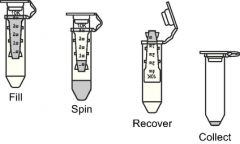
Use a molecular weight cut off filter in a tube - put proteins on top of filter in tube and centrifuge
Solute and buffer (unwanted) filter through the filter and is discarded |
|
|
What are the 2 reasons for concentrating a protein after using an electrophoretic technique?
|
To do different assays
For repeatability |
|
|
What is the concentration required for crytallisation? For EM? |
10 mg/ml for Crystallisation 0.1mg/ml for EM |
|
|
Lowry Assay is a way of measuring total _ _ _ _ _ _ _ concentration & relies on the _ _ _ _ _ _ _ _ _ of _ _^_ _ ions in Lowry reagent (phospho_ _ _ _ _ _ _ _ - _ _ _ _ _ _ _ _ acid) by peptide bonds, causing the _ _ _ _ _ _ _ _ _ of aromatic residues resulting in the _ _ _ _ _ _ _ _ _ of _ _ ^_ _ to _ _ ^ _ _ The colour changes are then detected in a visible light spectro photometer |
Protein Chelation |
|
|
Reduction of Lowry reagent results in a solution that has a _ _ _ _ colour, and absorbance can be measured at the _ _ _nm wavelength
The concentration of protein in solution can then be found by looking at a standard curve. |
Blue 760 |
|
|
What are the possible assays once you have purified the protein and checked it? (3) |
Amino acid composition Primary sequence of protein Structure of Protein |
|
|
What are the 3 steps involved in degrading a protein non-sequentially find the amino acid composition? |
Dissolve protein in 6M HCl and put solution in an evacuated ampoule (a glass capsule) Heat to 378-383K for 24 hours Separate using cation exchange column |
|
|
What is the downside of harsh hydrolysis of proteins? How is this resolved?
|
Trp, Ser, Tyr, Thr destroyed. by hydrolysis
Tyr, Thr, Ser protected by addition of phenol or thiol Trp can be detected by A280 |
|
|
What is the cation exchange column usually made of? What properties enables it to separate amino acids?
|
Made of sulphonated polystyrenes Separates amino acids in 2 ways: 1. negatively charged 2. The polystyrene is hydrophobic |
|
|
How do you find out the amount of amino acid in a protein after electrophoresis and purification without using fluorescence? |
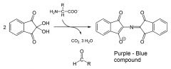
Ninhydrin reaction Add ninhydrin and heat to 373K To form purple compound |
|
|
What is an alternative to ninhydrin?
|
Phthalaldehyde and Beta mercaptoethanol - reacts with AA to give fluorescent complex (isoindole derivative of amino acid)
|
|
|
What are the 2 steps (each step has 1 requirement) for breaking down a disulphide linkage, and what is the product? |
Reductive alkylation Using beta-mercaptoethanol Requires excess thiol (drive equilibrium forward) Acetylation using iodoacetate Requires iodoacetic acid excess Produces acetylated cysteine residues |
|
|
How to separate the following using ion exchange chromatography?
V-S-L-A-G D-E-G-K-P-R |
Both have net 0 charge at physiological pH Run the chromatography at pH 1.9 At that pH V-S-L-A-G is +1 and D-E-G-K-P-R is +3 as COO- becomes neutralised |