Reading...
![]()
Play button
![]()
Play button
![]()
Use LEFT and RIGHT arrow keys to navigate between flashcards;
Use UP and DOWN arrow keys to flip the card;
H to show hint;
A reads text to speech;
84 Cards in this Set
- Front
- Back
|
What are cells composed of?
|
Mainly water (85%) & protein
A smaller fraction consists of lipids, carbohydrates, trace elements, and ions |
|
|
What roles do the lesser constituents of the cell play?
|
Catalysts & co-factors in the activity of enzymes, in transport systems, & in maintenance of osmotic and pH homeostasis
|
|
|
Basal lamina
|
Amorphous outer cell coating;
partially surrounding many/all cells; important for survival and for regeneration of cells following injury |
|
|
How big is the intercellular space?
|
100-200 Angstroms
|
|
|
What force maintains the intercellular space?
|
Electrostatic forces of repulsion
|
|
|
What are the constituents of the intercellular space?
|
A watery gel of protein, carbohydrates, & extracellular fluid
|
|
|
Types of Junctions between cells
|
Gap junctions
Desmosomes (macula adherens) Occluding junctions |
|
|
Gap junctions
|
clusters of protein channels & microfilaments that allow direct and rapid exchange of small ions and molecules between cells
|
|
|
Occluding junctions
|
Located where the plasma membranes of adjacent cells fuse
|
|
|
What are the general roles of junctions?
|
To regulate intercellular communication & fluid movement between cells; anchor cells together to create the stable organized structure of tissues
|
|
|
Eosin
|
A standard cytology dye; turns cells pink
|
|
|
Are cells basic or acidic?
|
Slightly basic and can retain acid dyes ("acidophilia")
|
|
|
Mitochondrial enzymes include:
|
oxidases, reductases, and dehydrogenases, as well as many enzymes involved in the Krebs cycle and fatty acid breakdown
|
|
|
Synonym for smooth endoplasmic reticulum (E.R.)
|
Agranular endoplasmic reticulum (E.R.)
|
|
|
Where are mixed function oxidases (ie. CYP 450) mainly located?
|
Smooth ER
|
|
|
Which cells have a large quantity of SER?
|
Hepatocytes, hormone secreting cells of testis and ovary, and cortical cells of the adrenal
|
|
|
Which ER is involved in metabolic degradation of drugs, hormones, and steroid metabolism?
|
Smooth ER
|
|
|
Which ER is involved in protein synthesis?
|
Rough ER because of its attached ribosomes
|
|
|
Protein synthesis occurs in:
|
ribosomes attached to ER, clusters of unattached ribosomes, polyribosomal granules (polysomes)
|
|
|
How do SER and RER adjust to the demands of the cell?
|
They can rapidly increase/decrease in abundance.
|
|
|
Roles of the Golgi complex:
|
package proteins for export from cell, aggregate and secrete products synthesized by SER & RER, glycoproteins and lipoproteins are formed here, many posttranslational modifications occurs here
|
|
|
What is a lysosome?
|
Vesicles containing hydrolytic enzymes that digest effete organelles & foreign particles, ie. bacteria.
|
|
|
What are the different lysosomes?
|
Primary lysosomes are poised to respond but have not yet been called to respond. Secondary lysosomes are fused with organelles and are actively digesting them. These are called cytosomes & cytosegresomes
|
|
|
3 lysosomal storage diseases associated with deficiency conditions:
|
Tay-Sachs disease (hexosaminidase A deficiency)
Gaucher's disease (glucocerebrosidase deficiency), Niemann-Pick disease (sphingomyelinase deficiency) |
|
|
What is a lethal lysosomal malfunction?
|
When lysosomes leak out of the cell.
|
|
|
2 Intracellular degradation systems & mechanisms
|
Lysosomal degradation system- acid hydrolases
Proteasomal degradation systems- proteases |
|
|
Residual bodies & lipofuscin
|
debris from destroyed targets of lysosomes contain a brown pigment termed lipofuscin
|
|
|
Parkinson's disease pathology
|
Abnormal proteosomal degradation of neurons
|
|
|
Ubiquitin
|
A molecular "tag" for a protein that needs to be degraded in the proteasome
|
|
|
Diameters of: Microfilaments
Intermediate filaments Microtubules |
Microfilaments - 5 nm
Intermediate filaments - 10 nm Microtubules - 22 nm |
|
|
Proteins in: Microfilaments
Intermediate filaments Microtubules |
Microfilaments: actin, myosin
Intermediate filaments: epithelia-keratins, mesenchyma-vimentin, muscle-desmin & troponin, glia-GFAP, nerve-neurofilament Microtubules: tubulin |
|
|
Functions of: Microfilaments
Intermediate filaments Microtubules |
Microfilaments- contraction, membrane reinforcement, cell scaffold
Intermediate filaments-structure, support Microtubules-structure, transport, cell division, motility |
|
|
The mitotic spindle is composed of which cytoskeletal filament?
|
Microtubules
|
|
|
The protein in epithelial intermediate filaments is
|
Keratins
|
|
|
The protein in mesenchymal intermediate filaments is
|
Vimentin
|
|
|
The 2 proteins in muscle intermediate filaments is
|
Desmin & troponin
|
|
|
The protein in nerve intermediate filaments is
|
Neurofilament
|
|
|
Is the nucleus acidic/basic?
|
The nucleus is acidic and stains with basic dyes "basophilic"
|
|
|
Hematoxylin
|
Basic dye to stain nucleus
|
|
|
Barr body
|
Inactive X chromosome; dense clump of chromatin attached to the inner surface of the nuclear membrane
|
|
|
Heterochromatin vs. Euchromatin
|
Heterochromatin-highly condensed & inactive transcription
Euchromatin-not condensed and actively transcriped |
|
|
The protein in glial cell intermediate filaments is
|
Glial fibrillary astrocytic protein (GFAP)
|
|
|
In what percentage of a female's cells contain a Barr body?
|
50%
|
|
|
What term means "irregular shrinkage and wrinkling of the nucleus?"
|
Pyknosis
|
|
|
What term means "fragmentation of the nucleus?"
|
Karyorrhexis
|
|
|
What term means " decrease in basophilia?"
|
"Fading"
|
|
|
In what percentage of a female's cells contain a Barr body?
|
50%
|
|
|
What term means "irregular shrinkage and wrinkling of the nucleus?"
|
Pyknosis
|
|
|
What term means "fragmentation of the nucleus?"
|
Karyorrhexis
|
|
|
What term means " decrease in basophilia?"
|
Karyolysis or nuclear fading
|
|
|
What are the 3 different nuclear changes? What do they indicate?
|
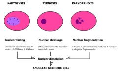
Karyolysis, Pyknosis, & Karyorrhexis, indicate cell death
|
|
|
Lesions of reversible injury is called
|
Degeneration
|
|
|
When can you see evidence of cell death under a microscope?
|
A few hours after the injured cell has actually died.
|
|
|
Adaptation of the liver cell in the chronic user of barbiturates
|
SER's hydroxylating enzymes detoxify the barbiturates, oxidative demethylation of the drug; high levels of drug induce SER synthesis of the enzyme; greater ability to detoxify; and drug tolerance ensues
|
|
|
Hypertrophy
|
Cell enlargement in size due to increased work demand
|
|
|
"Permanent" cells
|
Have lost their ability to divide and will hypertrophy in response to increased work loads
|
|
|
When a cell enlarges in size but not number, what happens to the mitochondria & nuclei?
|
Nuclei size is enlarged, mitochondria enlarges in size and number.
|
|
|
Decompensation
|
Patients w/ high b.p. & cardiac enlargement develop heart failure b/c reached maximum enlargement
|
|
|
Hyperplasia
|
Cells increase in number due to increased work demand; may or may not be controlled cell proliferation
|
|
|
Do the cells that undergo hyperplasia have the ability to undergo mitosis?
|
Yes, that explains why they increase in number.
|
|
|
Labile cells
|
Cells that undergo continuous renewal through mitosis (bone marrow, skin, gastrointestinal epithelia)
|
|
|
Atrophy
|
Decrease in cell size due to decreased work load, disuse (disuse atrophy), diminished blood supply (vascular atrophy) or loss of endocrine stimulation (endocrine atrophy)
|
|
|
Can a cell survive after atrophy?
|
Yes, but at a lower level of function
|
|
|
True or False?
Small organ size = Atrophy |
False, b/c it could also be hypoplasia, apalasia or agenesis
|
|
|
Hypoplasia
|
Developmental failure to achieve full size because of below normal number of cells. Less drastic than aplasia.
|
|
|
Aplasia & Agenesis
|
Total failure of the structure to develop
|
|
|
Do the hepatocytes of a normal uninjured adult liver undergo mitosis?
|
Rarely; liver cells usually live as long as its human host; it only mitoses if there's damage
|
|
|
Types of Hyperplasia
|
Physiologic hyperplasia
Endocrine hyperplasia Compensatory hyperplasia Pathologic hyperplasia |
|
|
Reparative proliferation
|
Controlled cell proliferation to replace lost cells
|
|
|
Physiologic hyperplasia
|
ie. Hyperplasia in the glandular epithelium of breast @ puberty, pregnancy, & lactation
|
|
|
Compensatory hyperplasia
|
ie. Enlargement of one kidney when the other is destroyed or removed
|
|
|
Endocrine Hyperplasia
|
ie. Postmenstrual regrowth of the uterine endometrium during a woman's reproductive life (also reparative proliferation)
|
|
|
Pathologic hyperplasias
|
many induced by excessive hormonal stimulation of target cells (ie. abnormal endometrial hyperplasia produced by excessive estrogen stimulation in pts with ovarian tumors); may or may not lead to uncontrolled neoplastic proliferation
|
|
|
Examples of controlled cell proliferation -> uncontrolled neoplastic proliferation
|
Endometrial hyperplasia -> endometrial cancer
|
|
|
Metaplasia
|
Adaptive substitution of one mature type of adult cell for another mature cell type more capable of meeting the stress; usu cell regresses in specializaton
|
|
|
What occurs in metaplasia of epithelial tissue? Is it reversible?
|
Columnar mucus-secreting cells replaced by stratified squamous epithelial cells, ie. in gallbladder, trachea, bronchi, etc. Yes, almost always reversible
|
|
|
What occurs in metaplasia of connective tissue?
|
Injury to soft tissue causes fibroblasts to be turned into osteoblasts, and bone may be formed.
|
|
|
What is myositis ossificans and is it reversible?
|
Muscle becoming bone is usu irreversible
|
|
|
Dysplasia
|
Most severe and disorderly non-neoplastic transformation in epithelia; loss of regularity in cells
|
|
|
Is dysplasia reversible?
|
Yes
|
|
|
True or false?
The more frequent mitoses occur, the higher the chances of cells to deter from normal homeostatis. |
Believed to be true because there's more chances for mutation
|
|
|
Atypical metaplasia
|
Bridge between the orderly patterns of metaplasia and disorderly patterns of dysplasia
|
|
|
Greek root for plasia
|
Plasis=shape, forming, molding
|
|
|
What does Vitamin A deficiency cause?
|
Epithelial metaplasia leading to keratinizing stratified squamous epithelium in the respiratory passages and the renal calyces and pelvis.
|