Reading...
![]()
Play button
![]()
Play button
![]()
Use LEFT and RIGHT arrow keys to navigate between flashcards;
Use UP and DOWN arrow keys to flip the card;
H to show hint;
A reads text to speech;
69 Cards in this Set
- Front
- Back
|
What is necrosis?
|
Death of large groups of cells followed by acute inflammation.
|
|
|
Necrosis is due to some underlying _________ process, it is never _________.
|
pathologic; physiologic
|
|
|
What is the morphologic hallmark of cell death?
|
Loss of the nucleus, which occurs via nuclear condensation (pyknosis), fragmentation (karyorrhexis), and dissolution (karyolysis).
|
|
|
The two mechanisms of cell death are ________ and ________.
|
apoptosis; necrosis
|
|
|
What are the patterns of necrosis?
|
(1) Coagulative necrosis
(2) Liquefactive necrosis (3) Gangrenous necrosis (4) Caseous necrosis (5) Fat necrosis (6) Fibrinoid necrosis |
|
|
A 67-yo man has had sharp chest pain not relieved by nitroglycerin for the last three hours. What do you expect to see on a CBC?
|
MI. Necrosis is followed by acute inflammation, which increases the white blood cell count.
|
|
|
What is coagulative necrosis?
|
Necrotic tissue that remains firm. The cellular proteins "coagulate", so that the cells remain their shape and the organ structure is preserved (because of denatured enzymes). However, the nucleus has disappeared.
|
|
|
Coagulative necrosis is characteristic of ischemic infarction of any organ except _________.
|
brain
|
|
|
An area of infarcted tissue is often _____ shaped and ______.
|
wedge; pale
Because, as vessels feed into an organ, they dichotomously branch and distribute blood to an area that looks wedge shaped. The wedge points to the area of occlusion. |
|
|
What is red infarction?
|
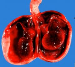
It's a type of infarction (other being pale) that occurs if blood re-enters a loosely organized tissue (so it can hold the blood, giving it a red color).
The picture shows a testicle that has undergone red infarction (e.g. testicular torsion). |
|
|
In cryptorchidism, there is an increased risk for the undescended testis to undergo __________.
|
testicular torsion
|
|
|
The treatment of testicular torsion is ___________. It can be performed as early as ___________ and should be completed by the age of ______.
|
orchiopexy; 6 months; 2 years
|
|
|
The MCC of testicular torsion is ____________ or ___________.
|
violent movement; physical trauma
|
|
|
Regarding torsion of the testicle, the majority occur between ________ years and ________ years old.
|
12; 18
|
|
|
A 15 year old boy comes in to the ER with a chief complaint of a "really bad pain" in his right testicle. It occurred suddenly. As a competent doctor, how would you confirm the diagnosis?
|
Diagnosis is by ultrasound
|
|
|
A 14 year old boy from the rural countryside comes in to your office complaining of a strong pain in his left testicle that has lasted for 10 hours. What should you do?
|
Confirm diagnosis with ultrasound. If torsion, perform surgery, it is imperative if the torsion has not remitted (which 1/3 spontaneously do) within 12 hours.
|
|
|
You note that the scrotum does not retract when you stroke the inner thigh of a 16 year old boy with a tongue blade. What is this called?
|
Absent cremasteric reflex, seen in testicular torsion.
|
|
|
What is liquefactive necrosis?
|
A type of necrosis where the necrotic tissue becomes liquefied. Enzymatic lysis of cells and proteins results in liquefaction.
|
|
|
Liquefactive necrosis is seen in three circumstances. What are they?
|
(1) Brain infarction
(2) Abscess (walled off area of dead tissue) (3) Pancreatitis • We activate the enzymes of the pancreas within the pancreas itself. |
|
|
Why do we see liquefactive necrosis in the brain?
|
Microglial cells (the "macrophages of the brain") release enzymes that digest the tissue after it has died.
|
|
|
What is gangrenous necrosis? What is it characteristic of?
|
It's not a specific pattern of necrosis... it is coagulative necrosis that resembles mummified tissue. It is characteristic of necrosis of the lower limb and GI tract.
|
|
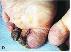
What is this?
|
Gangrenous (dry) necrosis. Seen often in diabetics who are often subject to ischemic necrosis of the lower limb.
|
|
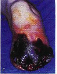
What is this?
|
Wet gangrene. Gangrene with superimposed infection.
|
|
|
Another word for Buerger's disease?
|
Thromboangiitis obliterans.
|
|
|
What is Buerger's disease?
|
A medium-sized vessel vasculitis with digital thrombosis.
|
|
|
What is the classic type of patients seen in Buerger's disease?
|
Men 25-5o years old who smoke cigarettes.
|
|
|
A 30 year old man with a smoking habit comes in to your office. He says he has lost sensation in his toes and fingers. What should you suspect?
|
Buerger's. It can damage neurons as well.
|
|
|
In contrast to the vascular insufficiency caused by atherosclerosis, in Buerger disease the insufficiency tends to be accompanied by _________, even at ________, related undoubtedly to the _________involvement.
|
severe pain; rest; neural
|
|
|
Often seen in Buerger's disease is pain in the ________ of the ________, brought on by exercise.
|
instep; foot
|
|
|
Regarding thromboangiitis obliterans, in addition to pain, ulceration and possible gangrene with loss of sensation of digits, _______________ may be seen.
|
Raynaud's phenomenon
|
|
|
How is Raynaud's phenomenon similar to thromboangiitis obliterans?
|
It's a medium sized vasculitis involving digital vessels in fingers and toes. May also affect tip of nose and ears in some cases.
|
|
|
Treatment of Buerger's disease.
|
Smoking cessation is essential. IV iloprost (prostaglandin analogue) may be given. Aspirin.
|
|
|
Raynaud's disease is classically seen in what patient group?
|
Young women
|
|
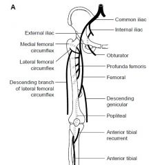
What artery would rank third as a preferred site of involvement in atherosclerosis?
|
Popliteal artery.
1. Abdominal aorta 2. Coronary artery 3. Popliteal 4. Internal carotid artery |
|
|
What is caseous necrosis?
|
It's basically liquefactive necrosis with something mixed in (like TB or fungus). Is in reality a combination of coagulative and liquefactive necrosis. Characterized by a soft, friable necrotic tissue with "cottage cheese-like" appearance.
|
|
|
How is the caseous material in caseous necrosis formed?
|
Caseous material is formed by the release of lipid from the cell walls of M. tuberculosis and systemic fungi (e.g.. Histoplasma) after immune destruction by macrophages.
|
|
|
What is fat necrosis?
|
Necrosis of adipose tissue. It has a chalky-white appearance due to deposition of calcium. When fat dies, FAs are released, they have the ability to bind with calcium (this is saponification).
|
|
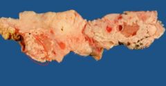
What is this?
|
Fat necrosis with white chalky deposits.
|
|
|
Fat necrosis is characteristic of what?
|
(1) Trauma to fat (e.g., breast)
(2) Pancreatitis-mediated damage of peripancreatic fat. |
|
|
A woman comes in with complaints of a breast mass. The skin is retracted. A biopsy is performed and shows giant cells, fat and calcification. What do you suspect?
|
Fat necrosis due to trauma. Skin retraction can be seen in this situation and may mimic cancer.
|
|
|
The most common cause of breast pain is ____________.
|
fibrocystic change
|
|
|
The mean age of breast cancer is _______.
|
64 years old
|
|
|
What other pathology of the breast may contain calcifications in addition to fat necrosis?
|
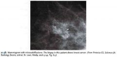
Ductal carcinoma in situ. Commonly contains microcalcifications. In fact, it cannot be detected by mammogram unless it contains microcalcifications. A third eventually invade.
|
|
|
Provide an example of saponification.
|
Dystrophic calcification. A dead or dying tissue becomes a nidus for calcium to deposit.
|
|
|
What is the serum calcium and serum phosphate in cases of dystrophic calcification?
|
Both normal
|
|
|
Provide examples of dystrophic calcification.
|

The cancer outgrows its blood supply, it dies and calcification occurs.
|
|
|
In metastatic calcification, the serum ________ or the serum _________ is high.
|
calcium; phosphate
|
|
|
Provide five causes of hypercalcemia.
|
(1) Malignancy-induced.
(2) Primary hyperparathyroidism (3) Hyperthyroidism (4) Vitamin D3 excess (5) Paget's disease |
|
|
What is a cause of hyperphosphatemia?
|
Hypoparathyroidism
|
|
|
Provide two examples of metastatic calcification.
|
(1) Massive calcification of renal tubular basement membranes in the collecting tlucls (nephrocalcinosis). This can produce nephrogenic diabetes insipidus and renal failure.
(2) Basal ganglia calcification in hypoparathyroidism. |
|
|
What is fibrinoid necrosis? How would it appear on histology?
|
Necrotic damage to blood vessel wall. It would appear pink due to leakage of proteins into the vessel wall.
|
|
|
Fibrinoid necrosis is characteristic of what?
|
(1) Malignant HTN
(2) Vasculitis |
|
|
Malignant HTN is defined as a BP of ___________.
|
(equal to or more than) 210/120 mm Hg
|
|
|
A pregnant woman in her third trimester presents with HTN of 150/90 and a proteinuria in the nephrotic range. In what vessels would you expect to see fibrinoid necrosis?
|
Placental vessels
|
|
|
What is apoptosis?
|
Energy-dependent, genetically programmed cell death. It involves single cells or small groups of cells.
|
|
|
Provide three examples of apoptosis.
|
(1) Endometrial cell breakdown after withdrawal of estrogen and progeslerone in the menstrual cycle (hormone-dependent atrophy). Decreased hormonal stimulation inactivates Bcl2.
(2) Loss of miillerian structures in a male fetus due to Sertoli cell synthesis of mullerian inhibitory factor (3) Removal of virally infected cells by CD8+'s |
|
|
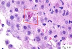
What is the morphology of apoptotic cells?
|
(1) The cell shrinks, and the cytoplasm becomes concentrated within the cell and becomes pink.
(2) Nucleus condenses and fragments. (3) Apoptotic body will fall from the cell and removed by macrophages. |
|
|
In apoptosis we see ________ (inflammation/no inflammation).
|
no inflammation
|
|
|
Apoptosis is mediated by ___________.
|
caspases
|
|
|
Corticosteroid destruction of B- and T-cells occur via _______.
|
apoptosis
|
|
|
Caspases activate ____________ and _____________.
|
proteases (degrades cytoskeleton, cell shrink) and endonucleases (break down DNA, nucleus shrinks)
|
|
|
What pathways activate caspases?
|
(1) Intrinsic mitochondrial pathway. Cellular injury, DNA dmg. Cytochrome c mediates this pathway.
(2) Extrinsic receptor-ligand pathway. FAS ligand bind FAS death receptor. TNF can bind TNF receptor as well. (3) Cytotoxic CD8+ pathway. Perforins and granzymes (activates caspases). |
|
|
Bcl2 __________ (stabilizes/destabilizes) the mitochondrial membrane.
|
stabilizes
|
|
|
FAS death receptor is also known as?
|
CD95
|
|
|
The BCL2 gene family is located on chromosome _____.
|
18
|
|
|
BCL2 gene family manufactures products that ___________.
|
inhibit apoptosis
|
|
|
TP53 suppressor gene __________ the cell cycle in the ____ phase to repair DNA damage.
|
arrests; G1
|
|
|
BAX gene products _______________ (activate/inactivate) the BCL2 antiapoptosis gene.
|
inactivate
|
|
|
Mammalian cells have developed protection to hypoxia. The best defined is ___________ which enhances what?
|
hypoxia-inducible factor-1; new blood vessel formation, enhanced anaerobic glycolysis
|