Reading...
![]()
Play button
![]()
Play button
![]()
Use LEFT and RIGHT arrow keys to navigate between flashcards;
Use UP and DOWN arrow keys to flip the card;
H to show hint;
A reads text to speech;
84 Cards in this Set
- Front
- Back
|
What is the first step of primary hemostasis?
|
Transient vasoconstriction of damaged vessel
|
|
|
What mediates initial vasoconstriction of vessel?
|
neural stimulation and ENDOTHELIN
|
|
|
Where does vWF come from?
|
Platelets and endothelial cells
Weibel-Palade bodies in endothelial cells Alpha granules of Platelets Weibel-Palade bodies have P-selectin speedbumps and vWF |
|
|
Describe what causes platelet degranulation...
|
adhesion of platelets to vWF cause shape change and subsequent degranulation with release of mediators.
ADP is released from dense granules which promotes exposure of GPIIb/IIIa receptors on platelets which is important for AGGREGATION. Also causes TXA2 to be synthesized by COX which also helps aggregation. |
|
|
Where is ADP stored in platelet?
|
dense granules
|
|
|
What is aggregation? how is it mediated?
|
Platelet aggregation is mediated with GPIIb/IIIa receptors that were upregulated in response to ADP release from platelet dense granules
Also, Thromboxane A2, synthesized by platelet cyclooxygenase and released, which promotes aggregation. |
|
|
What is adhesion? how is it mediated?
|
Platelet adhesion is mediated with vWF on basement membrane and GPIb on platelets
|
|
|
What is a generally true statement about QUANTITATIVE platelet reductions as opposed to QUALITATITVE platelet reductions?
|
quantitative reductions result in petechiae
|
|
|
What is a bone marrow biopsy used to assess?
|
Megakaryocytes, which produce platelets
|
|
|
Immune Thrombocytopenia caused by...
|
Antibodies binding to GPIIb/IIIa receptors on platelets. These antibodies are created in the spleen and the Ab-platelet complex is also consumed by macrophages in the spleen.
|
|
|
petechiae presents in child weeks after a viral infection or immunization
|
Immune Thrombocytopenia, is usually self-limited.
Antibodies bind to GPIIb/IIIa receptors on platelets |
|
|
ITP is A/W
|
SLE, Lupus. Since in Systemic lupus they can have Antibodies to almost anything, RBC's, WBC's or...PLATELETS.
|
|
|
Women with chronic ITP can have what complication...
|
During pregnancy, since IgG can cross membrane, Infant can have transient ITP until IgG's are consumed.
|
|
|
ITP lab findings
|
low platelet(<50,000)
PT and PTT normal Increase in Megakaryocytes to compensate for decrease in platelets |
|
|
One treatment for ITP is IVIG. Describe mechanism...
|
When you give IVIG, you throw in so many extra platelets that your hoping the spleen will want to eat up these IgG's instead of the one's that are attached to your platelets to cause a temporary increase in platelets. This effect is SHORT lived
|
|
|
Splenectomy does what in context of ITP
|
eliminates the PRIMARY SOURCE of antibody AND the site of platelet destruction
|
|
|
Microangiopathic Hemolytic Anemia
|
Microthrombi in vessels shear RBC's into schistocytes resulting in hemolytic anemia.
|
|
|
Thrombotic Thrombocytopenic Perpura is A/W what gene product?
|
ADAMTS13(von Willebrand factor-cleaving protease)
|
|
|
TTP Pathophys
|
ADAMTS13 normally cleaves vWF multimers. ADAMTS13 is reduced in TTP and thus the multimers persist and cause abnormal platelet adhesion resulting in microthrombi.
|
|
|
Common cause of TTP
|
Decreased ADAMTS13(von Willebrand factor-cleaving protease) is normally due to an ACQUIRED antibody that destroys ADAMTS13, commonly seen in adult women.
|
|
|
HUS A/W
|
Enterotoxigenic E.Coli(O157:H7) produces verotoxin that damages endothelial cells, particularly in kidney/brain, and that creates microthrombi which shear red blood cells.
|
|
|
undercooked beef, possibly in children
|
ETEC O157:H7
|
|
|
Most common clinical findings in HUS
|
Renal insufficiency - thrombi in vessels of kidney
|
|
|
Most common clinical findings in TTP
|
CNS abnormalities - thrombi in vessels of CNS
|
|
|
Lab findings in TTP/HUS
|
Thrombocytopenia with INCREASED bleed time
NORMAL PT/PTT Anemia with Schistocytes Increased megakaryoctes as a compensatory response(hyperplasia) |
|
|
Bernard-Soulier syndrome
|
genetic GPIB deficiency; platelet ADHESION impaired
blood smear shows thrombocytopenia with enlarged platelets. enlarged because they are slightly more immature |
|
|
Glanzmann's Thrombasthenia
|
genetic GPIIB/IIIA deficiency; platelet AGGREGATION is impaired
|
|
|
Aspirin MOA
|
blocks COX irreversibly. Blocks production of TXA2 which impairs AGGREGATION. Important chemoattractant for other platelets.
|
|
|
Uremia. How does it affect clot formation?
|
Uremia is a buildup of nitrogenous waste products due to poor kidney function - both adhesion and aggregation are impaired
|
|
|
what happens when fibrin is cross linked?
|
stable clot formed...
|
|
|
What three things are required to activate factors of coagulation cascade?
|
1.) Phospholipid surface - provided by platelets
2.) Activating substance - Tissue thromboplastin/Tissue Factor activates Factor VII or Subendothelial collagen activates factor XII. 3.) Calcium |
|
|
defective Secondary hemostasis clinical symptoms
|
deep bleeding, rebleeding
|
|
|
PT
|
extrinsic
|
|
|
PTT
|
intrinsic
|
|
|
Hemophilia A
genetic inheritance? |
Hemophilia EIGHT
X-linked recessive Factor VIII deficiency New mutation common |
|
|
describe a Mixing study and what it's used for
|
Mixing study used to distinguish between Hemophilia A and Coagulation Factor Inhibitor(antibody against Coagualation Factor)
Normal Plasma + Patient Plasma will yield normal bleed time if Hemophilia A. If CFI, then circulating Ab blocks factor VIII again(party pooper) |
|
|
Most common inherited coagulation disorder
|
Genetic deficiency of vWF
|
|
|
what secondary quality does vWF have? Relevance?
|
stabilizes factor VIII. As such, in vWF deficiencies, PTT goes up.
|
|
|
Ristocetin
|
Ristocetin causes platelets to bind to vWF via their GPIb receptor.
A test is abnormal in a vWF deficiency since the platelets cannot bind, which causes lack of adhesion/aggregation |
|
|
Desmopressin
|
increases release of vWF from Weibel Palade bodies in endothelial cells.
|
|
|
what is the physiological function of Vitamin K?
|
allows gamma-carboxylation of 2,7,9,10, Protein C and Protein S.
|
|
|
MOA of Warfarin/Coumadin
|
Blocks epoxide reductase in liver, which activates vitamin K which then gamma carboxylates the necessary coagulation factors(2,7,9,10,C,S) to allow their normal function.
If epoxide reductase blocked, vitamin K cannot be activated and PT goes up. |
|
|
How do you follow liver function?
|
Check PT
|
|
|
Large volume transfusion?
|
Causes a relative dilution of coagulation factors
|
|
|
How does liver function affect coagulation cascade?
|
--Liver synthesizes most of the coagulation factors.
--It is also the main site of epoxide reductase, which activates vitamin K. If your liver fails, then not only can you not MAKE the factors, but you can't activate Vit K to make the factors functional via Vit K dependent gamma carboxylation. |
|
|
Fat malabsorption
|
Bad for vitamin absorption, particularly fat soluble ones
Vitamins D, A, K, E |
|
|
Dense core granules of platelets contain?
|
ATP, calcium and serotonin
|
|
|
alpha granules of platelets contain?
|
everything else except calcium, serotonin and ATP
vWF, fibrinogen, coagulation factors V and XIII, fibronectin, platelet factor 4 |
|
|
Most common coagulation factor inhibitor?
|
Factor VIII
|
|
|
What complication do you give vitamin K to newborns to prevent?
|
hemorrhagic disease of the newborn
|
|
|
Heparin induced thrombocytopenia
|
Heparin causes a decrease in platelet count because of the formation of a complex with platelet factor 4
IgG antibodies against platelet-heparin complex that the spleen proceeds to destroy |
|
|
Platelet factor 4 stored in what granule in platelet?
|
alpha granule
|
|
|
Cause of DIC in pregnancy
|
TF(thromboplastin) in amniotic fluid activates coagulation
|
|
|
Acute Promyelocytic Leukemia
|
Contain auer rods which can enter circulation and cause DIC
|
|
|
Rattlesnake bite
|
venom enters circulation causing DIC
|
|
|
Lab findings of DIC
|
decreased platelets
increased PT/PTT decreased fibrinogen Microangiopathic Hemolytic Anemia(Schistocytes) Elevated fibrin split products(D-dimer) D-dimer is an indicator of lysed cross-linked fibrin. Elevated D-dimer is the BEST test for DIC. |
|
|
What hallmark lab finding is found in DIC?
|
elevated D-dimer
|
|
|
tPA
|
converts plasminogen to plasmin
plasmin cleaves cross-linked fibrin(clot), fibrinogen(ability to form new clots), destroys coagulation factors, and blocks platelet aggregation produced by endothelial cells |
|
|
What inactivates plasmin?
Where is this protein produced? |
alpha-2-antiplasmin.
Liver |
|
|
What diseases are a/w overactivity of plasmin
|
radical prostatectomy - release of urokinase activates plasmin
liver cirrhosis - reduced production of alpha-2-antiplasmin |
|
|
Lab findings of disordered fibrinolysis. What lab value is normal?
|
-- increased PT/PTT
-- increased bleeding time--since plasmin blocks platelet aggregation -- increased fibrinogen split products WITHOUT D-dimers. -- NORMAL platelet count |
|
|
blocks activation of plasminogen
|
aminocaproic acid
|
|
|
Features distinguishing from post-mortem clot
|
1.) Lines of Zahn
2.) attachment to vessel wall |
|
|
Thromboxane A2
|
it stimulates activation of new platelets as well as increases platelet aggregation
produced by platelets |
|
|
Prostoglandin I2
|
protects against thrombosis, blocks platelet aggregation
produced by endothelial cells |
|
|
Heparin like molecules
|
augments anti-thrombin III which inactivates thrombin and coagulation factors(notably factor Xa)
produced by endothelial cells |
|
|
thrombomodulin
|
Modulates activity of thrombin. Thrombomodulin redirects activity of thrombin from activating fibrinogen to fibrin towards activating Protein S to Protein C which proceeds to inactivate factors V and VIII
|
|
|
increase of homocysteine does what?
|
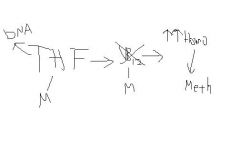
damages endothelium. This can be caused by Vitamin B12 deficiency OR a folate deficiency(THF)
can also be caused by Cystathione Beta Synthase deficiency which leads to vessel thrombosis, mental retardation, lens dislocation and long slender fingers. |
|
|
What causes high levels of homocysteine?
|
Vitamin B12 or folate deficiency
or Cystathionine Beta Synthase(CBS) CBS converts homocysteine to cystathione; enzyme deficiency leads to buildup of homocysteine |
|
|
Vessel thrombosis, mental retardation, lens dislocation and long slender fingers(arachnodactyly)
|
Cystathionine Beta Synthase clinical symptoms
high serum homocysteine causes vessel thrombosis. |
|
|
recurrent DVT's in children
|
hypercoagulable state
|
|
|
Protein C and S deficiency a/w
|
Warfarin/Coumadin skin necrosis
MOA Warfarin blocks epoxide reductase in liver which activates Vitamin K. No vitamin K disables 2,7,9,10, protein C and protein C. Protein C and S are first to degrade with loss of Vitamin K, therefore these anti-coagulants are lost first while the coagulation factors 2,7,9,10 are still around leading to a relative hypercoagulable state. As such, these patients are simultaneously given heparin to reduce temporary levels of 2,7,9,10. |
|
|
Factor V Leiden
|
Most common hypercoagulable state
mutated factor V that is resistant to Protein C and Protein S cleavage. |
|
|
Prothrombin 20210A
|
hypercoagulable
mutation that leads to increased levels of prothrombin and thus levels of thrombin. |
|
|
ATIII Deficiency
|
ATIII when activated, normally inactivates thrombin and factor Xa
ATIII deficiency means that heparin-like molecules secreted by endothelial cells are useless. NOTABLY this renders heparin completely fucking useless, since the MOA of heparin works via ATIII. As such, PTT does NOT rise in response to standard Heparin treatment. |
|
|
MOA heparin
|
activates ATIII which inactivates thrombin and factor Xa.
|
|
|
Why are oral contraceptives associated with hypercoagulable state?
|
Estrogen increases production of coagulation factors
|
|
|
What is characteristic of atherosclerotic embolus?
|
cholesterol clefts in the embolus
|
|
|
risk after fracture of bone shortly after repair
|
fat embolus
|
|
|
who is at risk for Gas embolus
|
Divers and laparoscopic surgery
|
|
|
Caisson disease. What disease will this cause?
|
Characterized by multifocal ischemic necrosis of bone
due to gas embolus |
|
|
What notable and relevant substance is in amniotic fluid?
|
Tissue Factor/Thromboplastin
causes DIC if it enters maternal circulation |
|
|
Amniotic fluid embolus clinical symptoms
|
Shortness of breath, neurologic symptoms and DIC
|
|
|
What notable lab finding is present in PE?
|
D-dimer elevated because of the cleavage of the fibrin in DVT as well as PE.
|