Reading...
![]()
Play button
![]()
Play button
![]()
Use LEFT and RIGHT arrow keys to navigate between flashcards;
Use UP and DOWN arrow keys to flip the card;
H to show hint;
A reads text to speech;
4 Cards in this Set
- Front
- Back
|
"Granulation tissue" refers to the soft, pink granular appearance of the wound's surface, as shown in the large skin ulcer. The abundant underlying capillaries give the surface this appearance.
Macrophages migrate into the granulation tissue and phagocytose bacterial contaminants and any necrotic cellular debris. Fibroblasts synthesize and secrete collagen into the extracellular space. With time there is a loss of vascularity and maturation of the collagen resulting in a scar. Eventually the scar may have tensile strength that approximates that of the tissue prior to injury. |
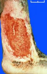
Describe wound surface. Why does it have this appearance?
|
|
|
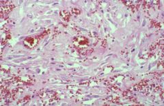
The main components of granulation tissue are budding capillaries filled with erythrocytes and fibroblasts.
FYI: Initially, the invading fibroblasts secrete collagen type III. Later, fibroblasts secrete collagen type I while macrophages digest the type III collagen, thus resolving the wound and restoring normal connective tissue components. |
?
|
|
|
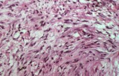
Collagen is initially randomly arranged within an area with numerous small blood vessels and abundant fibroblasts in the proliferative stage of granulation tissue.
|
?
|
|
|
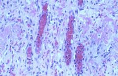
Later, the proportion of collagen to fibroblasts increases, and the capillaries become less numerous.
|
?
|