Reading...
![]()
Play button
![]()
Play button
![]()
Use LEFT and RIGHT arrow keys to navigate between flashcards;
Use UP and DOWN arrow keys to flip the card;
H to show hint;
A reads text to speech;
8 Cards in this Set
- Front
- Back
|
Several factors predispose to pathological clotting:
|
1. endothelial damage
2. disturbances in blood flow, stasis or turbulence 3. hypercoagulability |
|
|
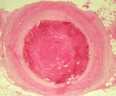
Thrombosis within an artery.
|
?
|
|
|
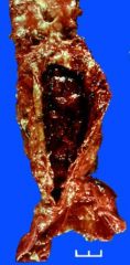
A large aneurysm of the abdominal aorta just above the iliac bifurcation.
A large mural thrombus is seen filling the lumen so that the caliber of the aorta is less than normal despite the dilatation. |
?
|
|
|
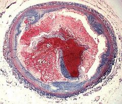
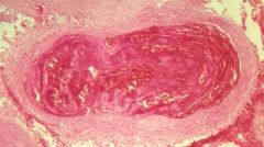
A cross section of a coronary artery with advanced athero- sclerosis (Masson connective tissue stain). The atheroma (light red) has ruptured and a thrombus (dark red) has formed and completely occluded the lumen which was already compromised by the atheroma.
|
?
|
|
|
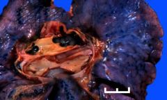
Notice the large black clot [thrombus] in the lung block at right. The clot does not fit within the artery as if it were formed there. It is neither a thrombus that formed at this site nor a postmortem clot
|
?
|
|
|
Large thrombus within a deep vein of the patient's thigh.
Diminished physical activity in this quadriplegic led to stasis of the blood within the veins of the leg which predisposes to thrombosis. "Lines of Zahn" represent layering of blood elements within the thrombus. |
?
|
|
?
|
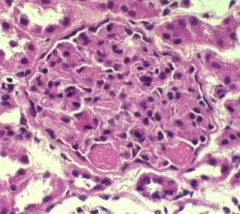
Fibrin microthrombi are shown obstructing the glomerular capillaries. This finding is characteristic of disseminated intravascular coagulation (DIC).
|
|
|
Notice that the articular cartilage has largely been eroded away. Bleeding into the joint space has caused accumulation of hemosiderin with proliferation of hemosiderin-containing reddish-brown synovial tissue seen between one tibial condyle and the distal femur (far right).
|
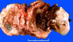
View of knee joint looking toward the head of the tibia. Pt has hemophilia, explain findings.
|