Reading...
![]()
Play button
![]()
Play button
![]()
Use LEFT and RIGHT arrow keys to navigate between flashcards;
Use UP and DOWN arrow keys to flip the card;
H to show hint;
A reads text to speech;
14 Cards in this Set
- Front
- Back
|
definition of hypertrophy
|
Hypertrophy (from Gr. hyper = excessive & trophe = nutrition) refers to an increase in the size of individual cells which may in turn produce a concomitant increase in the size of an organ. Hypertrophy is independent of the general growth of the body.
Hypertrophy which can be physiologic or pathologic, is a response to a stimulus such as increased functional demand or excessive hormonal stimulus. |
|
|
The myocardium of the left ventricle is substantially thicker than normal. This excessive growth characterizes hypertrophy. What circumstances may lead to this change?
|
Hypertrophy of the left ventricle reflects an increased work load. This may result from increased resistance, as in systemic hypertension, aortic valvular stenosis, or from significantly increased volume, as with anemia, aortic valvular regurgitation or a hypermetabolic state, e.g. hyperthyroidism.
|
|
|
microscopic manifestations and causes of hypertrophic myocardium
|
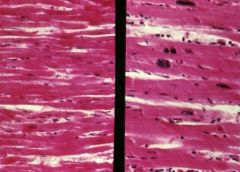
The increase in the size of hypertrophic heart cells is usually due to an increase in both the cytoplasmic volume and the size of the nuclei.
The larger nuclei have an increased chromatin content, but have not divided; therefore, they are tetraploid or octaploid. In the hypertrophic myocardium, the individual myocardial fibers are widened in comparison to normal and the nuclei are larger. The nuclear shape is sometimes termed "box-like". |
|
|
Causes of organ atrophy
|
Atrophy can be caused by:
1) decreased workload 2) loss of innervation 3) diminished blood supply 4) inadequate nutrition 5) loss of endocrine stimulation 6) aging |
|
|
Atrophic tubules in testis microscopic characteristics, causes
|
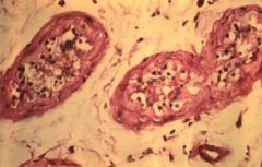
Atrophic tubules in an undescended testis with no spermatogenesis are smaller than the normal (equivalent magnification). The cells lining the tubules are mostly Sertoli cells rather than germinal epithelium.
One cause of testicular tubular atrophy is failure of the testis to move from the abdominal cavity to the scrotal sac. Atrophy may also result in decreased cell mass of all cells of an affected tissue without loss of specific cell types (e.g., myocardial atrophy). |
|
|
Hyperplasia definition
|
absolute increase in the number of cells per unit of tissue or organ
|
|
|
hyperplastic gland microscopic characteristics
|
In the hyperplastic gland, there is an increased ratio of glandular cells to adipose tisse. The cells are also slightly larger than normal.
|
|
|
Secondary hyperplasia of parathyroid glands description
|
occurs when there is chronic loss of calcium. The most common cause is chronic renal disease. In this circumstance, all four parathyroid glands will become hyperplastic with an increased production of parathormone attempting to maintain the appropriate serum calcium level.
|
|
|
tertiary parathyroid hyperplasia
|
in patient w/ secordary hyperplasia of parathyroid glands, if normal kidney function is restored (i.e. transplant), hypercalcemia may result from the hyperfunctioning parathyroid glands
|
|
|
primary hyperplasia of parathyroid
|
hyperplasia of a single parathyroid and will have hypercalcemia without a known stimulus.
|
|
|
Metaplasia
|
denotes the change of one mature tissue into another mature tissue. This change may involve either epithelial or mesenchymal tissues.
|
|
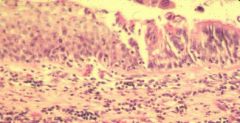
morphological alterations in this bronchus epithelium
|

metaplastic squamus epithelium.
infiltration of lymphocytes in the lamina propria, an indication of chronic inflammation |
|
|
Dysplasia definition
|
(from Gr. dys = hard, ill, bad; plasia = to form or mold) connotes deranged development, such as that found in cells that have undergone proliferation and atypical cytologic alterations involving cell size, shape and organization.
|
|
|
aplastic anemia, the marrow space is virtually devoid of hematopoietic elements. Most of that space is occupied by fat cells. Such severe aplasia (or extreme hypoplasia) of the bone marrow, resulting in inadequate production of hematopoietic elements, characterizes acquired aplastic anemia. This disorder is often of unknown cause (idiopathic), but may result from exposure to certain toxic chemicals or to chemotherapeutic agents used for cancer treatment which not only halt proliferating neoplastic cells, but also affect the normal marrow cells.
|
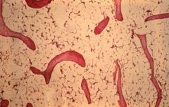
what is shown in this slide? What are possible causes?
|