![]()
![]()
![]()
Use LEFT and RIGHT arrow keys to navigate between flashcards;
Use UP and DOWN arrow keys to flip the card;
H to show hint;
A reads text to speech;
202 Cards in this Set
- Front
- Back
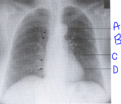
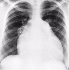
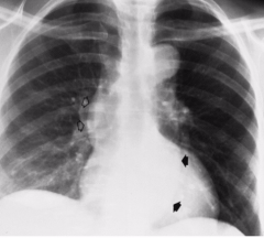
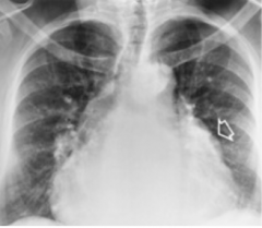
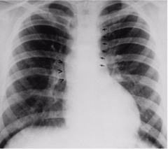
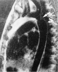
Label. |
A: Aortic knob B: Main and left pulmonary arteries C: Left atrial appendage D: Left ventricle |
|
|
What do you look at first on a chest x-ray? |
Start with the edges to make sure everything's included and go from there. |
|
|
What is the left atrial appendage? |
Tissue that creates a outpouching continuous with the left atrium. |
|
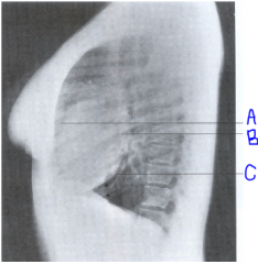
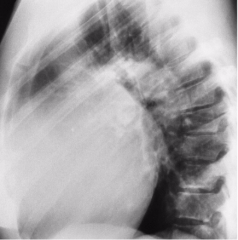
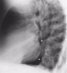
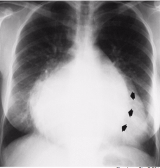
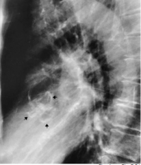
Label. |
A: Right ventricle B: Left atrium C: Left ventricle |
|
|
What is the cardio-thoracic ratio? |
The transverse diameter of the cardiac silhouette should NOT exceed 50% of the transverse diameter of the thoracic cage. |
|
|
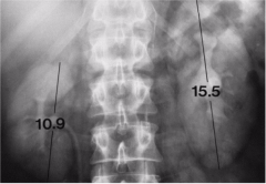
How is the cardio-thoracic ratio measured? |
From widest part of heart side to side, compare to widest part of internal diameter of chest. |
|
|
Why is it important to mark a chest x-ray erect or supine? |
When the patient is supine it widens the mediastinum. Don't want radiologist to think it's because of a disease process. |
|
|
What is congestive heart failure? |
The inability of the heart to pump enough blood to meet the body's metabolic needs due to impaired pumping ability and increased workload. (Heart can no longer keep up with venous return eventually resulting in blood damming back into the venous system). |
|
|
What are some things that can cause impaired cardiac function? |
Myocardial disease, valvular heart disease, congenital heart defects, constrictive pericarditis, cardiac tamponade. |
|
|
What are some things that cause excess work demands on the heart? |
Increased pressure (hypertension), increased volume, increased perfusion. |
|
|
Describe Left sided failure: |
When the left ventricle can no longer pump all the blood into the aorta and blood starts to back up into the pulmonary circuit leading the lungs to become congested (edema). Causes difficulty breathing and interferes with movement of O2 from lungs into tissue. |
|
|
What are some manifestations of Left sided heart failure? |
Diminshed cardiac output, fatigue/exertional dyspnea, tachypnea and increased HR to compensate, orthopnea (difficulty breathing while lying down), CYANOSIS, chronic dry cough, paroxysmal nocturnal dypnea (sudden severe shortness of breath that wakes you up), blood tinged sputum. |
|
|
Describe Right sided heart failure: |
Right ventricle can't empty properly so blood backs up into the systemic circulation and increases the venous pressure in the venous circuit. Can happen if Left sided failure goes untreated. |
|
|
What are some manifestations of Right sided failure? |
Fatigue, dependent edema, distention of the jugular veins, liver engorgement, ascites, GI distress, CYANOSIS, increased peripheral venous pressure (1st impacted). |
|
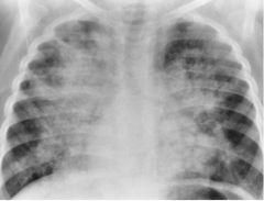
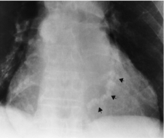
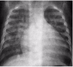
What sided heart failure is this? |
Left sided; Pulmonary vasculature is saturated with fluid. Shows diffuse peri-hilar alveolar densities. |
|

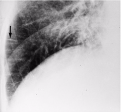
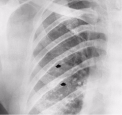
What sided heart failure is this? What does the image demonstrate? |
Left sided. Shows fluid in the interstitial space causing loss of normal sharp definition of lungs. |
|
What radiographic hallmark is seen in this image and the one above? |
Kerley B lines: thin horizontal lines at the bases (this image also shows interstitial pulmonary edema) |
|
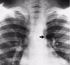
What sided heart failure is this? What appearance does this image have? |
Right sided; global cardiomegaly (both sides of heart are over worked). |
|
What sided heart failure is this? What appearance does this image have? |
Right sided; Shows right ventricular enlargement and no retro-sternal air space left. |
|
|
What is done to treat congestive heart failure? |
Low sodium diet: won't retain as much water so it decreases amount of blood heart needs to pump Diuretics: also decreases blood volume Digitalis: increases heart muscle strength and regulates HR Swan Ganz catheter Correct underlying problem: angioplasty (balloon), stenting, CABG |
|
|
What is digitalis also known as? |
Foxglove |
|
|
What does a Swan Ganz catheter measure? |
Lots. Mainly assesses pulmonary venous pressure to assess what kind of heart failure the patient is in. (Wedge pressure) |
|
|
What is wedge pressure? |
Tip of catheter placed just inside pulmonary valve. Balloon on catheter is inflated and blocks flow momentarily. The tip of the catheter measures pressure and function to see whether patient in L sided heart failure or R sided heart failure. |
|
|
How many blockages would be necessary to require a stent? How many require a CABG? |
2 blockages need stent, 3 or more need CABG. |
|
|
Where does the left coronary artery branch from? |
The left side of the aorta. |
|
|
The circumflex artery feeds? It branches off of? |
Left atrium and left ventricle. The left main coronary artery. |
|
|
What artery runs down the left side to the apex of the heart and feeds both ventricles? |
The Left Anterior Descending artery (LAD)/Anterior interventricular artery. |
|
|
What will happen if there's a blockage in the left main coronary artery? |
The left ventricle won't be getting any blood which greatly increases the risk of death. |
|
|
What does the right main coronary artery feed? |
The right atrium. |
|
|
What does the marginal artery feed? |
The right ventricle. |
|
|
What artery feeds both ventricles on the right side? |
The Posterior Descending Artery (PDA) or Posterior Interventricular Artery. |
|
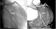
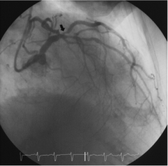
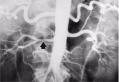
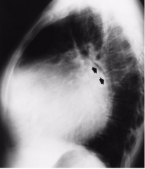
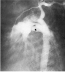
What is this image demonstrating? |
The bifurcation of the Circumflex artery and Left Anterior Descending artery from the left main. |
|
|
Coronary artery disease is the name for three forms of cardiac disease that result from insufficient cardiac flow, they are: |
1. Atherosclerotic heart disease 2. Angina pectoris 3. Myocardial infarction |
|
|
What is arteriosclerosis? |
A GROUP of disorders resulting in thickening and inelasticity of arterial walls. (hardening of arteries). |
|
|
What is atherosclerosis? |
The most common form of arteriosclerosis. Affects large and medium sizes arteries and is a common cause of CAD. The coronary arteries slowly narrow over years slowly depriving the myocardium of blood. |
|
|
Describe the pathogenesis of atherosclerosis: |
Begins in childhood, fatty streaks/lipids deposited in the intima of the vessels and stays there. Fibrous encapsulation of the lipids creates fibrous plaques that can become thrombi or emboli if they become detached. |
|
|
What happens as the plaque builds up? |
It elevates the lining of the vessel, roughening the surface and narrowing the lumen. The stricture causes eddy currents to form which can increase emboli formation. |
|
|
What are some non-modifiable risk factors for CAD? |
Age Sex: below 50 more common in men, above 50 more common in women Heredity |
|
|
What are some modifiable risk factors for CAD? |
Cholesterol, BP, smoking, physical inactivity, diabetes, obesity, stress/personality. |
|
|
What is angina pectoris? |
Chest pain that occurs when there is a deficit of oxygen for the heart muscle. (i.e. impaired blood oxygen supply to myocardium). Heart is working harder than it needs to to get proper oxygen. |
|
|
What are the symptoms of angina? |
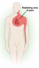
Substernal pain may radiate down the medial aspect of the left arm. Can mimic indigestion and is brought on by activities that increase O2 demand. |
|
|
How is angina treated? |
Primarily by Nitroglycerine, a coronary vasodilator drug. |
|
|
What effect does prolonged ischemia have? |
Over 30-35 minutes causes irreversible cellular damage and muscle death (necrosis). No amount of perfusion will bring the tissue back. |
|
|
What are the signs of a myocardial infarction? |
Severe prolonged "crushing" chest pain that is substernal and radiates to the left arm. Can also cause sweating, nausea, and vomiting. Increased serum levels of cardiac enzymes released by necrotic myocardial cells.
|
|
|
What are some complications associated with myocardial infarction? |
CHF (Part of heart not functioning) - pulmonary congestion, hypertension, edema - pleural effusion - peripheral edema from right failure Cardiogenic shock Papillary dysfunction Acquired septal defects (pressure difference) Cardiac rupture (flimsy necrotic tissue) Thrombo-embolism Pericarditis Arrythmia/Tachycardia/Bradycardia |
|
What is being demonstrated in this image? |
Calcified Circumflex artery which is highly suggestive of CAD. |
|
|
What would you see on a chest x-ray to suggest CAD? |
Enlarged heart, pulmonary effusion (blunted costophrenic angles), pulmonary congestion (interstitial densities). Chest x-rays aren't always helpful only really for diagnosing based off of calcification. |
|
|
How is Selective Coronary Arteriography used to diagnose CAD? |
Definitive study that demonstrated stenosis and occlusion of vessels and collateral circulation. (Go in through femoral artery up to aortic arch) |
|
|
How is CT used to diagnose CAD? |
Used to image coronary circulation. Uses houndsfield units to grade lesions BEFORE an invasive angio procedure. |
|
|
How is Nuc Med Stress Test used to diagnose CAD? |
Identifies the amount of residual damage to the myocardium. |
|
|
How occluded does a vessel have to be before an invasive procedure is performed? |
40% occluded |
|
What is being demonstrated in this image? How is it fixed? |
A lesion in the left main coronary artery. Usually fixed with balloon angioplasty or a stent. |
|
|
Describe an angioplasty: |
A balloon tipped tube is inserted into the coronary artery. Markers at the beginning and end of balloon to make sure the whole length of the lesion is ballooned. The markers also help decide stent length. |
|
|
What is a drug eluding stent? |
A stent that has medication on it that helps prevent plaque build up on and around the stent. |
|
|
BONUS QUESTION: What is Full Metal Jacket? |
When you have to stent a large component of a vessel or the vessel in its entirety. (a stent can cost around $4,000 - $12,000) |
|
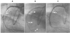
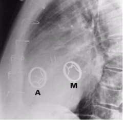
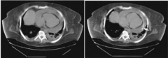
What is being demonstrated in each image? |
A: The Right Coronary Artery (C shape, runs between ventricles) with an obvious stenosis. B: Guide wire inserted into the artery, M stands for the markers on the balloon that are used to choose the right stent size. C: Balloon inflated and possibly stent inserted (hard to tell), artery has normal, even diameter. |
|
|
What is blood pressure (BP)? |
The measure of force exerted by blood on the arterial walls during contraction (systole) and relaxation (diastole) of the LEFT VENTRICLE. |
|
|
What are some things that affect BP? |
Stress, weight, age, gender, time of day, lying down or standing up. |
|
|
What are some things that help maintain BP? |
Peripheral resistance Pumping action of heart (first will decrease because of a problem then increase to compensate) Blood volume Blood viscosity Elasticity of vessel walls |
|
|
What is hypertension known as and why? |
The silent killer because it's the leading cause of strokes and CHF. |
|
|
Relate risk of hypertension to BP: |
For every 10 years past 40 you can increase the pressures by 10mmHg as you age it increases without disease. |
|
|
Primary hypertension is also known as: |
Idiopathic aka not related to disease processes just age. |
|
|
Describe the benign form of primary hypertension: |
GRADUAL onset related to: stress, obesity, heredity, salt intake, sex (F over 50, M under 50). |
|
|
Describe the malignant form of primary hypertension: |
ABRUPT onset, leads to renal failure and stroke. |
|
|
How is hypertension treated? |
Drugs: anti-hypertensive (Beta blockers) and diuretics Modify lifestyle: increase physical activity Remove factors: stress, obesity, salt, smoking and alcohol |
|
|
What percent of hypertension is secondary? |
Only 6% of hypertension overall. |
|
|
What causes secondary hypertension? |
Results from another disease, usually renal disease. Can also come from endocrine imbalances that upset H2O regulation (Cushing's and pheochromocytoma). Coarctation of aorta increases BP to neck and arms. |
|
|
How is secondary hypertension treated? |
Surgery or angioplasty. |
|
|
What are the 3 main effects of hypertension? |
HEMORRHAGE: due to rupture of weakened vessles and aneurysms ARTERIOSCLEROSIS: accelerated by propelling blood high in cholesterol into the vessel walls roughening the lumen and increasing the chance something will get stuck HYPERTENSIVE HEART DISEASE: constriction of peripheral vessels from plaque increases resistance to flow causing L ventricular enlargement and CHF |
|
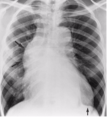
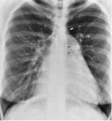
What is demonstrated in this image? |
Hypertensive heart disease. Tortuosity and elongation of ascending and descending aorta. Black arrows: Widening of aorta White arrows: dilation of SVC |
|
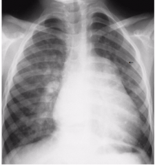
What is demonstrated in this image? |
Hypertensive heart disease with prominent left ventricle. |
|
|
What treatment for hypertensive heart disease? |
Treatment just prolongs life by preventing complications. |
|
|
What are the 3 main causes of death in hypertensive heart disease? |
WEAR OUT: 45% die of cardiac failure, heart stops working BLOW OUT: 19% die of strokes (thrombus/embolus) RUN OUT: 9% die of renal failure, secondary hypertension is small 6% this is a portion of that |
|
|
What 4 things can cause renovascular hypertension? |
1. Primary Hypertension: damages glomerulus leading to renal disease OR 2. leaves plaque deposits in the renal artery -> stenosis 3. Severe chronic renal disease: obstructive process increases blood volume because of increased intake of water, pressure causes destruction of parenchyma 4. Fibromuscular dysplasia: narrows renal artery |
|
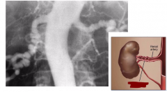
What is being demonstrated in this image? |
Affected kidney appears smaller because of destruction of parenchyma. |
|
What is being demonstrated in this image? |
Stenosis of the Right renal artery. Stenosis is most often found in the most proximal segment close to the aorta. |
|
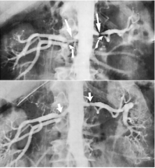
What is being demonstrated in this image? |
Bilateral stenosis of the renal arteries in the first image and bilateral stenting on the second image. Stenting usually not done if patient is asymptomatic. |
|
What is being demonstrated in this image? |
Fibromuscular Dysplasia: alternating segments of dilation and stenosis aka a disease process of the vessel walls that's most common in women. |
|
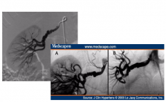
What is procedure is being demonstrated in this image? |
Stenting/Ballooning to clear the fibromuscular dysplasia. |
|
|
What is happening when the heart valves open? |
The pressure in the chamber proximal to the valve exceeds the pressure in the chamber distal to the valve. The valve opens and blood flows. |
|
|
What is happening when the heart valves close? |
The pressure in the chamber beyond the valve exceeds the pressure in the proximal chamber and the valve closes. |
|
|
Valvular disorders are mainly the result of: |
Rheumatic fever. An inflammatory disease that develops from inadequate treatment of a hemolytic strep infection, becomes autoimmune. Causes platelets to build up on leaflets of valves (called nodules/vegetation). |
|
|
Stenosis and Regurgitation are best demonstrated using what modality? |
Echocardiography |
|
|
What are 2 other things that can cause valvular disorders? |
Bacterial endocarditis: bacteria gets direct route to heart from IV drug abuse Papillary muscle rupture: valve won't open or close the way it's supposed to |
|
|
What valve is most commonly affected by valvular disorders? |
Mitral valve |
|
|
What valve is second most commonly affected by valvular disorders? |
Aortic valve; results in greater pressure on left side of heart |
|
|
Valvular dysfunction always causes: |
Increased cardiac workload |
|
|
Describe regurgitation: |
Valve leaflets fail to close securely and permit backflow. Forces the heart to pump the additional regurgitated volume. Heart pumps faster. |
|
|
Describe stenosis: |
Valve opening is narrowed and restricts outward flow and blood is not moving through the heart. Increased pressure is required to overcome the resistance to flow. Heart pumps harder. |
|
|
How does the myocardium respond to stenosis and regurgitation? |
Ventricles hypertrophy to deal with increased load. Atria dilate for additional blood volume. |
|
|
What is cardiomyopathy? |
Increased thickness of muscle but functionality is compromised; heart doesn't have same pumping capacity as it used to. |
|
|
What is mitral stenosis? |
The valves of the mitral valve become thickened and fuse along the leaflet margins. Not enough blood is leaving the atrium so increased back pressure and starved peripheral arteries occur. Full ventricle of blood is never pumped out. |
|
|
What is the most common cause of mitral stenosis? |
rheumatic disease |
|
What is being demonstrated in this image? |
Mitral stenosis: Obstruction of blood flow from left atrium during diastole increases pressure causing chamber enlargement. This is causing posterior displacement of the esophagus. The atrium is also calcified in this image. |
|
What is being demonstrated in this image? |
Mitral stenosis: Double contour sign caused by projection of enlarged left atrium through the normal silhouette of the right atrium. |
|
|
If someone has mitral valve stenosis you can also expect them to have? |
Chronic venous congestion: increased left atrial pressure which increases pressure in pulmonary veins |
|
|
What is mitral regurgitation? |
The valve leaflets do not close properly allowing reflux of blood from left ventricle to left atrium during ventricular systole. |
|
|
What is causes mitral regurgitation? |
Most often caused by rheumatic disease but can also be caused by rupture of the papillary muscles or chordae tendinae. |
|
|
What heart chamber becomes larger than it does in mitral stenosis? |
Left atrium. |
|
What is being demonstrated in this image? |
Mitral regurgitation: Gross cardiomegaly from enlargement of left atrium and left ventricle, pulmonary edema; Double contour, left atrium seen through left ventricle. Causes downward displacement of cardiac apex. |
|
What is being demonstrated in this image? |
Mitral regurgitation: Elevation of mainstem bronchus due to left atrial enlargement. |
|
|
What is used to treat mitral stenosis and regurgitation? |
Surgical valve replacement or correction of stenosis. |
|
|
Name 3 things that can cause aortic stenosis? |
Rheumatic disease, congenital deformity, aging process. |
|
|
What happens with aortic valve stenosis? |
Obstruction to outflow of left ventricle increases workload. Patient experiences syncope due to decreased flow to cerebral arteries, and angina because cardiac vessels aren't perfused. |
|
What is this image demonstrating? |
Aortic stenosis: Left ventricle hypertrophied, lateral bulging of the ascending aorta (post stenotic dilation). This happens because blood getting through valves has increased velocity and is hitting the lateral wall causing it to bulge. |
|
What is this image demonstrating? |
Aortic stenosis: Calcification of leaflets |
|
|
What are 5 things that can cause aortic valve regurgitation? |
1. Rheumatic disease 2. Syphilis 3. Infective endocarditis 4. Dissecting aneurysm 5. Marfan's syndrome |
|
|
What happens during aortic valve regurgitation? |
Reflux of blood from the aorta during diastole causes volume overload and dilation of left ventricle, left atria becomes enlarged because of back up. Leads to LHF and pulmonary edema. (also sees angina and syncope) |
|
What is being demonstrated in this image? |
Aortic regurgitation: Left ventricular enlargement and downward/lateral displacement of cardiac apex. The ascending aorta has lateral bulging but descending/knuckle is normal. |
|
|
What is infective endocarditis? |
Infection of endocardial heart surfaces. Nodules/bacterial vegetations on heart valves tend to break off and go into the blood stream causing the patient to become septic. |
|
|
What is it caused by? |
IV drug abusers and vascular procedures give bacteria direct route to heart. Usually the bacteria is a staph infection. |
|
|
What is the best modality to image infective endocarditis? |
Echocardiography |
|
What pathology is shown in this image? |
Pericardial effusion. |
|
|
What radiographic hallmark is shown on this image? |
Mateus bottle heart: gross enlargement of the cardiac silhouette. |
|
|
What is pericardial effusion? |
Accumulation of fluid/blood within the pericardial space, secondary to pericarditis. Cardiac tamponade can compress heart decreasing cardiac output. |
|
|
What causes pericardial effusion? |
Idiopathic, hydropericardium (CHF), neoplastic tumours, infections, trauma (vessel rupture). |
|
|
What modality best demonstrates pericardial effusion? |
Echocardiography; shows amount of fluid accumulated in sac around heart |
|
What is being demonstrated in this image? |
First image: massive pericardial effusion stopping heart from pumping properly Second image: drainage inserted to drain fluid from pericardial sac, called pericardial centesis. |
|
|
What is the foramen ovale? |
The shunt between the left and right atriums that bypasses the lungs. Becomes the fossa ovalis after 1 year. |
|
|
What is the ductus arteriosus? |
Some blood flows regularly into the pulmonary circuit but does nothing and goes through shunt right into the aorta. When baby is born it becomes ligamentum arterioles. |
|
|
What happens when blood is shunted right to left? |
It doesn't enter the pulmonary circuit so no oxygen is getting into the blood leading to CYANOSIS. |
|
|
What happens when blood is shunted left to right? |
A larger than normal volume goes to the lungs but patient will not become cyanotic until later when it becomes too much for the right side of the heart. |
|
What pathology is shown in this image? |
Atrial septal defect: Pulmonary vasculature seen because of back up. White arrow: Aortic knuckle not seen because extra pressure in left ventricle doesn't have enough force going through the knuckle. Clear arrow: pressure being put on pulmonary arteries from the right ventricle |
|
|
What is atrial septal defect? |
The most common congenital cardiac lesion caused by lack of closure of the foramen ovale. Free communication occurs between the atria. |
|
|
What direction is blood shunted with atrial septal defects? |
Left to right because there is greater pressure on the left side. Pulmonary circuit becomes overloaded. |
|
|
Does atrial septal defect cause cyanosis? |
NO!!!!! |
|
What pathology is demonstrated in this image? |
Ventral septal defect. |
|
|
What is a ventral septal defect? |
An opening between the ventricles allows some blood goes into normal route and some will move into the right ventricle. Left to Right shunting. |
|
|
What do ventral septal defects cause? |
Increased pulmonary flow and increased venous return. Shunting happens during systole so increased pressure in the pulmonary trunk, left atrium and left ventricle. NO CYANOSIS. |
|
|
What complications are seen on the above image? |
Enlarged triangular heart and large pulmonary trunk. The pulmonary trunk is enlarged because extra blood is immediately pushed into the pulmonary trunk. |
|
What pathology is demonstrated on this image? |
Patent ductus arteriosus. (aortogram showing communication between aorta and pulmonary tree). |
|
|
What is patent ductus arteriosus? |
The ductus arteriosus remains open after birth so blood is shunted from the aorta into the pulmonary artery. (left to right). |
|
|
What does this result in? |
Increased blood volume in the pulmonary circuit, and left atrium/ventricle. NO CYANOSIS. |
|
What complications are shown in this image? |
Cardiomegaly and enlarged L atrium and ventricle. |
|
What pathology is shown in this image? |
Coarctation of the aorta. |
|
|
What radiographic hallmark is shown in the image? |
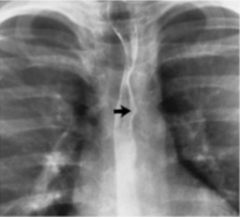
Figure 3 sign. Arrow points to the middle. Top is pre-stenotic bulge, bottom is post-stenotic bulge. (this image shows backwards 3) |
|
|
What is coarctation of the aorta? |
Narrowing of the aorta most often after the branches that go to the head and arms. Always distal to the left subclavian artery. |
|
|
What is caused by coarctation? |
Decreased blood flow to the abdomen and legs and development of progressive collateral circulation. |
|
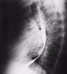
What is demonstrated in this image? |
Rib notching of ribs 4-8. Result of erosion by dilated intercostal and mammary arteries that are trying to help with transmission of blood to areas that need it. The pulsing eats away at the bone. |
|
What is being shown in this image? |
A lateral view of a coarctation of the aorta. You can clearly see the pre and post stenotic bulges. |
|
What pathology is shown in this image? |
Tetrology of fallot. |
|
|
What radiographic hallmark is seen in the above image? |
Upward and lateral tilting of the cardiac apex, right ventricular enlargement. |
|
|
What are the 4 abnormalities associated with tetrology of fallot? |
1. High Ventricular Septal Defect 2. Pulmonary stenosis 3. Overriding of the aortic orifice (above VSD; blood can't get through stenosis so it's moving into left ventricle and out through the aorta) 4. Right ventricular hypertrophy |
|
|
Does tetrology of fallot cause cyanosis? |
YES!!!! it's the only one that does. Deoxygenated blood is getting pumped into the body by the aorta, only small amount getting through stenosis. |
|
|
Describe transposition of great vessels: |
Aorta emerges from Right Ventricle. Pulmonary artery emerges from Left Ventricle. |
|
|
Is transposition compatible with life? |
No and it's very rare. |
|
|
What is levocardia? |
Apex to the left (normal). |
|
|
What is solitus? |
Left chamber of heart on left side of body (normal). |
|
|
What is dextrocardia? |
Apex of the heart is to the right side. One of the main parts is flipped so it causes more symptoms. |
|
|
What is situs inversus? |
Everything is normal just flipped. |
|
|
What is portal hypertension? |
Increased resistance of blood flow through the portal system causes overloading the portal circuit. This causes collateral channels around the obstruction. |
|
|
What are 4 things that can be caused by back pressure in the portal vein? |
1. Splenomegaly 2. Ascites 3. Esophageal varices -> PV -> Lt gastric vein -> eso ver -> azygos vein -> SVC 4. Hepatic coma (liver no longer filters toxins) |
|
|
What is TIPS? |
Transjugular Intrahepatic Portosystemic Shunt |
|
|
What does it do? |
It's the formation of a tract between the hepatic veins and portal veins that shunts blood away from the sinusoids relieving pressure. |
|
|
Where is the access point for TIPS? |
The right internal jugular vein. |
|
|
What veins does the catheter run through? |
RIJV -> SVC -> R Atrium -> IVC -> RHV |
|
|
How is the puncture made? Where does the catheter go? |
From RHV into RPV. Catheter fed into MPV. |
|
|
What are the last steps? |
Dilation of tract with angioplasty balloon then stenting of the new tract. Venogram to demo new bypass. |
|
|
What is an aneurysm? |
Abnormal localized dilation/ballooning caused by a weakening in the vessel wall or heart chamber. (decreased amount of elastin, increased collagen) |
|
|
What are 5 things that can cause weakness in the vessel walls? |
Atherosclerosis Infection (affects integrity of vessels) Trauma (tear or stenosis) Congenital (marfan's) Hypertension |
|
|
What are 3 consequences of aneurysms? |
Depending on location and size 1. Rupture related to size and dilation 2. Thrombus formation due to stasis and eddy currents 3. Pressure on adjacent structures |
|
|
Describe fusiform aneurysms. |
Involve the entire circumference of the vessel. Gradual and progressive dilation. |
|
|
Where are they most common? |
Thoracic/Ascending Aorta or Abdominal Aorta. |
|
|
Describe saccular aneurysms. |
The weak area is on one side of the vessel (maybe due to trauma). Blood is stagnant in the pouch creating a thrombus, blood travels between wall and thrombus causing more dilation. |
|
|
Describe dissecting aneurysms. |
Disruption of the intima (inner layers) due to increased BP allows blood to travel between the layers of the vessel wall. This causes excruciating chest pain. |
|
|
Where are dissecting aneurysms usually found? |
In the aortic arch because of the velocity of blood being pumped out of the LV. |
|
|
What is a pseudoaneurysm? |
Disruption of the two outer layers of the vessel wall. |
|
|
What is the candy cane sign? |
On a CT wherever the tear is you'll see a bright line, tear spirals around the vessel making the aorta look like a candy cane. |
|
|
Describe berry aneurysms. |
A small spherical dilation of the vessel at a bifurcation. Usually the Circle of Willis. |
|
|
What is a symptom of a berry aneurysm? |
Excruciating headache. Happens when there's a rupture causing a subarachnoid hemorrhage. |
|
|
How is a berry aneurysm treated? |
Coil placed into the aneurysm. Wire is straight and coils up as it's pushed into the aneurysm, when the coil is detached it winds up tighter packing the area so there won't be more room for blood to enter and dilate. |
|
|
What are some radiographic appearances of an aneurysm? |
Widening of the mediastinum, thickened para-tracheal stripe, intimal flap, large thrombus around normal lumen. |
|
|
What is the para-tracheal stripe? |
A natural finding on a normal chest x-ray; white area between trachea and lung. Thickened stripe is indicative of disease process or hematoma. Less than 4cm is normal. |
|
|
What is a stent? |
A metal scaffold used to support a vessel wall. |
|
|
When is it used? |
To treat stenosis and occlusive disease. |
|
|
What is a stent graft? |
A stent covered with graft material. |
|
|
When is it used? |
To treat aneurysms and arterial rupture. (trying to create a new lumen). |
|
|
Describe surgical clipping. |
Clip placed on external surface of vessel if there's a neck on aneurysm the clip keeps blood from entering the segment. |
|
|
Describe stent assisted coiling. |
If the neck of a berry aneurysm is too wide there's nothing holding the coils in so they'll use a stent to back it up. |
|
|
What causes peripheral vascular disease? |
Atherosclerosis causes progressive narrowing of the peripheral vessels. |
|
|
What is the most common affect of peripheral vascular disease? |
Intermittent claudication. |
|
|
What is intermittent claducation? |
Intermittent leg/calf pain due to ischemia that stops with rest. (when old people have to sit at the mall) |
|
|
What are some other things caused by peripheral vascular disease? |
Gangrene in the toes and feet Aneurysm formation in the iliac arteries Thrombus formation at stenotic sites |
|
|
What are 4 factors that affect the outcome of a peripheral atherosclerotic lesion? |
1. Site of the lesion (foot not as bad as leg) 2. Severity of lesion 3. Metabolic needs of the tissue beyond the lesion 4. Extent of collateral circulation |
|
|
What does collateral circulation look like on an image? |
Lots of very twisty and small vessels. |
|
|
How are peripheral artherosclerotic lesions treated? |
With a bypass graft, usually from the saphenous vein. |
|
|
What are 3 causes of intravascular thrombosis? |
1. Stasis of blood (usually venous) 2. Lesions of the endothelial lining (from trauma or infection) 3. Hypercoagulability of blood (dehydration, polycythemia) |
|
|
If a clot forms in the leg it'll most likely get trapped: |
In the lungs. |
|
|
What are 3 things that can happen if a thrombus is left alone? |
Canalization: channel formed through the clot and blood flows again Enlargement: can become a permanent occlusion Embolus: if venous goes into pulmonary circuit; if arterial interrupts flow to affected tissue causing infarct; if in the heart affects the vessels that feed myocardium |
|
|
What is the best way to image a DVT? |
Doppler ultrasound. |
|
|
How is intravascular thrombosis treated? |
Blood thinners-> heparing, coumadin Thrombolysis -> bolus amount given in emergency IVC filter -> captures clots coming up from extremities |
|
|
What are varicose veins? Where are they most common? |
Dilated tortuous vessels that are most common in the superficial leg veins. |
|
|
Who is predominantly affected by varicose veins? |
Women are 5x more common. Increased risk if you're pregnant or stand and sit for long periods of time. Also hereditary. |
|
|
Stasis of blood in a varicose vein can lead to? |
Phleboliths (calcified clot) |
|
|
What do varicose veins look like in an image? |
Static blood calcified in the tissue; body lays down new bone triggered by venous stasis. |
|
|
What is cor pulmonale? |
An alteration in the structure and function of the right ventricle (RV) of the heart caused by a primary disorder of the respiratory system. |
|
|
What is arrhythmia? |
A condition in which the heart beats with an irregular or abnormal rhythm. |
|
|
What is bradycardia? |
Slow heart beat. |
|
|
What is tachycardia? |
Fast heart beat. |
|
|
What is fibrillation? |
The upper chambers of the heart, or atria, fibrillate. This means that they beat very rapidly and irregularly. |
|
|
What is heart block? |
An abnormal heart rhythm where the heart beats too slowly (bradycardia). In this condition, the electrical signals that tell the heart to contract are partially or totally blocked between the upper chambers (atria) and the lower chambers (ventricles). |
|
|
What is a pacemaker? |
A small device that's placed in the chest or abdomen to help control abnormal heart rhythms. This device uses electrical pulses to prompt the heart to beat at a normal rate. |