Reading...
![]()
Play button
![]()
Play button
![]()
Use LEFT and RIGHT arrow keys to navigate between flashcards;
Use UP and DOWN arrow keys to flip the card;
H to show hint;
A reads text to speech;
49 Cards in this Set
- Front
- Back
|
Pigment definition
|
Substances that are inherently colored.
|
|

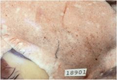
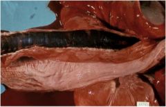
What abnormal pigmentation do you see here?
|

This is canine pulmonary anthracosis.
|
|
|
What is carbon anthracosis? Who is prone to getting it?
|
Carbon accumulated in macrophages in the lung, lymph nodes mainly. Common to individuals in smokey cities or coal mines.
|
|
|
What two general types of pigments are there? What subtypes are within those categories?
|
Exogenous and Endogenous pigments
Exogenous: carbon, dusts, metals, tattoos, carotenoids, chlorophyll. Endogenous: phenolic, hematogenous, and lipofuscin pigments |
|
|
What are the five kinds of hematogenous pigments?
|
Hemoglobin, bile, porphyrin, hemosiderin, and hematins/parasitic pigments
|
|
|
What are some examples of visible particles left by mineral dusts in the lungs?
|
Pneumoconiosis (anthracosis)
silicosis asbestosis |
|

What kind of pigment is this in the sheep brain? What would it be called in a cat or dog brain?
|
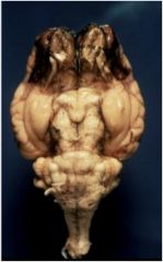
It's a melanoma in the meninges of ovine forebrain.
In a cat or dog brain, it would be called melanosis. |
|
|
What post-mortem pigmentary change can melanosis be confused with?
|
Pseudomelanosis.
|
|
|
What is the correlation between tattoos and lymph nodes?
|
The lymph nodes that drain the area of tattoos will be colored the same color of the tattoo.
|
|
|
Are carotenoids fat-soluble or water-soluble? What other pigment can they be confused with? What is the differentiator?
|
Fat-soluble
Carotenoids cause the same yellow staining as jaundice. They can be differentiated by checking areas without fat, like sclera, or the mucous membranes - if that area is yellow, it's not carotenoids. |
|
|
What is the main cause of staining the GI tract?
|

Chlorophyll
|
|
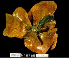
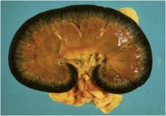
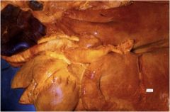
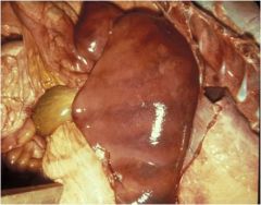
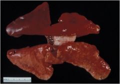
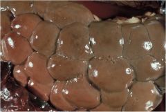
What color does this liver look to you? What might cause that pathology?
|

This liver is jaundiced, which could be caused by a post-hepatic obstruction.
|
|
What color does this equine mesentery look to you? What might be causing this pigmentation?
|

This could be jaundice or carotenoid pigment. It is from carotenoids.
|
|
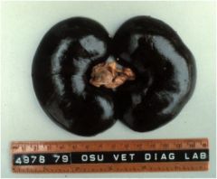
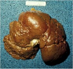
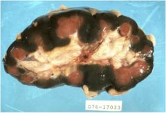
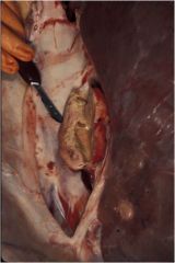
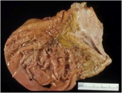
What pigmentation do these kidneys have? What could cause this?
|
These dark brown to black kidneys are caused by copper toxicosis. You can see on this inside shot that there is jaundice - both hemolytic and hepatic.
|
|
|
What is the difference between conjugated and unconjugated bilirubin?
|
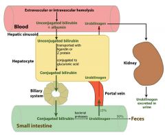
Erythrocytes (red blood cells) generated in the bone marrow are disposed of in the spleen when they get old or damaged. This releases hemoglobin, which is broken down to heme as the globin parts are turned into amino acids. The heme is then turned into unconjugated bilirubin in the reticuloendothelial cells of the spleen. This unconjugated bilirubin is not soluble in water. It is then bound to albumin and sent to the liver.
In the liver it is conjugated with glucuronic acid by the enzyme Glucuronosyltransferase, making it soluble in water. Much of it goes into the bile and thus out into the small intestine. Some of the conjugated bilirubin remains in the large intestine and is metabolised by colonic bacteria to urobilinogen, which is further metabolized to stercobilinogen, and finally oxidised to stercobilin. This stercobilin gives feces its brown color. Some of the urobilinogen is reabsorbed and excreted in the urine along with an oxidized form, urobilin. |
|

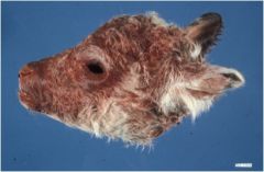
What type of pigmentation is they likely to be on this old grey mare?
|
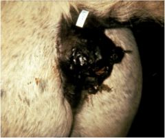
Perineal melanoma
|
|
What is likely to have caused this black pigmentation of the trachea?
|

Anthracosis
|
|

What pigment might this be a histo of?
|
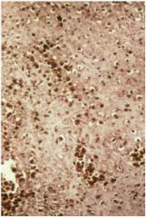
melanoma
|
|
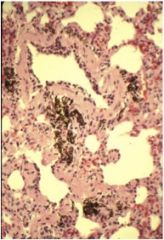
What pathology causing pigmentation of these dog lungs could this be?
|

Pulmonary anthracosis
|
|
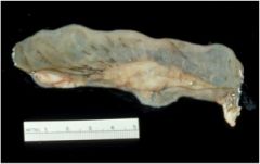
This perihepatic lymph node has a pigment - what kind is it likely to be?
|

Parasitic hematin
|
|

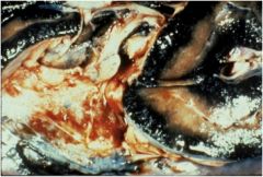
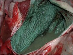
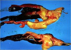
Here's a camelid stomach. What is likely to have caused this?
|

This is hydrochloric acid hematin due to an ulcer in the 3rd compartment of a camelid stomach.
|
|
|
Finished pigmentscom.ppt. and post-mortemchangescom.ppt. Still need to figure out how much the pigments and PM change handout there is to input - I think that the beginning of the doc is in here. Might need to cross-reference with slides made into flashcards and those mentioned in the hand-out from in-class notes.
Will this ever end? |
no.
|
|
What is Santa's favorite pigment? Is the pigment endogenous or exogenous?
|

exogenous pigment - copper-based diarrhea treatment
|
|

What is the black pigment that corresponds with production of hydrogen sulfide in rotting tissues?
|
Pseudomelanosis
|
|
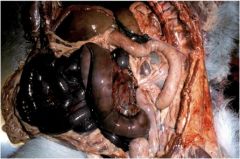
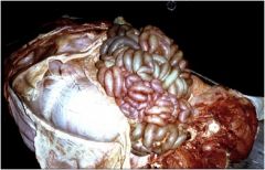
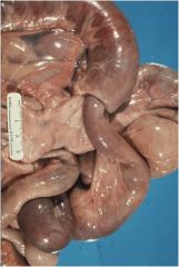
What causes the color changes due to altered blood flow? What has happened in this intestinal volvulus?
|

The discoloration is due to the distribution of hemoglobin pigment. The intestinal volvulus has blood pooling in the loops of the intestine.
|
|
|
What are the seven hues of hemoglobin?
|
Oxyhemoglobin
Reduced hemoglobin Methemoglobin Suflhemoglobin Sulfur Methemoglobin Carboxyhemoglobin Hemoglobin Imbibition |
|
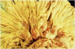
What does this lesion look like? Where is this lesion most easily seen? What are types of this leasion?
|

Jaundice (icterus). Most easily seen in sclera, mucous membranes. The Bedlington terrier suffers from a copper metabolism that damages the liver (hepatic jaundice). The yellow pluck is an example of isoimmune hemolysis of foals (prehepatic jaundice).
|
|

What brown/yellow pigment is this?
|
lipofuscin/ceroid
|
|

What pigment is this in kidney of a neonatal calf?
|
Biliverdin
|
|

What is the green pigment found in the placenta of dogs?
|
Uteroverdin
|
|

What is the hemoglobin hue that is green?
|
Sulfur methemoglobin
|
|
|
What is the principle of self-digestion?
What is it important to distinguish from? |
autolysis
Important to distinguish from antemortem lesions, especially necrosis. (in essence, somatic death vs cell death) |
|
|
How can autolysis by minimized?
|
Minimized by prompt necropsy and collection of tissues into fixative.
|
|
|
What things could effect rot?
|
Environmental temperature
Antemortem hyperthermia Size External insulation Nutritional status Species variation Tissue variation |
|
What post-mortem change does this look like?
|

Rib imprints
|
|
What type of post-mortem change is this? How can you tell if its pre- or post-mortem?
|

Post-mortem intussusception
You can tell because in the post-mortem, you can easily pull it out. |
|
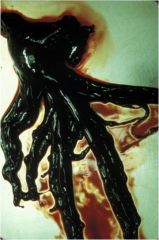
What is this?
|

A currant jelly blood clot
|
|
The solid, laminated appearance of this differentiates it from a blood clot. What is this?
|

A true thrombus in the vena cava for comparison.
|
|
What is this?
|

A chicken fat clot
|
|

What post-mortem change is this?
|
Hemoglobin imbibition
|
|
What post-mortem change is this? What is the clinical relevance?
|

Bile imbibition. This can get over the stomach organs.
|
|

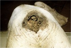
What post-mortem change is this?
|
Raven strike
|
|

Describe this kidney.
|
Putty-like, friable kidney with foci of emphysema.
|
|
What post-mortem change is this?
|

Raven strike
|
|
|
What are the other features of post-mortem change?
|
Odor
Rigor Mortis Livor Mortis Sloughing Epithelium Histologic changes |
|
|
How long does rigor mortis last? How long after death does it start?
|
Lasts 1-2 days
Starts in 1-6 hours |
|
|
What is livor mortis?
|
Hypostatic congestion of skin
|
|
|
What are types of epithelium that slough post-mortem?
|
Gall bladder, urinary bladder, ruminant forestomachs
|
|
|
What are examples of post-mortem histologic changes?
|
Cadaver bacilli, gas bubbles, nuclear fragmentation/loss, erythrocyte lysis
|