![]()
![]()
![]()
Use LEFT and RIGHT arrow keys to navigate between flashcards;
Use UP and DOWN arrow keys to flip the card;
H to show hint;
A reads text to speech;
150 Cards in this Set
- Front
- Back
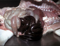
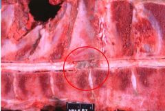
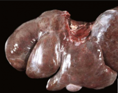
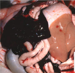
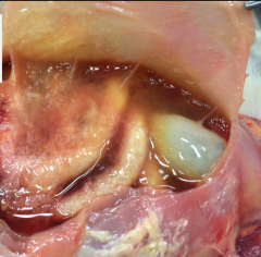
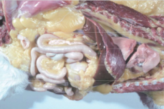
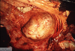
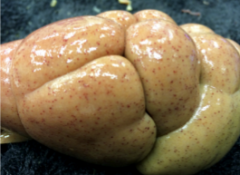
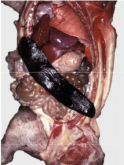
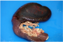
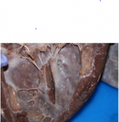
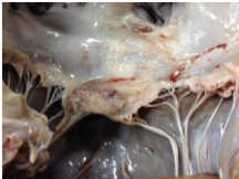
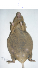
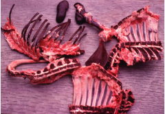
what is this lesion and what is a possible cause? |
Venous infarction due to torsion of the small intestine occluding the veins |
|
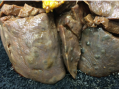
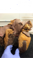
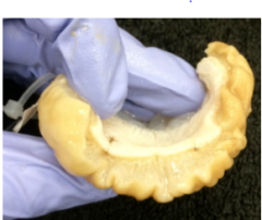
What type of azotemia could this problem cause and why? |
Pre-renal azotemia due to dehydration causing decreased renal blood flow |
|
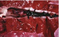
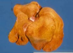
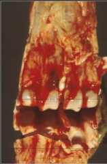
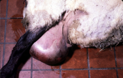
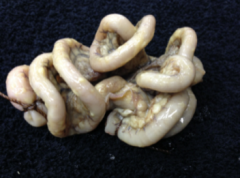
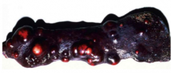
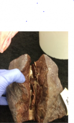
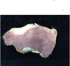
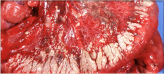
what is this lesion called? |
Hematoma |
|
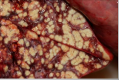
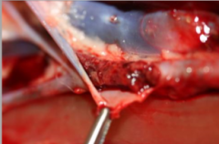
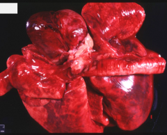
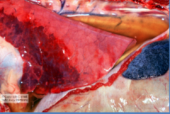
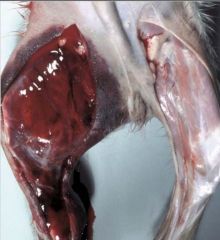
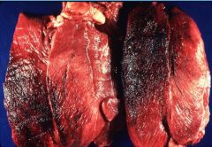
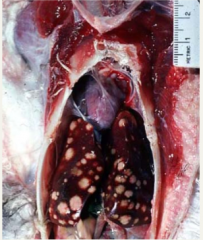
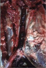
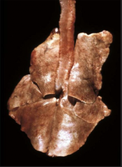
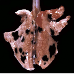
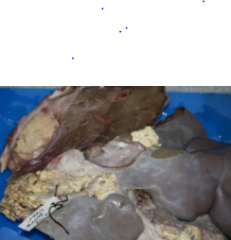
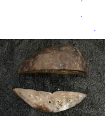
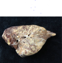
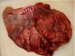
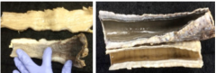
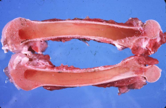
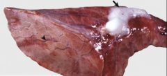
What is the cause of this necrosis of the lung? |
oxygen deficiency |
|
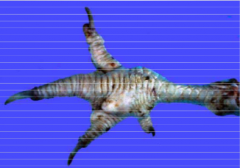
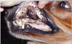
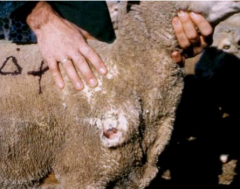
what is this lesion? Which of the four mechanisms of oxygen deficiency will this cause? |
aorto-iliac (saddle) thrombosis Loss or reduction of blood supply to an area/body part |
|
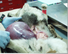
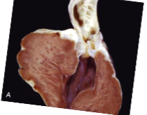
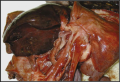
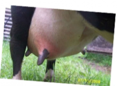
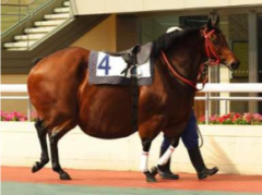
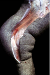
What is the lesion and what is it causing? |
Uterine torsion resulting in ischemia and oxygen deficiency |
|
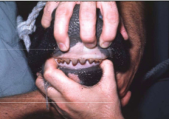
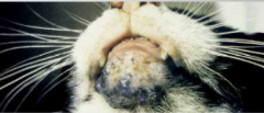
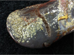
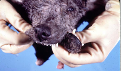
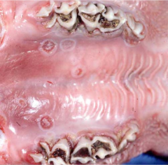
What is this pigmentation called and what group of animals is it most common in? |
Porphyria (accumulation of porphyrins) Young animals |
|
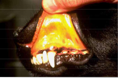
what are the two names of this pigmentation? |
Jaundice or icterus |
|
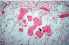
what type of gout is this? |
articular |
|
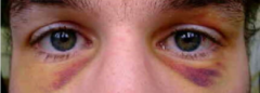
What causes each of the different colours present in this lesion? |
Hemoglobin = red-blue bilirubin and bilverdin = blue-green hemosiderin = golden brown |
|
If lesions like this are seen throughout the body what could this be? |
Disseminated Intravascular Coagulation (DIC) |
|
What is visible in this photo that tells you this is an arterial thrombus? |
lines of Zahn |
|
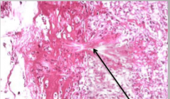
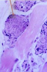
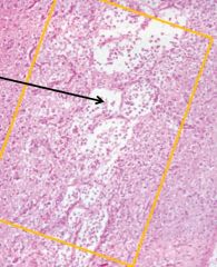
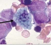
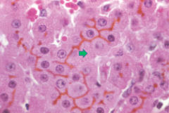
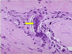
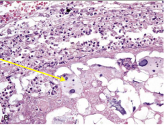
what is accumulated where the arrow is pointing and what disease is this causing? |
Sodium urate trophi (crystal aggregate) gout |
|
Is this chronic or acute inflammation? How can you tell? |
Chronic Accumulation of fibrous connective tissue |
|
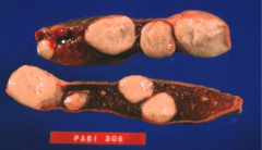
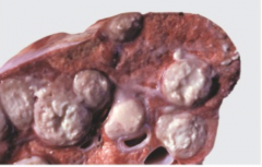
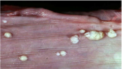
what are the possible differential diagnosis for these lesions? |
NAG (for any nodules) - Neoplasia - Abscess - Granuloma |
|
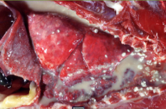
Is this an exudate or a transudate? What is this disease called? |
Exudate Pyothorax |
|
what stage of acute cell swelling are these arrows pointing to? |
ballooning degeneration |
|
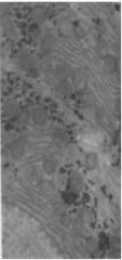
what are the lines shown in this image and what disease process do they occur during? |
myelin figures acute cell swelling |
|
what is the generic term for this abnormality? |
hepatomegaly
|
|
what type of necrosis is this? |
coagulative |
|
Is this an exudate or a transudate? What is this disease called? |
transudate Hydrothroax |
|
what is the primary cell type in the fluid from this lesion? What stage of inflammation is this? |
neutrophils acute |
|
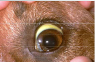
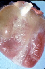
what is accumulating to make the sclera yellow? |
bilirubin |
|
what disease process is caused by accumulation of what the arrow is pointing to? |
silicosis |
|
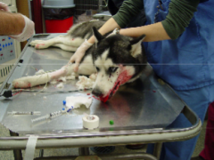
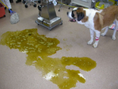
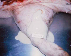
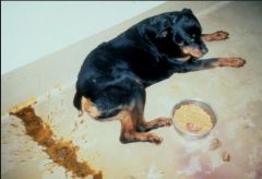
what space is this dog losing fluid from? What type of shock might this dog experience? |
intravascular hypovolemic shock |
|
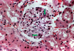
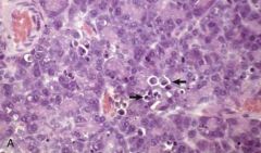
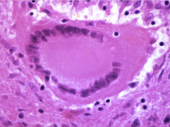
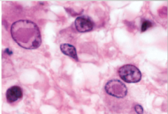
what structure is this and what are the arrows pointing to? |
glomerulus intranuclear viral inclusions |
|
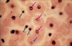
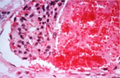
what stage of necrosis of the nuclei is each colour of arrow pointing to? |
purple = karyorrhexis blue = karyolysis green = pyknosis |
|
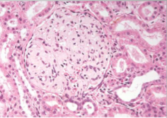
what is accumulated in this glomerulus? |
amyloid |
|
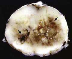
what type of necorsis is indicated by the arrow and what is this called in the CNS? |
Liquefactive malacia |
|
Is this hypertrophy or hyperplasia? What are cells called that can only undergo the above process? |
Hypertrophy permanent |
|
what type of hemorrhage is this? |
suffusive |
|
Which two spaces could this dog be losing fluid from? |
interstitial intracellular |
|
what is this an accumulation of? Given that this is a lung what is this called? |
Carbon Anthrocosis |
|
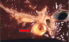
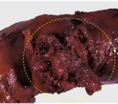
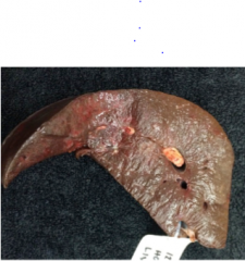
what is this lesion? (This is a liver) |
Portal vein embolism |
|
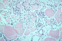
Is this dystophic or metastatic calcification? |
dystrophic (can see necrotic muscle fibres) |
|
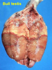
What type of necrosis is seen in this image? |
caseous |
|
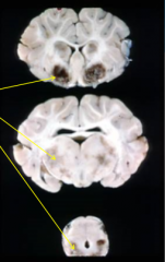
Briefly describe this lesion? |
malacia of the spinal cord due to liquefactive necrosis |
|
what process are the cells indicated by the arrows undergoing? |
apoptosis |
|
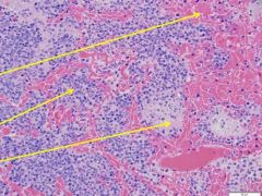
What is indicated by each arrow (different for each)? This is lung tissue |
top arrow = hemorrhage middle arrow = neutrophils bottom arrow = fibrin accumulation |
|
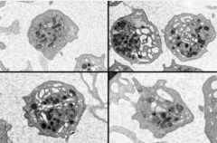
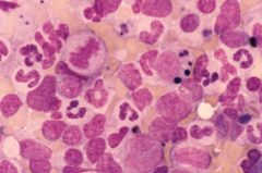
what type of cells are the large ones? What are the two main functions of these cells? |
eosinophils Parasite killing and histamine release from macrophages and basophils |
|
what lesions are shown on this slide? |
thromboembolisms |
|
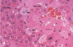
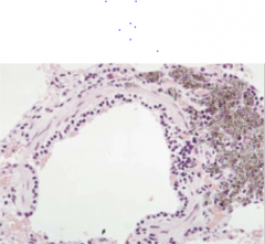
What pathology is seen on the right side of the image? Is this process still reversible in the cells with the arrows? |
Acute cell swelling Yes (still have nuclei visible) |
|
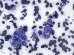
Given that this is lymph tissue what type of abnormal cells are shown? What electrolyte imbalance can this cause? |
Neoplastic lymphocytes Hypercalcemia |
|
What is this lesion and what are the components that it is made up of? |
A thrombus Platelets, fibrin, trapped red and white blood cells |
|
Is this venous or arterial? How can you tell? |
Venous It is red, soft, and gelatinous looking |
|

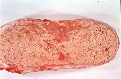
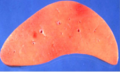
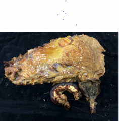
What pathology is seen in this image of a liver? How would this liver feel if you could touch it?
|
Hepatic lipidosis friable and greasy |
|
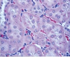
What are the darker pink dots around these renal tubules called? What are they an accumulation of? |
hyaline droplets protein |
|
What type of necorsis is seen in this image?
|
gas gangrene |
|
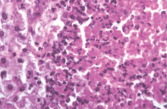
Given that this is liquefactive necrosis what cells are the arrows pointing to? |
Macrophages (also called gitter cells) |
|
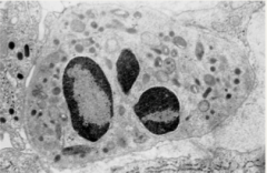
Given that these are plasma cells what is the arrow pointing to within the cell? What does this indicate? |
Russel Bodies Immune system activation |
|
Other than hepatic lipidosis what could this be and how could you tell the difference? |
Glycogen accumulation or steroid induced hepatopathy This liver would not feel greasy |
|
What process causes the change in thickness seen here? What are cells called that can only undergo this process?
|
Hyperplasia Labile cells |
|
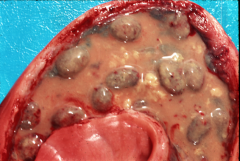
What disease is seen in this kidney? What are the two forms of this disease and which is most common in domestic animals?
|
Amyloidosis Primary = immunocyte dyscrasias/neoplasias Secondary = reactive systemic amyloidosis from chronic inflammation Secondary is most common in domestic animals |
|
What type of necrosis is this? Is cell shape and tissue architecture preserved? |
dry gangrene Yes |
|
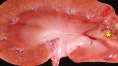
What type of lesion is indicated by the star and what is a probable cause? |
coagulative necrosis renal infarct |
|
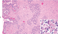
what disease is shown here? |
suppurative endometritis |
|
What type of cell is this? What stages of inflammation are these most commonly found in? |
Lymphocytes subacute and chronic inflammation |
|
Given that this is from a horse what is the most likely cause of this pigmentation? |
Carotenoid accumulation in fat |
|
What is the pigmentation shown here and what is its pathological significance? |
Congenital melanosis No pathological significance |
|
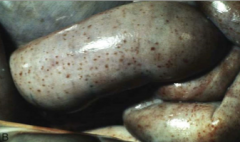
What is the term for this size of hemorrhage? |
petechial hemorrhage |
|
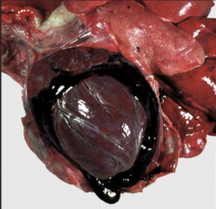
What disease is shown here and what will this cause that results in death? |
Hemopericardium cardiac tamponade |
|
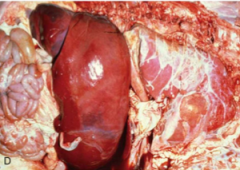
What disease process causes this liver's colour? |
Chronic congestion |
|
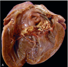
What disease is this and what are these accumulations made up of? |
vegetative valvular endocarditis thrombi and bacteria |
|
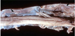
What are the arrows pointing to? |
Fibrocartilaginous embolism in the spinal cord |
|
Given that these lesions are not tumors or abcesses what are they and what defining cell type would be found in them? |
Granulomas epitheliod macrophages |
|
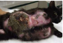
What disease is seen in this photo? What is this called in a cat? |
Saddle thrombus (saddle thromboembolism) Feline aortic thromboembolism (FATE) |
|
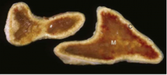
Is this venous or arterial infarction or both? How can you tell? |
Both There is congestion as well as ischemic necrosis |
|
What are the pale pink cells in this image (enlarged in the corner) and what type of inflammation are they associated with? |
epitheliod macrophages granulomatous inflammation |
|
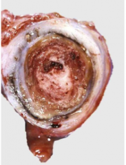
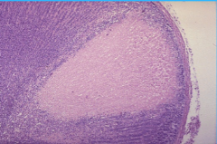
What type of granuloma is shown here? What is the defining feature? |
Complex granuloma Can see white necrotic tissue in the centre |
|
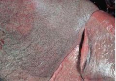
What is the colour of these mucous membranes indicative of? |
oxygen deficiency |
|
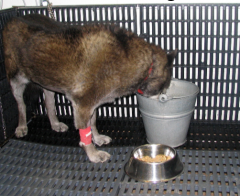
What is abnormal about this dog's requirements and what is a probable cause? |
Excessive water requirement - polydipsia hyperglycemia |
|
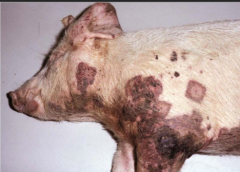
What are these lesions? |
cutaneous infarcts |
|
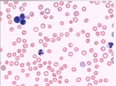
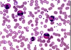
Given that this is a blood smear what is abnormal about it and what is this indicative of? |
Very low concentration of red blood cells anemia |
|
What is this disease called? |
hemarthrosis |
|
What are two names for this disease? |
hemoabdomen hemoperitoneum |
|
What type of cell is this and what is its defining feature? |
Langhan's type giant cell Multinucleated with nuclei arranged around the outside of the cell |
|
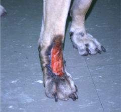
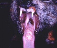
What is this lesion? |
lick granuloma |
|
What are two names for this disease? In this case is the cause more likely heart failure or infection? |
ascities and hydroperitoneum more likely infectious because the fluid is gelatinous indicating higher protein content and probable vascular damage |
|
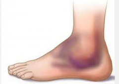
What is this type of edema called and what tissue layer is the fluid in? |
Pitting edema subcutaneous tissue |
|
What causes this colouring of the lungs? |
Chronic congestion |
|
What type of cells are shown here? |
Platelets |
|
What type of necrosis is this? |
caseous (tissue architecture is lost) |
|
What type of necrosis is this? How is this modified from the dry version of this? |
wet/moist gangrene Has some liquefactive necrosis as well as coagulative necrosis due to saprophytic bacteria |
|
what type of necorsis is this? How can you tell? |
coagulative tissue architecture is maintained |
|
What disease is shown here? What type of inflammation is this? |
lymphadenitis reactive inflammation that can be acute, subacture, or chronic
|
|
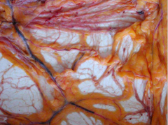
What is abnormal in this photo? What causes this? |
Can clearly see the lymph vessels (white squiggly things) Thickening of the serosa of the vessels due to chronic lymphangitis |
|
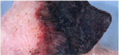
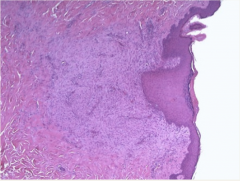
What is this disease? |
Melanoma |
|
What is the brown stuff in this image? What is excessive accumulation of this called? |
hemosiderin hemochromatosis
|
|
If this disease is caused by increased excessive breakdown of red blood cells what is is called? What is a common cause? |
Prehepatic icterus hemolytic anemia |
|
Given that this is liver tissue what are the brown lines called and where are they accumulating? |
Bile casts Accumulating in bile canaliculi |
|
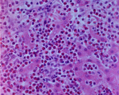
What type cellular infiltration is seen here? What might this be indicative of? |
Eosinophil infiltration Allergies, parasites infection, or mast cell tumors in dogs |
|
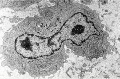
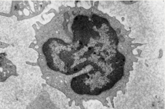
What type of cell is this? What are its four main functions? |
Macrophage 1) phagocytosis 2) modulation of inflammation and repair 3) regulation of immune response 4) Production of interleuikin 1 (inflammatory mediator)
|
|
What disease is seen here? What is being incorrectly metabolised to cause this |
Visceral gout purines |
|
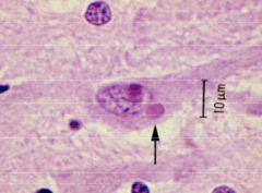
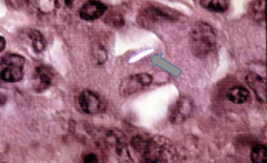
What is the arrow pointing to in the cell? Where can these be located within the cell? |
Viral inclusion body - specifically a Negri body Cytoplasm, Nucleus, or both |
|
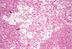
Given that this is liver what is accumulated in these cells? What is a differentiating feature? |
Glycogen Outlines are more irregular and less distinct than if this were fat |
|
Given that this is a canine spleen what is the disease shown? What is the clinical significance? |
Siderotic Plaques There is no clinical significance (normal, common finding in canines) |
|
Which of the four outcomes of acute inflammation is shown here? |
Healing by formation of scar tissue |
|
What type of cells is the arrow pointing to? What is a defining feature? |
Foreign-body type giant cells Multiple nuclei aggregated towards the middle of the cell |
|
Which of the four outcomes of acute inflammation is shown here? |
Abscess formation |
|
Is this chronic or acute inflammation? What are the hallmarks of this type of inflammation? |
Chronic Infiltration of mononuclear cells Proliferation of fibroblasts Increased connective tissue (fibrosis) Tissue destruction |
|
What process is seen on this heart? |
Serous atrophy of fat |
|
What is accumulated in this organ? What four organs does this most commonly accumulate in? |
Amyloid Liver, Kidneys, lymph nodes, and spleen |
|
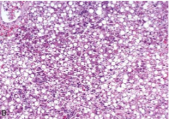
What is accumulated in these hepatocytes? What disease does this cause? |
Triglycerides Hepatic lipidosis |
|
What organ has undergone hyperplasia to cause this? What is this disease most commonly secondary to? |
Parathyroid gland Renal failure |
|
What disease is seen here? What are the three main causes of this? |
Adrenocotical atrophy 1) iatrogenic 2) Destructive pituitary gland lesion 3) idiopathic |
|
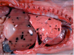
Describe the gross lesion? What species is this from? |
petichial hemorrhage bovine |
|
What disease is this? |
Metastatic hemangiosarcoma |
|
Given that this is a cat what disease is this? |
Feline infectious peritonitis |
|
What pigment has accumulated at the black patches? What four organs does this most often occur on? |
Melanin Pleura, liver, lining of the aorta, meninges |
|
What disease is shown here? |
splenomegaly |
|
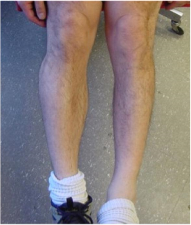
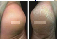
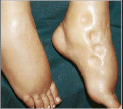
What process has the leg on the right of the image undergone? Is this process normally reversible? |
Atrophy Yes |
|
What process causes this enlargement? When this organ later shrinks what is that process called? |
Hyperplasia Involution |
|
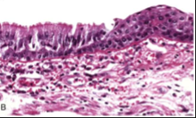
Given that this is part of the upper airway what process is occurring here? What type of cells should be lining the airway? |
Metaplasia Pseudo-stratified columnar epithelium with goblet cells |
|
What process or processes results in this condition? |
hyperplasia and hypertrophy of adipocytes |
|
Given that this is a canine what disease is this? |
Osseous metaplasia of the dura mater |
|
Given that this is an older dog what disease is this? What is its clinical significance? |
Nodular hyperplasia of the spleen No significance unless the nodules burst then dog can bleed to death |
|
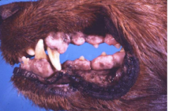
What is this disease? |
Gingival hyperplasia |
|
Given that this from the lungs of an older dog what is this disease? |
Osseous metaplasia |
|
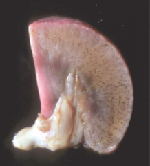
What disease is seen here? |
splenic infarction |
|
What disease is this? What age group does it usually affect? |
Fibroadenomatous hyperplasia of the mammary glands Young cats |
|
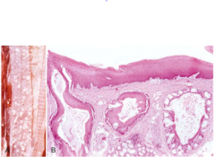
Given that this is the wall of a trachea what process is shown here? What causes this in birds? |
Squamous metaplasia Vitamin A deficiency |
|
What disease is shown here? |
Myocardial infarction |
|
What is the disease shown here? What would be the immediate cause of death? |
Ruptured aorta Hemopericardium resulting in cardiac tamponade |
|
What disease is shown here? |
pulmonary thrombosis |
|
What disease is shown here? |
Vena cava thrombosis |
|
What disease is shown here? |
valvular endocarditis |
|
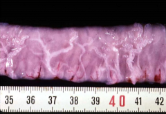
What disease is shown here? (intestine) What are the four underlying mechanisms of this? |
severe submucosal edema 1) increased hydrostatic pressure 2) increased vascular permeability 3) decreased plasma osmotic pressure 4) decreased lymphatic drainage |
|
What is this colouring called? How does this occur? |
Livor mortis After death blood pools in the downward part of the animal but any pressure points will remain white as blood cannot flow into those areas |
|
What disease is seen here? |
Acute, passive congestion of the liver |
|
What disease is shown here? What are the three inciting causes of this category of disease? |
Venous thrombosis 1) endothelial injury 2) abnormal blood flow 3) hypercoagulability |
|
What disease is shown here? (lung) |
suppurative broncho-pneumonia and thrombosis |
|
What disease is shown here? How can hemorrhage cause this? |
Icterus Break down of hemoglobin by macrophages results in higher production of bilirubin, the liver cannot metabolize the bilirubin fast enough so it accumulates in tissue |
|
What disease is shown here? What is the more general term for dust (not specifically carbon) accumulation in this organ? |
Anthrocosis Pneumoconiosis |
|
What are two names for this disease? |
hydroperitoneum ascities |
|
What disease is this? Which of the four mechanisms of oxygen deficiency would this result in? |
Pneumonia Inadequate oxygenation of blood |
|
What type of necrosis is seen here? What is often deposited in this type of necrosis? |
Fat necrosis Cholesterol crystals and basophilic calcium |
|
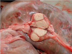
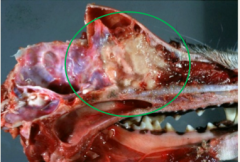
What disease is shown here? What type of inflammation is involved? |
Caseous lymphadenitis granuloma inflammation |
|
What lesion is shown here? What process is responsible for the darkness of the tissue? |
Bloat line Congestion (veins occluded from pressure from the bloated rumen) |
|
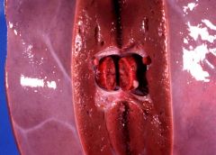
What disease is shown here? |
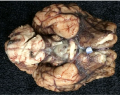
bacterial meningitis |
|
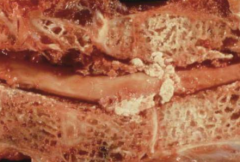
What substance is the arrow pointing to? What are the two components of this substance? |
Saponified fat Free fatty acids combined with calcium |
|
What disease is this? What is the inciting cause of this type of inflammation? |
Granulomatous sinusitis Stimuli resistant to phagocyte killing and degradation
|
|
If this lesion is found throughout the body what is the disease? What is the pathogenesis of this disease? |
Disseminated Intravascular Coagulation Widespread microthrombosis causes diffuse circulatory insufficiency and rapid consumption of clotting factors. This causes consumptive coagulopathy and widespread hemorrhage |
|
What disease is seen here? What is this indicative of? |
Serous atrophy of fat Starvation |
|
What disease is seen here? What cellular change is causing these growths? |
viral papilloma hyperplasia |
|
What type of cell is this? What are its main roles? |
Neutrophil Eliminate microorganisms, tumor cells, and foreign material |
|
What is accumulated in all of this tissue? |
Porphyrins |
|
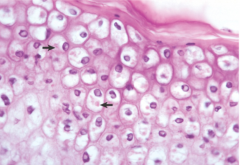
What is abnormal about these cells? What is this an accumulation of? |
Contain viral inclusions viral proteins |
|
What is this disease called? |
pulmonary edema
|
|
What disease is this? what would this skin feel like to touch? |
Subcutaneous edema doughy and fluctuant |
|
what type of inflammation is this? Are fibrosis and neovascularization features of this type? |
Subacute No |