![]()
![]()
![]()
Use LEFT and RIGHT arrow keys to navigate between flashcards;
Use UP and DOWN arrow keys to flip the card;
H to show hint;
A reads text to speech;
30 Cards in this Set
- Front
- Back
|
Define Adaptation |
Adaptations are reversible changes in the number, size,phenotype, metabolic activity, or functions of cells in response to changes in their environment. |
|
|
Write out the ADAPTATION MECHANISMS |
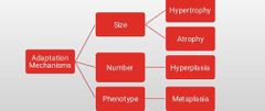
ADAPTATION MECHANISMS • Hyperplasia • Hypertrophy • Metaplasia • Atrophy |
|
|
An increase in the number of cells in an organ or tissue thus an increase in size. Is called |
Hyperplasia |
|
|
Only labile or stable cells capable of cell division become hyperplastic. T/f |
Non-dividing cells do not (e.g. myocardium and skeletal muscle) |
|
|
Cell proliferation ceases when stimulus stops unlike in neoplasia. T/f |
True |
|
|
Cell proliferation is initiated by _________ |
Growth factors, hormones andcytokines • Cytokines: TNF, IL-6 • Growth factors • Transforming growth factor (TGF) • Epidermal growth factor • Hepatocyte growth factor • Platelet-derived growth factor |
|
|
Mention two examples of physiological Hyperplasia |
• Hormonal, eg -Breast at puberty, -Pregnancy and lactation; uterus in pregnancy • Compensatory, eg following partial hepatectomy. TFG-ά andhepatocyte growth factor stimulate the cell proliferation |
|
|
Give two examples of pathological Hyperplasia |
• Excess hormone: -Endometrial hyperlasia due to oestrogen/progestorone imbalance. -BPH due to androgen. • Chronic irritation/ Injury: Connective tissue response in keloidform |
|
|
Increase in the size of the tissue or organ due to increase in the size of cells. Is termed what |
Hypertrophy |
|
|
Pathological hyperplasia can act as a fertile soil for cancer |
True |
|
|
Hypertrophy Occurs in tissues incapable of cell division. T/f |
True Occurs in non-dividing cells such as cardiac, skeletal and smooth muscle as a response to increased workload. |
|
|
Hypertrophy is induced by:
|
Growth factors:• Transforming growth factor-β (tgf-β)• Insulin-like growth factor (IGF)• Fibroblast growth factor (FGF) Hypertrophy agonists:• a-adrenergic agonists• Endothelin-i• Angiotensin-ii• Nitric oxide (NO)• Bradykinin
|
|
|
Mention 3 causes of hypertrophy |
Hypertrophy: • 1. Increased synthesis of contractile proteins • 2. Induction of embryonic/fetal genes • 3. Increased production of growth factors |
|
|
List the physiological causes of hypertrophy |
Physiologic • Uterus during pregnancy (hypertrophy & hyperplasia) • Skeletal muscle hypertrophy e.g. athletes and manual labourers |
|
|
Hypertrophy could be |
• Functional- Exercise • Compensatory- kidney • Obstructive- GIT • Hormone mediated- Anabolic steroids |
|
|
List the pathological causes of Hypertrophy |
Pathologic: • Myocardial hypertrophy in hypertension • Smooth muscle hypertrophy e.g. - Cardiac achalasia (in oesophagus)• Pyloric stenosis (in stomach) |
|
|
Shrinkage in the size of cell by loss of cell substance is called ______ |
Atrophy |
|
|
List major causes of atrophy |
Atrophy is due to: • Decreased workload • Loss of innervation • Diminished blood supply • Inadequate nutrition • Loss of endocrine stimulation • Aging- senile atrophy |
|
|
Cellular atrophy results from a combination of decreased protein synthesis and increased protein degradation. T/f |
True |
|
|
In many situations, atrophy also is associated with autophagy, with resulting increases in the number of autophagic vacuoles. T/f |
True |
|
|
List the examples of Physiological atrophy:
|
• During fetal development: e.g. atrophy of embryonic structures such as thyroglossal duct. • During adult life: e.g. involution of thymus, atrophy of brainand heart due to aging (senile atrophy). |
|
|
List the examples of pathological atrophy |
1. Local - Disuse atrophy (decreased workload): Immobilization/ prolonged bed rest. - Denervation atrophy: Poliomyelitis - Ischemic atrophy: Brain atrophy produced by ischemia due to atherosclerosis of the carotid artery. - Pressure atrophy: Renal parenchyma atrophy in hydronephrosis due to increased pressure. 2. Generalized - Starvation atrophy: Protein-calorie malnutrition |
|
|
A reversible change in which one adult cell type (eg epithelial) is replaced by another adult cell type is termed |
Metaplasia |
|
|
Metaplasia in certain cases may progress to ______&_______ |
Dysplasia and Neoplasia |
|
|
Substitution of more sensitive cells by cell types better able to withstand stress and adverse conditions is also termed metaplasia. T/f |
True |
|
|
_______ mechanism is Due to re-programming of stem cells or undifferentiated mesenchymal cells to adifferent cell type • Both extra- and intra-cellular signals induce transcription of genes controlling differentiation along new pathway. |
Metaplasia |
|
|
Mention two examples of metaplasia |
1. Epithelial Metaplasia • Squamous metaplasia: • Respiratory tract: e.g. chronic irritation due to tobaccosmoke • Cervix: associated with chronic infection. • Columnar metaplasia: • Squamous to columnar: Barrett esophagus • Intestinal metaplasia
2. Connective Tissue Metaplasia • Osseous metaplasia: New bone at sites of tissue injury.Myositis ossificans- usually following intramuscular hemorrhage. |
|
|
DYSPLASIA • Deranged development • Proliferation with associated cytological atypia (size, shapeand organization) • Not an adaptive process • Sometimes called atypical hyperplasia• Usually accompanies adaptive changes• A precursor of neoplasia.
T/f |
True |
|
|
HYPOPLASIA • Reduction in size of cells and tissue • Mech: Failure of growth to normal size T/f |
True |
|
|
APLASIA• Also called ________
|
Agenesis
• The complete failure of growth of an organ or part atembryogenesis• Seen in genetic disorders e.g amelia |