![]()
![]()
![]()
Use LEFT and RIGHT arrow keys to navigate between flashcards;
Use UP and DOWN arrow keys to flip the card;
H to show hint;
A reads text to speech;
14 Cards in this Set
- Front
- Back
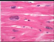
|
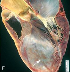
Boxcar nuclei indicative of LV hypertrophy |
|
|
Tet spells |
Tetralogy of Fallot; relieved by squatting to improve O2 circulation. Children suddenly develop deep blue skin, nails and lips after crying, feeding, having a bowel movement, or kicking his or her legs upon awakening |
|
|
1) ASD a/w ___________ 2) PDA a/w ___________ 3) Mitral valve prolapse a/w __________ 4) Hypertrophic cardiomyopathy a/w __________ |
1) Down's Syndrome 2) Turner Syndrome 3) Marfan's Snydrome 4) Freidrich's ataxia |
|
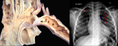
|
Patent Ductus Arteriosus (PDA), specifically the adult form, where the obstruction is proximal to the aorta. Collateral circulation to the internal thoracic arteries causes rib notching. |
|
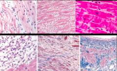
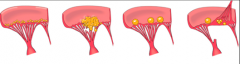
Progression of MI A) B) C) D) E) |
A) Coagulative necrosis with wavy fibers (removes nucleus) - 1 day old B) PMN infiltrate; risk for fibrinous pericarditis - few days to 1 week old C) Removal of necrotic debris by macrophages; risk for ventricular wall rupture and cardiac tamponade - 1 week old D) Granulation tissue characterized by loose CT and many capillaries -- few weeks old E) Healed infarct with dense collagenous scar; risk of aneurysm or Dressler Syndrome - 1 month old |
|
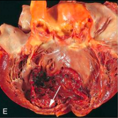
Complication from MI |
Recent expansion anteroapical infarct with wall stretching and thinning and mural thrombus; may lead to system emboli to brain. |
|
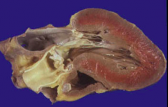
Complication from MI |
Apical LV aneurysm which will likely lead to CHF |
|
LV hypertrophy due to calcific aortic stenosis 1) Congenital relationship? 2) _____________ murmur 3) Abnormal heart sound? |
1) A/w bicuspid aortic valve 2) Crescendo-decrescendo mid-systolic ejection murmur 3) Paradoxically split S2 |
|
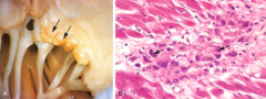
Vegetations a/w heart diseases 1) 2) 3) 4)
|
1) Rheumatic heart disease (along line of closure of affected valve) 2) Infective endocarditis (large, irregular, destructive masses extending from valve leaflets to adjacent structures) 3) Nonbacterial thrombotic endocarditis (small'ish non-destructive vexations along line of closure) 4)Libman-Sacks endocarditis (a/w SLE) are seen on either side of the affected valve |
|
In RF, vegetations alone line of closure of affected valve is called ________. Myocarditis with focal inflammatory lesions are called _______. Modified myocytes that look like "caterpillar cells" are called _________. |
Verrucae; Aschoff bodies; Anitschkow cells. |
|
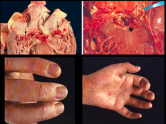
Infective endocarditis. Describe clinical signs seen. LL, LR |
LL: nailbed splinter hemorrhades LR: Janeway lesions + "Roth spots" which are retinal hemorrhages + Painful fingertip nodules which are Osler nodes |
|
|
1) Subacute endocarditis a/w what bug? 2) Acute endocarditis a/w what bug? 3) IV drug users are susceptible to pathology in ______ valve with _______ (what bug?). |
1) Strep viridans on previously myxomatous MV 2) Staph aureus 3) Tricuspid; staph aureus |
|
|
_____________ is associated with primary tumor of the small intestine that secretes 5-HT. Classic sx include facial flushing, cramps, diarrhea, asthma-like bronchoconstriction. |
Carcinoid heart disease |
|
|
_____________ myopathy causes a banana-shaped ventricular lumen and possible sudden death in young athletes. |
Hypertrophic cardomyopathy with asymmetric septal hypertrophy. |