![]()
![]()
![]()
Use LEFT and RIGHT arrow keys to navigate between flashcards;
Use UP and DOWN arrow keys to flip the card;
H to show hint;
A reads text to speech;
106 Cards in this Set
- Front
- Back
- 3rd side (hint)
|
What groups are considered at-risk for getting a parasitic infection? |
Immunosuppressed (children under 2; elderly) Immunocomprimised (HIV/AIDS) |
|
|
|
What are the risk factors for parasitic infections? |
Poor hygiene Use of human feces as fertilizer (fecal-oral transmission) World travel/trade- endemic regions, shipping of contaminated goods |
|
|
|
Symbiosis |
"living together" typically 2 different organisms benefit from relationship, metabolic dependence |
|
|
|
Mutualism |
two different organisms benefit from the relationship |
|
|
|
Commenalism |
one organism benefits at no cost/harm to the other |
|
|
|
Parasitism |
one organism benefits at the cost/harm of the other, smaller organism is metabolically dependent on larger host |
|
|
|
Endoparasites |
lives inside a host |
|
|
|
Ectoparasites |
lives outside host |
|
|
|
Obligate parasites |
must spend a part or all of their life cycle in a host |
|
|
|
Facultative parasites |
typically free living but if get into a human via ingestion or wounds they can become parasitic and cause harm to human host |
|
|
|
Accidental parasites |
live on or in non-human host but accidentally get into humans, but do not survive long in humans |
|
|
|
Definitive hosts |
one in which parasite reaches reproductive maturity (adult phase and/or sexual reproduction) |
|
|
|
Intermediate hosts |
one in which parasite passes through its larval or asexual reproductive phase |
|
|
|
Transport hosts |
harbors a parasite that does not reproduce but can infect another individual at a new location |
|
|
|
Reservoir hosts |
an organism in which a parasite that is pathogenic for some other species lives and multiplies without damaging its host |
|
|
|
Accidental hosts |
one which is not the normal host species, parasite may or may not be able to complete its life cycle |
|
|
|
Zoonosis hosts |
a disease of animals that can be transmitted to humans, originally used in those cases transmitted by domestic animals |
|
|
|
What are some ways hosts are susceptible to infections? what are some methods of infections (general)? |
boring larvae, penetrate, migrate entrance into mouth as cysts, eggs, larvae bite of a vector |
|
|
|
Cyst |
have a protective membrane or thickened wall non-motile non-feeding infective found in environment identification--> size/shape, # of nuclei, other inclusion bodies |
|
|
|
Trophozoite |
motile reproducing found in definitive host feeding identification: nuclear structure, cytoplasmic inclusions, size, motility unicellular, eukaryotic large nucleus dark, irregular thickened ring of chromatin around nuclear periphery eccentric nucleus contains large karysome/endosome (nucleolus in humans) |
|
|
|
General features of protozoans |
eukaryotic, unicellular simple life cycle--> cyst, trophozoite that serve as diagnostic stage reproduction via binary fission (asexual) |
|
|
|
What are the modes of transmission for protozoans? |
ingestion arthropod vector sexual intercourse |
|
|
|
Karyosome |
small mass of chromatin within the nucleus also called endosome |
|
|
|
Chromatoid bars/bodies |
rod shaped structure composed of condensed RNA found in cytoplasm of some amoebic cysts |
|
|
|
What is the method of infection for amoebae? |
cyst is infective stage for humans that are usually ingested from fecal contaminated food/water they move to the lower intestine and excyst then begin to multiply (reproduce) as feeding trophozoites |
|
|
|
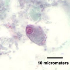
How can Entamoeba histolytica be diagnosed? |
ingested RBCs on a wet mount, stains of trophozoites |
|
|
|
What can Entamoeba histolytica cause? |
dysentery intestinal ulceration extraintestinal disease- liver, lungs, other organs |
|
|
|
What are the characteristics of motility for Entamoeba histolytica? |
thin, pointed pseudopodia active motility in wet mounts of fresh specimen |
|
|
|
Entamoeba histolytica is considered what? |
considered an STD may be fatal if left untreated pathogenic |
|
|
|
What is the life cycle of Entamoeba histolytica? |
1. resistant, infective cysts that are passed in feces (trophs found in soft/fluid feces) 2. human ingests infective cyststransmitted by feces, fingers, food, fomites, flies 3. cyst passes to small intestine where excystation occurs 4. trophs in large intestine multiplying asexually by binary fission (10-20 um) |
|
|
|
What is the diagnostic stage of Entamoeba histolytica? |
resistant, infective cysts that are passed in feces (trophs found in soft/fluid feces) |
|
|
|
What is the infective stage of Entamoeba histolytica? |
resistant, infective cysts that are passed in feces (trophs found in soft/fluid feces) |
|
|
|
What is the method of infection for Entamoeba histolytica? |
human ingests infective cysts transmitted by feces, fingers, food, fomites, flies |
|
|
|
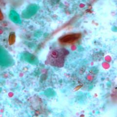
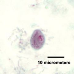
What are the characteristics of Entamoeba histolytica trophozoite? |
10-65 um Dark RBC inclusion Finely granular cytoplasm 1 nucleus Thin, uniform, chromatin ring Small central karysome |
|
|
|
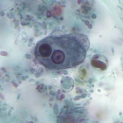
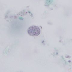
What are the characteristics of Entamoeba histolytica cyst? |
10-20 um 1-4 nuclei chromatoid bodies (cigar shaped) |
3 nuclei chromatoid bodies |
|
|
Entamoeba coli is considered what? |
non-pathogenic intestinal amoeba |
|
|
|
How would you describe the motility of Entamoeba coli? |
sluggish due to blunt pseudopodia |
|
|
|
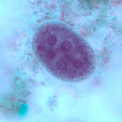
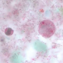
What are the characteristics of Entamoeba coli cyst? |
8-35 um 8-10+ nuclei Presence of chromatoid bodies (splinter like with pointed ends) in cytoplasm |
|
|
|
What are the characteristics of Entamoeba coli troph? |
15-50 um Vacuoles present in coarsely granular cytoplasm 1 nucleus Eccentrically located karyosome Irregular thickened chromatin ring |
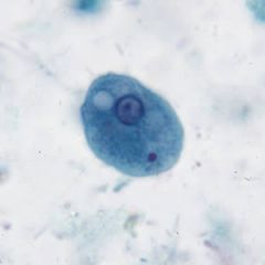
Vacuole present |
|
|
Entamoeba hartmanni |
non-pathogenic small cyst (less than 10 um) similar to E. histolytica |
|
|
|
Entamoeba dispar |
non-pathogenic morphologically identical to E. histolytica except does not ingest RBCs non-invasive |
|
|
|
Entamoeba polecki |
pigs, monkeys, occasionally humans differentiate from E. histolytica by the cyst only having 1 nucleus with abundant and pointed chromatoid bodies |
|
|
|
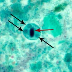
Endolimax nana |
non-pathogenic no peripheral chromatin nucleus has ball and socket appearance only 6-12 um both cyst and troph |
Cyst |
|
|
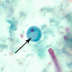
Iodamoeba butschlii cyst |
large glycogen vacuole only 1 nucleus |
|
|
|
Iodamoeba butschlii troph |
can contain bacteria, yeast, one nucleus |
|
|
|
Entamoeba gingivalis |
non-pathogenic mouth, occasionally sputum, not in the intestinal tract has no cyst stage only amoeba known to ingest WBCs fecal-oral route |
|
|
|
Naegleria fowleri |
brain eating amoeba facultative human parasites (don't need us) free-living amoebaflagellate |
|
|
|
Where are Naegleria fowleri parasites found? |
fresh/salt water, moist soil, decaying vegetation, swimming pools, tap water and air conditioning units cysts resistant to chlorination/drying |
|
|
|
What are the characteristics of disease for Naegleria fowleri? |
Primary amoebic meningoencephalitis (PAM) rapid, usually fatal disease- diagnosis made on autopsy typical onset (flu-like) progressing to irrational behavior, coma, death purulent CSF- increased WBCs, no bacteria |
|
|
|
Where are Naegleria fowleri ameboid trophozoites found?
|
in host tissues (flagellate trophozoite and cyst form are not found in human but are in the environment) |
|
|
|
How do Naegleria fowleri enter the host? |
Ameboid trophozoites penetrate nasal mucosa and penetrate brain via nerves through ostia *treatment usually unsuccessful |

|
|
|
Acanthamoeba spp. causes what? |
Granulomatous amebic encephalitis (GAE) - chronic form of meningioencephalitis |
|
|
|
What are the two methods of infection of human infection for Acanthamoeba? |
1. isolated from respiratory tract as cysts and trophozoites, can be inhaled can enter blood and spread to CNS 2. directly into the eye from contaminated saline Keratitis- poor contact lens care Lives in saline not water- most common in home-made saline solutions |
|
|
|
Acanthamoeba cyst |

|
|
|
|
Acanthamoeba trophozoite |

|
|
|
|
What is the life cycle for Naegleria fowleri? |
1. Free-living trophozoites (cyst or biflagellate form) 2. trophozoite enters nasal cavity from infected water while swimming 3. trophozoite migrates to CNS via olfactory lobes (7-10 days) 4. invades frontal cortex (1-2 days) 5. active trophozoites in spinal fluid; trophozoite found in brain at autopsy: PAM usually ends in death (3-6 days) |
|
|
|
What is the life cycle for Acanthemoebia spp. |
1. Free-living forms trophozoite (cyst) 2. trophozoite enters through respiratory tract, broken skin or mucous membranes; directly invades eye 3. reaches CNS and other tissues, including bone via bloodstream (weeks-months) 4. trophozoites and cysts recovered from brain or skin biopsy or corneal scraping- rarely seen in CSF |
|
|
|
How does Balantidium coli move? |
By cilia |
|
|
|
Balantidium coli |
only pathogenic ciliate to infect humans largest parasitic protozoa (40x60 um trophozoite) can cause dysentery may invade tissues/produce lesions have a cyst and trophozoite stage |
|
|
|
What are the characteristics of Balantidium coli cyst and troph stages? |
small micronucleus large bean shaped macronucleus cytosome- rudimentary mouth cilia around the periphery asexual reproduction via binary fission sexual reproduction via exchange of micronuclei |
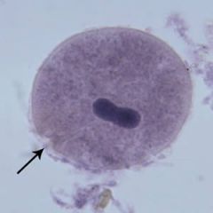
Trophozoite |
|
|
Morphological features of flagellates |
flagella axostyle axoneme costa |
|
|
|
Axostyle |
rod-like support structure in some flagellates |
|
|
|
Axoneme |
Internal cytoskeletal support structure of flagella |
|
|
|
Costa |
rod-like structure located at the base of undulating membrane connecting it to the body of some flagellate trophozoites |
|
|
|
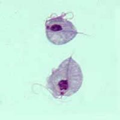
Giardia lamblia (intestinalis) general info |
most common parasite in US pathogenic symptoms range from mild diarrhea to overt malabsorption syndrome severe cases may have copious light colored, fatty stools world wide distribution- associated with contaminated water increases being recorded in homosexual males |
|
|
|
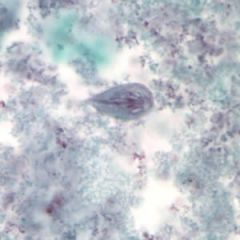
Giardia lamblia troph |
old man/monkey face/tennis racket/pear ventral sucking disk attaches to GI mucosa bilateral symmetrical paired structures median bodies (microtubules component of cytoskeleton) 8 flagella 2 anterior nuclei falling leaf motility |
|
|
|
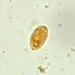
Giardia lamblia ovoid cyst |
4 nuclei (2 in immature cysts) and median bodies clustered nuclei and axoneme (intracellular portion of flagellum) gives little old lady appearance cyst wall set off from cytoplasm in fixed preparations |
|
|
|
What is the infective stage of Giardia lamblia? |
resistant, infective cysts passed in feces (trophozoites may be found more commonly in soft or fluid feces) |
|
|
|
What is the diagnostic stage of Giardia lamblia? |
resistant, infective cysts passed in feces (trophozoites may be found more commonly in soft or fluid feces) |
|
|
|
What is the method of infection of Giardia lamblia? |
human ingests infective cysts transmitted by feces, fingers, food, fomites, flies and infected water |
|
|
|
What is the life cycle of Giardia lamblia? |
1. resistant, infective cysts passed in feces (trophozoites may be found more commonly in soft or fluid feces) 2. human ingests infective cyststransmitted by feces, fingers, food, fomites, flies and infected water 3. cyst passes to small intestine; excystation occurs 4. trophozoites in small intestine; multiply asexually by binary fission (15x10 um) |
|
|
|
Dientamoeba fragilis |
pathogenic life cycle poorly understood no cyst stage binucleate troph stage no peripheral nuclear chromatin (4-8 chromatin granules in central mass) |
|
|
|
What is diagnostic of Dientamoeba fragils? |
In wet preps, cytoplasmic granules exhibit Brownian motion known as Hakansson phenomenon |
|
|
|
Dientamoeba fragilis troph |
binucleate |
|
|
|
Chilomastix mesnili |
pear-shaped flagellate with bilateral symmetry falling-leaf motility sucking disk on ventral side has cyst and troph stages non-pathogenic |
|
|
|
Chilomastix mesnili cyst |
|
clear knob on top cytosome (mouth) |
|
|
Chilomastix mesnili troph |
|
curved posterior cytosomal groove --> non-rudimentary mouth 3 anterior flagella and nucleus |
|
|
Trichomonas hominis |
No cyst stage found in fecal specimen undulating membrane extends the entire length of the body Found in GI tract |
|
|
|
Trichomonas vaginalis |
Found in female/male lower reproductive tract- considered STD and in wet prep of discharge/urine or PAP smear Motile trophozoites No cyst stage |
|
|
|
What pathologies does Trichomonas vaginalis cause in females and males? |
Considered STD Females: vaginitis, discharge, dysuria Males: asymptomatic |
|
|
|
Trichomonas tenax |
found in oral cavity considered non-pathogenic |
|
|
|
hemoflagellates |
ALL SPECIES REQUIRE ARTHROPODS AS INTERMEDIATE HOST! multiply in human blood/tissues pathogenic forms: Trypanosoma sp. and Leishmania sp. |
|
|
|
kinetoplast |
accessory body consisting of large mitochondrion next to basal granule of anterior undulating membrane flagellum containing mitochondrial DNA |
|
|
|
amastigote |
small, ovoid, non-flagellated form with notable structure including mitochondrial kinetoplastid and large nucleus |
|
|
|
epimastigote |
flat, spindle-shaped, flagellated forms seen in the gut or salivary glands of insect vectors in trypanosomes life cycles have an undulating membrane extending from flagellum to small kinetoplast just anterior to nucleus |
|
|
|
promastigote |
flagellate form where kinetoplast is located at the anterior end of the organism with no undulating membrane seen in the midgut and pharynx of vectors in life cycle of leishmania |
|
|
|
trypomastigote |
flagellate form with kinetoplast located at posterior end and undulating membrane extending entire body from flagellum to posterior end seen in blood of humans infective stage transmitted by insect vectors |
|
|
|
What is the infective stage for Trypanosoma rhodesiense and Trypanosoma gambiense? |
Tsetse fly bites human, trypomastigotes from salivary glands deposited |
|
|
|
What is the method of infection for Trypanosoma rhodesiense and Trypanosoma gambiense? |
Tsetse fly bites human, trypomastigotes from salivary glands deposited |
|
|
|
What is the diagnostic stage for Trypanosoma rhodesiense and Trypanosoma gambiense? |
Trypomastigotes multiply in peripheral blood early in disease and later in lymph nodes and CNS T. rhodesiense is more acute process, earlier CNS involvement (~1 month) T. gamiense is more of a chronic process that takes ~1 year for CNS involvement |
|
|
|
What is the intermediate host of Trypanosoma rhodesiense and Trypanosoma gambiense? |
Tsetse fly |
|
|
|
African Trypanosomiasis (Sleeping Sickness) |
bite site lesion present enlarged lymphnodes (neck, cervical area) WINTERBOTTOM'S SIGN fever, sweats, headache, joint/muscle pain CNS involvement lethargy/motor changes daytime sleepiness, night time sleep disturbances (CNS involvement) possibly coma/death if untreated |
|
|
|
How do you come to a diagnosis for African Sleeping Sickness? |
trypomastigotes in blood- thick and thin smears using Giemsa stain with a good clinical history to help distinguish subspecies level |
|
|
|
How do you treat African Sleeping Sickness? |
various drugs depends on early/late in disease cure IS achievable if caught early enough |
|
|
|
What is the life cycle of African Sleeping Sickness? |
1. trypomastigotes ingested by tsetse fly; epimastigote forms multiply in fly gut; infective form moves to salivary gland 2. tsetse fly bites human, trypomastigotes from salivary glands deposited 3. trypomastigotes multiply in peripheral blood early or lymph nodes and CNS later |
|
|
|
Where can you find Trypanosoma cruzi (Chagas' disease)? What does it affect in humans? |
Mexico, Central and South America Amastigotes parasitize heart muscle, liver, CNS |
|
|
|
What is the Chagas' disease acute condition characteristics? |
fever, weakness, enlarged spleen, liver, lymph nodes In children- rapid death due to cardiac involvement may progress to chronic infection and death due to cardiac involvement |
|
|
|
What is the life cycle of Chagas' disease? |
1. reduviid bug bites human and ingests trypomastigotes 2. epimastigote forms multiply in midgut of bug 3. trypomastigotes in feces of bug 4. reduviid bug bites and fecally contaminates wound 5. trypomastigotes are rubbed into wound or conjunctiva, invade various tissues cells, and become amastigotes 6. amastigotes found in heart muscle, liver or CNS in macrophages; trypomastigotes occasionally found in blood smear |
|
|
|
What is the diagnostic stage of Trypanosoma cruzi? |
amastigotes found in heart muscle, liver or CNS in macrophages; trypomastigotes occasionally found in blood smear |
|
|
|
What is the infective stage of Trypanosoma cruzi? |
trypomastigotes in feces of bug |
|
|
|
What is the method of infection for Trypanosoma cruzi? |
reduviid bug bites and fecally contaminates wound |
|
|
|
What is the intermediate host for Leishmania? |
Sandfly- Phlebotomus spp |
|
|
|
What is the life cycle of Leishmania? |
1. vector bites human and regurgitates promastigotes 2. promastigotes invade tissue at wound site 3. macrophages engulf promastigotes, which convert to amastigote form 4. amastigote forms multiply in macrophages 5. amastigotes form in macrophages tropica, mexicana, braziliensis invade skin-lesion macrophages only; donovani also invades bone marrow, liver, and spleen macrophages 6. amastigotes multiply by longitudinal division in macrophages 7. biting sandfly ingests infected macrophages containing amastigotes 8. promastigote form multiplies in gut |
|
|
|
What is the diagnostic stage of Leishmania? |
amastigotes form in macrophages tropica, mexicana, braziliensis invade skin-lesion macrophages only; donovani also invades bone marrow, liver, and spleen macrophages |
|
|
|
What is the infective stage of Leishmania? |
promastigote form multiplies in gut |
|
|
|
What is the method of infection for Leishmania? |
vector bites human and regurgitates promastigotes |
|