![]()
![]()
![]()
Use LEFT and RIGHT arrow keys to navigate between flashcards;
Use UP and DOWN arrow keys to flip the card;
H to show hint;
A reads text to speech;
46 Cards in this Set
- Front
- Back
|
Aspiculuris tetraptera |
common name: mouse pinworm
host: mouse pathology: proximal loop of colon, w/ large infection: rectal prolapse, enteritis, sticky stool and puritis diagnostics: eggs on flotation *females do not migrate to deposit eggs* |
|
|
Syphacia obvelata, Syphacia muris |
common name: mouse and rat pinworms host: mouse and rats pathology: reside in cecum, gravid female in colon to deposit eggs on perianal area, female dies after laying, large infection: enteritis, sticky stool, rectal prolapse, prutitis diagnostics: banana shaped eggs (obvelata), football shaped eggs (muris) |
|
|
Trichosomoides crassicauda |
rat bladder worm - adults parasitize bladder wall,associated with bladder tumors, male worm resides in female - eggs in urine sediment |
|
|
neotenic means what |
male resides in female genital tract and lives attached to female solely for reproduction |
|
|
Dentostomella translucida |
gerbil pinworm - parasitize stomach and proximal 1/3 of intestine, no clinical signs (mild inflammation and irratation) - eggs in fecal float but only intermittently shed *males have unique cuticular inflation cranial to cloaca* |
|
|
Paraspidodera uncinata |
Guinea pig ascarid - in cecum and colon, generally nonpathogenic, heavy infection can cause diarrhea and weight loss - eggs in fecal float |
|
|
Passalurus ambiguous |
Rabbit pinworms - in cecum and colon, nonpathogenic, adults feed on bacteria within intestinal content, do not disturb intestinal lining - pass in feces, immediately infective |
|
|
Obeliscoides cuniculi, Trichostrongylus calcaratus |
Rabbit Trichstrongyle - Common in wild rabbits -Adult O. cuniculi are found in the stomach (hemorrhagic gastroenteritis) - Adult T. calcaratus are found in the small intestine (anemia) -Eggs can be recovered on fecal flotation |
|
|
Ascaridia species |
Bird Ascarid -Common in poultry and waterfowl, uncommon in pet and aviary birds -With large infections, intestinal obstruction within the smallintestine -Eggs can be seen on flotation and direct smear |
|
|
Spiroptera incestaDyspharynx nasuta& Tetrameres species |
BirdSpirurids - Australian finches - Incesta - Finches - Nasuta - Pigeons - Tetrameres -Uncommon in pet and aviary birds -Adults in the ventriculus/proventriculus -Detection is difficult: histopath examination of the tissue |
|
|
Capillaria species |
Poultry Capillaria - Pheasants, peafowl, and other poultry Adults within crop and upper alimentary tract(cause inflammation and GI disturbances) -Diagnosis: eggs from fecal flotationor direct smear of a sample from the crop |
|
|
Gongylonema pulchrum |
RuminantEsophageal Worm - embedded within the submucosa/mucosa - have unique zigzag appearance within the tissue Eggs can be identified on fecal flotation |
|
|
Bunostomum, Chabertia, Cooperia, Haemonchus, Oesophagostomum, Ostertagia, Trichostrongylus |
Trichostrongyles of Ruminants - Adults in the abomasum, small intestine & large intestine -Oval, thin-shelled eggs with a morula -Mixed infections are common and the eggs are too similar to identify specific genus/species*Record on a fecal as a strongyle type egg* |
|
|
Nematodirus |
-Camelids, Cattle, Goats, and Sheep -Similar characteristics as the other ruminant trichostrongyles except eggs are the largest ofthe trichostrongyles |
|
|
Strongyloides papillosus |
Bovine threadworm - Adults in small intestine, feed on host’s blood -Heavy infections: diarrhea, anorexia, blood and mucus in thefeces, weight loss -Only female is parasitic -female produces larvated eggs - Diagnosed via fecal flotation |
|
|
Trichuris ovis |
Ruminant Whipworm - Adults in cecum and colon -Heavy infections: dark feces, anorexia, anemia - Fecal flotation to identify eggs |
|
|
Elaeophora schneideri |
Sheep Artery Worm - Adults infect carotid arteries of sheep, females produce microfilariae -microfilariae can produce dermatitis on face, poll and feet - Diagnosis is made by characteristic skin lesions and identificationof the microfilariae - treat based on clinical signs |
|
|
Dictyocaulus viviparous, Dictyocaulus filaria |
Ruminant Lungworms - Adults infect bronchi and cause coughing -Larvae found on fecal float, float ofsputum/mucus discharge, or the Baermann technique - Larvae have characteristic brown food granules in their intestinal cells |
|
|
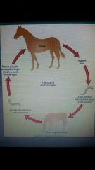
Dictyocaulus viviparous,Dictyocaulus filaria life cycle |
|
|
|
Muellerius capillarisProtostrongylusspecies |
Sheep/Goat Lungworms - Adults infect the bronchioles where they can lead to bronchopneumonia -First-stage larvae can be found on standard fecal flotation |
|
|
Stephanofilaria stilesi |
Ruminant Skin worm - adults and microfilariae reside in skin, they produce dermatitis along the ventral midline -lesions begin as red papules, progress to pruritic areaswith alopecia and thick, moist crusts -Microfilariae found in deep skin scrapings after the crust havebeen removed from the lesions |
|
|
Thelazia rhodesii, Thelazia gulosa |
Ruminant Eye worm - Adults live within conjunctival sac and lacrimal duct where theycause inflammation -Diagnosed by observing ova or L1 larvae in lacrimal secretions |
|
|
Setaria cervi |
Ruminant Abdominal Worm - Adults are found free in abdominal cavity -Often an incidental finding during abdominal surgery -Microfilariae noted in peripheral blood smear |
|
|
Habronema microstoma& Habronema muscae |
Horse Stomach Worms - Adults reside within mucosa of stomach - Heavy infection causes colic and diarrhea -Intermittent host is muscid fly -Summer Sores- fly deposits larvae in skin wound,the larvae become an aberrant parasite,cutaneous habronemiasis -Larvated eggs or larvae onfecal flotation - The eggs are elongated, thin-walled |
|
|
Trichostrongylus axei |
Large Animal Stomach Strongyle - Adults in the stomach, suck host’s blood -Can infect cattle, sheep, and swine if they ingest the infectivethird-stage larvae - Diagnosed on standard fecal flotation as a strongyle type egg |
|
|
Parascaris equorum |
Horse Roundworm/Ascarid - Adults in small intestine of young foals - Heavy infections cause unthriftiness, depression, pot-bellied appearance, anorexia, colic, and coughwith nasal discharge -Diagnosed on standard fecal flotation -eggs are sticky -Adults largest of equine nematode have large,distinctive lips- adults in feces after dewormed |
|
|
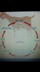
Parascaris equorum lifecycle |
|
|
|
Strongylus vulgaris, Strongylus edentaus, Strongylus equinus |
Large strongyles - Adults parasitize large intestine -Clinical signs related to migration of the larvae through mesenteric arteries and liver -Clinical signs include colic, weight loss, lethargy, fever, and poorappetite -S. vulgaris often causes thrombi within the anterior mesenteric artery |
|
|
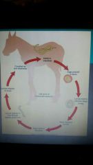
Large strongyles life cycle |
|
|
|
Equine Small strongyles-Over40 different species |
- all live in cecum and intestines, do not undergo migration as the large strongyles do - Diagnosed on fecal flotation -eggs of large and small strongyles are identical and should be recorded as “strongyle type egg”-3rd stage larvae may becomeencysted in the intestinal wallslarval cyathostominosis occursas a mass emergence of 4th stage larvae -High fatality rate despite the best standard of care given to affectedhorses |
|
|
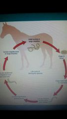
Small strongyles-Lifecycle |
|
|
|
Strongyloides westeri |
Horse Threadworm - Adults in the small intestine, feed on blood -Only parthenogenetic female is parasitic -Larvae enter the host through the skin or transmitted from mare tofoal via colostrum - Symptoms include: diarrhea, weight loss, anemia, and poor appetite -Larvated eggs on standard fecalflotation |
|
|
Oxyuris equi |
Equine Pinworm - Adults in the cecum, colon, and rectum - Adult female worms attach eggs to the exterior of the anus with sticky, gelatinous material -eggs cause inflammation and anal pruritus -eggs rubbed off anus into rubbed area - Adults worms often observed protruding from rectum - Diagnosis: finding eggs on microscopic exam of tapeimpressions |
|
|
Dictyocaulus arnfieldi |
EquineLungworm - Adults in bronchi and bronchioles -ova and larvae found on standard fecal flotation -larvae hatch within few hours after feces is passed -Identification of the eggs must be made from fresh feces |
|
|
Onchocerca cervicalis |
Equine Filarial Skin Worm - Adults in the Nuchal ligament -adult females produce microfilariae that migrate to the skin - neckand ventral midline -Many infected horses are asymptomatic -microfilariae cause cutaneous onchocerciasis and leavepermanent scars and white hairs -microfilariae can also migrate inside the eye and cause blindness |
|
|
Thelazia lacrymalis |
Equine Eyeworm - Adults in conjunctival sac and lacrimal duct where theycause obstruction of lacrimal duct -Examination of the lacrimal secretions may reveal eggs or first-stagelarvae |
|
|
Setaria equina |
Horse Abdominal worm - Adults found freely within abdominal cavity -Adults may be noted during abdominal surgery -Microfilariae can be diagnosed in peripheral blood smears |
|
|
Ascarops strongylina, Physocephalus sexalatus |
Pig Stomach Worms - Adults attached to stomach mucosa, suck host’s blood -Clinical signs include anemia, diarrhea, and weight loss - Thick-walled, larvated eggs can be recovered on fecal flotation |
|
|
Hyostrongylus rubidus |
Pig Red Stomach Worm - Adults suck host’s blood and cause dehydration, weight loss,diarrhea, and anemia - Trichostrongyle type egg can be recovered onfecal flotation -Diagnosis of individual genus/species cannot be made -ID should be recorded as “strongyle type of egg” |
|
|
Ascaris suum |
Pig Roundworm - Largest intestinal nematode in pigs -small intestine of pigs (intestinal obstruction) -Clinical signs: reduced growth, weight loss, unthriftiness, Respiratory symptoms due to larval migration -Adults noted in the feces -Eggs on flotation and have prominentprojections, give them lumpy, bumpy appearance |
|
|
Strongyloides ransomi |
Pig Threadworm - Only parthenogenetic female is parasitic -Adult females located in small intestine where suckthe host’s blood and can cause diarrhea, anemia, and weight loss -Transmission occurs through the colostrum or percutaneous -Eggs recovered in flotation of fresh feces |
|
|
Oesophagostomum dentatum |
Pig Nodular Worm - larval stages & adults are in large intestine -Larval stages form nodules within wall of the large intestine -Cause intestinal disturbances and obstruction -Clinical signs: anorexia, diarrhea, weight loss, & GI disturbances -Strongyle type egg onstandard fecal flotation |
|
|
Trichuris suis |
Pig Whipworm - Adults infect the cecum and colon -Symptoms of anemia, bloody diarrhea, anorexia, stunted growth -Eggs can be recovered on fecal flotation |
|
|
Trichinella spiralis |
Pig Trichinella Worm -pig is intermediate and definitive host -Definitive host in small intestine -Intermiediate host in striated muscle fibers -Pigs usually asymptomatic -Significant zoonotic concerns -Diagnosed by finding encysted larvae within muscle tissue |
|
|
Metastrongylus elongatus |
Pig Lungworm - Intermediate host is earthworm -Adults parasitize bronchi and bronchioles -Clinical signs: persistent cough, reduced growth rate, unthiftiness from partial airway obstruction -eggs heavier then other nematode eggs |
|
|
Stephanurus dentatus |
Pig Kidney Worm - Adults in cystic spaces that connect to kidneys,ureters, and perirenal tissues -Much of damage caused by larval migration to these tissues -Symptoms include: anorexia, decreased growth rate, & weight loss -Eggs recovered from urine sedimentation |