Reading...
![]()
Play button
![]()
Play button
![]()
Use LEFT and RIGHT arrow keys to navigate between flashcards;
Use UP and DOWN arrow keys to flip the card;
H to show hint;
A reads text to speech;
81 Cards in this Set
- Front
- Back
|
Which week in development does the dental lamina form?
|
Week 5
|
|
|
Dental lamina budding is induced by________________?
|
mesenchymal neural crest cells
|
|
|
The dental lamina is formed by the involution of what type of cells?
|
epithelial
|
|
|
The bud stage is in what week of development?
|
Week 8
|
|
|
The cap stage is in what week of development?
|
Week 9
|
|
|
The vestibular lamina develops (adjacent to/within) the dental lamina?
|
adjacent
|
|
|
The dental papilla is formed by an unequal growth of what cells?
|
epithelial cells
|
|
|
Which structure extends from the dental lamina during the cap stage?
|
the enamel organ
|
|
|
The _________________ surrounds the enamel organ.
|
dental sac
|
|
|
____________ occurs in the bell stage.
|
Cytodifferentiation
|
|
|
The enamel organ is surrounded on the inside by the ________________ and on the outside by __________________.
|
inner enamel epithelium; the outer enamel epithelium
|
|
|
Which layer lies next to the inner enamel epithelium?
|
stratum intermedium
|
|
|
The _____________ comprises the center of the enamel organ.
|
stellate reticulum
|
|
|
What is the purpose of the stellate reticulum?
|
To protect the developing tooth
|
|
|
What is the purpose of the successional lamina?
|
To form the secondary tooth
|
|
|
From what is the successional lamina formed?
|
from the dental lamina
|
|
|
The cervical loop is formed at the junction of which two structures?
|
The inner and outer enamel epithelia.
|
|
|
What is the purpose of the stratum intermedium and where is it located?
|
It is located adjacent to the inner enamel epithelium and serves to nourish pre-ameloblasts and ameloblasts.
|
|
|
The inner enamel epithelium will become which cells?
|
ameloblasts
|
|
|
Preameloblasts in the inner enamel epithelium are (more/less) differentiated towards the cervical loop?
|
less
|
|
|
What initiates the differentiation of odontoblasts?
|
preameloblasts
|
|
|
Odontoblasts arise from the (dental papilla/dental sac)?
|
dental papilla
|
|
|
What is an odontoblast called before it begins to secrete dentin?
|
preodontoblast
|
|
|
What is the difference between predentin and dentin?
|
predentin is not mineralized
|
|
|
T or F: Like predentin, enamel is non-mineralized when it is formed?
|
False, it is partially mineralized
|
|
|
The secretion of predentin causes differentiation of what cells?
|
preameloblasts to ameloblasts
|
|
|
The reduced enamel epithelium is formed by the collapse of what structure?
|
the enamel organ
|
|
|
the reduced enamel epithelium covers the tooth through ____________?
|
eruption
|
|
|
T or F: The reduced enamel epithelium is avascular?
|
False, it is surround by many capillaries
|
|
|
What is immature enamel?
|
It is the enamel which is secreted and is only partially mineralized.
|
|
|
What structure thickens between the preodontoblasts and the preameloblasts?
|
basement membrane
|
|
|
Preodontoblasts send out __________ which will come in close contact with _____________?
|
processes; preameloblasts
|
|
|
The nuclei of preameloblasts and preodontoblasts move (towards/away from) the secretory end of the cells?
|
away from
|
|
|
What portion of the ameloblast cell is responsible for the secretion of ename.?
|
tome's process
|
|
|
Ameloblasts secrete (dentin/enamel)?
|
enamel
|
|
|
T or F: When the ameloblast stops secreting enamel, it loses its tome's process and replaces it with a striated border?
|
True
|
|
|
During the maturation stage of enamel, what is lost?
|
organic material and water
|
|
|
What is the function of the striated border of the ameloblast?
|
To resorb organic material and water from immature enamel
|
|
|
T or F: In a primary tooth, the incisal portions of the tooth are more calcified at eruption?
|
False, there is an even distribution of calcification in primary teeth.
|
|
|
In permanent teeth, there is an (even/uneven) distribution of calcification?
|
Uneven, incisal edges are more calcified (this is NOT true for primary teeth)
|
|
|
Hydroxyapatite crystals run (obliquely/parallel) in the rod core and (obliquely/parallel) in the rod sheath?
|
parallel;obliquely
|
|
|
In maturing enamel, hydroxyapatite crystal become (thicker/thinner)?
|
thicker
|
|
|
When does root formation begin?
|
After the crown is fully competed
|
|
|
The epithelial root sheath is an extension of what structure?
|
the enamel organ
|
|
|
The epithelial root sheath induces the differentiation of what cells?
|
odontoblasts to form root dentin (remember that crown odontoblasts are induced by preameloblasts)
|
|
|
The inward turning of the apical portion of the epithelial root sheath is called __________________?
|
the epithelial diaphragm
|
|
|
The dental papilla become the ___________?
|
pulp
|
|
|
The epithelial root sheath is composed of what two layers?
|
inner and outer enamel epithelia
|
|
|
Upon induction of the odontoblasts and root dentin formation, the epithelial root sheath does what?
|
disintegrates and forms epithelial rests
|
|
|
What are epithelial rests?
|
small groups of epithelial cells which remain around the root
|
|
|
As the epithelial root sheat disintegrates, cells from the dental sac migrate into this space and become _______________?
|
cementoblasts
|
|
|
The first layer of dentin is called ___________ dentin, while subsequent layers are called ______________ dentin.
|
mantle,circumpulpal
|
|
|
_____________ dentin is a small layer between _____________ dentin and _______________ dentin?
|
globular, mantle, circumpulpal
|
|
|
Globular dentin is (more/less) mineralized than other types of dentin?
|
less
|
|
|
Enamel and dentin are first formed in the (cuspal/cervical) region of the tooth?
|
cuspal
|
|
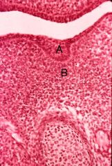
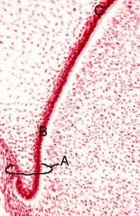
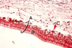
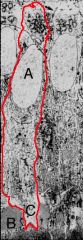
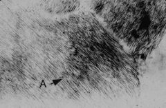
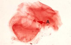
What are A and B?
|
A - dental lamina
B - mesenchymal neural crest |
|
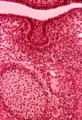
What stage?
|
Bud stage
|
|
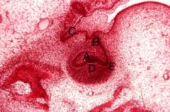
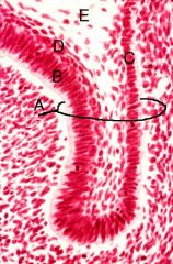
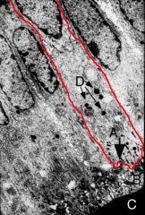
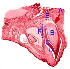
What are A-E?
|
A-enamel organ
B- dental lamina C- vestibular lamina D- dental papilla E- dental sac |
|
What stage is this?
|
Bell
|
|

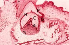
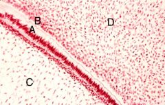
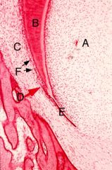
What are A-G?
|
A- inner enamel epithelium
B- outer enamel epithelium C- stellate reticulum D- successional lamina E- dental lamina F- dental papilla G- dental sac |
|
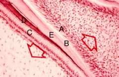
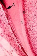
What are A-E?
|
A- cervical loop
B- inner enamel epithelium C- outer enamel epithelium D- stratum intermedium E- stellate reticulum |
|
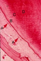
What are A-C?
|
A- cervical loop
B- least differentiated ameloblasts C- most differentiate ameloblasts |
|
What are A-D?
|
A- preameloblasts
B- preodontoblasts C- stellate reticulum D- dental papilla |
|
What are A-E?
|
A- odontoblasts
B- predentin C- ameloblasts D- enamel E- dentin |
|
What are A-C?
|
A- reduced enamel epithelium
B- mature/protective ameloblasts C- capillary |
|
What are A and B?
|
A- ameloblasts
B- immature enamel |
|
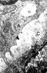
What are a-c?
|
a- preodontoblast
b- preameloblast c- basement membrane |
|

What are a-c?
|
a- odontoblast nucleus
b- secretory end of odontoblast c- predentin |
|

What are a-c?
|
a- ameloblast nucleus
b- enamel c- tome's process |
|
What kind of cells is this?
|
preodontoblast
|
|
What kind of cell is this?
|
ameloblast
|
|
What kind of cells is this?
|
maturative ameloblast (dense granules present)
|
|

What are a-d?
|
a- striated border
b- immature enamel c- mature enamel d- dense granules |
|

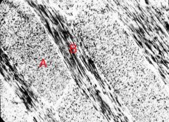
what are a and b?
|
a- enamel
b - dentin |
|
What is a?
|
hydroxyapatite crystal
|
|
what are a and b?
|
a - rod sheath
b - rod core |
|
what are a-f?
|
a- epithelial diaphragm
b- radicular pulp cavity c- dentin d- enamel space e- alveolar bone f- root |
|
what are a-f?
|
a- radicular pulp cavit
b- dentin c- dental sac d- point of epithelial root sheath disintegration e- epithelial diaphragm f- epithelial rests |
|
what are a-c?
|
a- cementoblasts
b- odontoblasts c- predentin |
|
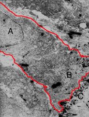
what are a-d?
|
a- epithelial rests
b- mantle dentin c- globular dentin d- circumpulpal dentin |
|
what is a?
|
dentin deposition on mesiobuccal cusp
|