![]()
![]()
![]()
Use LEFT and RIGHT arrow keys to navigate between flashcards;
Use UP and DOWN arrow keys to flip the card;
H to show hint;
A reads text to speech;
37 Cards in this Set
- Front
- Back
|
Oral mucous membrane = oral mucosa Oral mucoperiosteum (has a strong lamina propria) 4 functions of oral mucosa? |
Protection Sensation Taste Secretion |
|
|
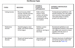
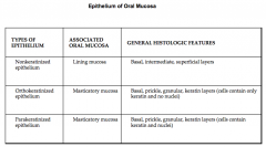
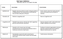
Organization - 3 types: Masticatory Lining Specialized |
–Masticatory - gingiva & hard palate –Lining - lips, cheeks, floor of mouth, under tongue, alveolar mucosa –Specialized - red area of lip, dorsum of tongue |
|
|
- |
|
|
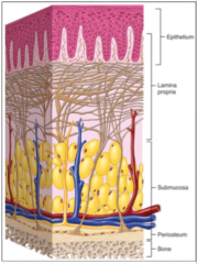
Components of oral mucosa: Mucosa has what kind of epithelium? Submucosa -loosely arranged irregular connective tissue , vessels, nerves, minor salivary glands, & occasional sebaceous glands Either bound tightly (hard palate) orloosely (cheek |
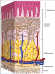
Stratified squamous supported by lamina propria with irregular CT •The junction between the submucosaand lamina propria is not distinct. |
|
|
What glands can be found in the oral mucosa and appear as yellow spots called Fordyce's granules? |
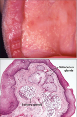
sebaceous glands |
|
|
Theinterface between the oral epithelium and the lamina propria is called the__ . Note the interdigitationbetween connective tissue papilla and the epithelial ridges or __. This increases the __ and increasesthe strength between the two |
Basal lamina Rete pegs Surface area |
|
|
Deepto submucosa in hard palate and gingiva is the __ Thistight attachment is characteristic of masticatory mucosa |
mucoperiosteum |
|
|
Epithelium of oral mucosa can be what 3 things? What cells attach to the underlying basal lamina? |
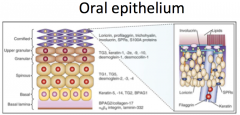
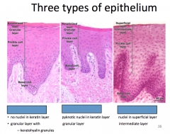
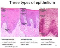
Non-keratinized (wet) Keratinized (orthokeratinzed) Parakeratinized (partially) Basal layer of cells |
|
|
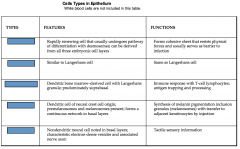
What are the 2 types of basal cells? |
Mitotic cells Amplifying cells –increasethe number of cells that can mature •TurnoverEstimates: –Cheek~ 15 - 25 days –Gingiva~ 40 - 50 days –Gut~ 4 – 14 days –Skin~ 52 – 75 days |
|
|
The next layer after the basal layer is the? What is abundant in this layer? |
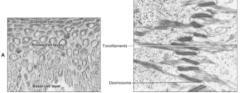
Spinous or prickle cell layer abundantdesmosomes in the prickle cell layer |
|
|
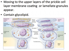
After the spinous layer is the? •Tonofilaments appear in bothkeratinized and non keratinized epithelia. -Theseare intermediate filaments found in epithelia - In keratinized epithelia thetonofilaments aggregate to form tonofibrils - Innon-keratinized epithelia the tonofilaments remain dispersed |
Granular layer –uppergranular layer and cornified layer appear in keratinized epithelium –anintermediate and upper layer appear in non keratinized epithelium |
|
|
–Asthe lamellate granules or bodies reach the surface the lipids accumulate aroundthe plasma membrane. Important in establishing a__ |
Permeability layer |
|
|
•In keratinized epithelium__ appear.Give the basophilic appearance tothe granular layer |
keratohyaline granules |
|
|
•As cells reach the keratinizedlayer: –Nucleidisappear –Organellesdisappear –Keratohyalingranules disappear –Whats left are __ cross linked by disulfide bonds |
cells packed with filaments |
|
|
The superficial cells will have their corneodesmosomes break down they are called? |
Squames |
|
|
Keratinizedepithelium is impermeable except in the area __ |
Under the tongue (non-keratinzed region to permit the absorption of some drugs) |
|
|
- |
|
|
|
|
|
|
|
|
Increase area for secure attachment Greatest in masticatory mucosa where shearing forces are greatest |
CT junction Hemidesmosomes and anchoring fibrils interlock CT and epithelium |
|
|
Notethat the lamina propria can be subdivided into a __ and a __ |
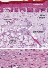
papillary layer reticular layer Alsothe submucosa has minor salivary glands |
|
|
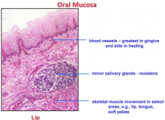
Bloodsupply in the floor of the mouth and the cheek |
|
|
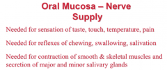
__ are important for sensing pain and temperature. They can be found in the lower and middleregions of the epithelium |
Free nerve endings |
|
|
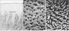
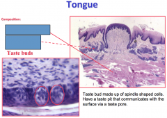
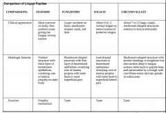
** The tongue and pharynx have specialsensory receptors for taste. They arefound on what 3 papilla? |
fungiform, foliate and circumvallate |
|
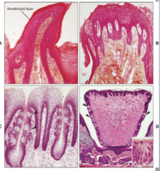
Identify A, B, C, D Skeletal muscle in 3 planes in the tongue |
Filiform (A), fungiform (B), foliate (C) circumvallate papilla (D). |
|
Von ebner glands are what kind of gland? |
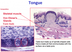
Serous secretin glands |
|
|
- |
|
|
3 cell types for taste |
- Bitter/sweet/umami -Sour -Salt |
|
|
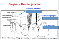
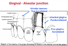
Lining mucosa: tongue, floor of mouth, soft palate, epithelium,lamina propria, submucosa Masticatory mucosa: hard palate, attached gingiva - Epithelium: Thick, orthokeratinized orparakeratinized in parts) stratified squamous epithelium -Longpapillae; thick, dense collagenous tissue - Densecollagenous connective tissue attaches mucosa to periosteum (mucoperiosteum) |
fix |
|
|
|
|
|
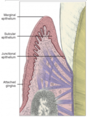
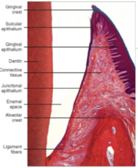
Where epithelium meets tooth? Site of potential weakness wherebacteria can cause inflamation |
Dentogingival junction |
|
|
|
|
The sulcular epithelium continues towardthe junctional epithelium which is in contact with the enamel and perhaps latercementum |
|
|
|
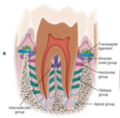
PDL |
|
|
gingival ligament |
|
|
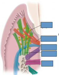
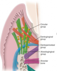
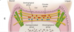
Fibers are found in the laminapropria of the gingiva and collectively form the gingivalligament: - Dentogingival group - Alveologingival group - Circular group - Dentoperiosteal group - Transseptal fiber system |
|
|

|
- |