![]()
![]()
![]()
Use LEFT and RIGHT arrow keys to navigate between flashcards;
Use UP and DOWN arrow keys to flip the card;
H to show hint;
A reads text to speech;
78 Cards in this Set
- Front
- Back
|
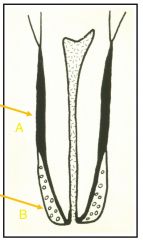
Cellular and Acellular cementum A= Acellularcementum next to dentine B= Cellular cementum around apical region |
|
|
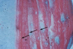
Incremental lines of salter |
|
|
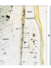
AEFC and CIFC |
|
|
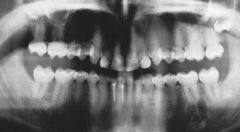
Gemination and fusion |
|
|
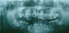
Root dilaceration |
|
|
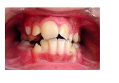
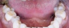
Trauma to deciduous teeth |
|
|
Sometimes there is a change in variety and shape of tooth |
|
|
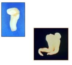
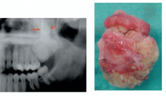
Odontoma |
|
|
Odontoma |
|
|
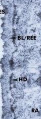
Amelongenesis imperfecta |
|
|
Dentinogenesis imperfecta |
|
|
Dentinogenesis imperfecta |
|
|
Fluorosis and tetracycline staining |
|
|
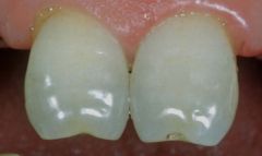
Enamel pearls |
|
|
Hypercementosis |
|
|
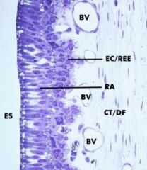
Reduced enamel epithelium Derived from ameloblast and stratum intermedium layers of the enamel organ May include remnants of the stellate reticulum and outer enamel epithelium |
|
|
Primary enamel cuticle Basal lamina of the reduced enamel epithelium |
|
|
abc |
|
|
The epithelium becomes shorter and is eventually replaced with the secondary junctional epithelium. The gingival sulcus forms during the replacement at a stage where further recession would expose the cementum SJE contains some cells derived from the oral epithelium |
|
|
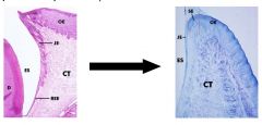
Coatings of teeth during eruption As the tip of the crown emerges the reduced enamel epithelium disintegrates and fuses with the oral epithelium. During this fusion apoptosis occurs This becomes the primary junctional epithelium |
|
|
Cleidocranial dysplasia/ Dystosis |
|
|
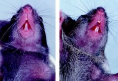
Osteopetrotic mice 'stone bone' or 'marble bone disease' |
|
|
Primary failure of tooth eruption |
|
|
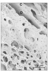
Alveolar bone crypt of mouse molar |
|
|
Osteoclasts on alveolar bone surface |
|
|
|
|
|
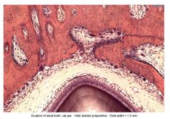
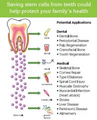
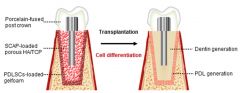
Teeth that can be used for stem cell use |
Impacted third molars in young, middle and aged populations Teeth extracted for ortho reasons Primary teeth exfoliated Teeth extracted due to irreversible perio Gingiva thats been resected due to aesthetic or hyper plastic reasons |
|
|
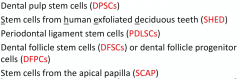
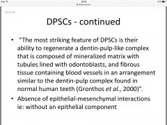
DPSCs Dental papilla stem cells |
|
|
|
|
|
|
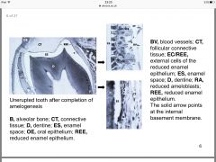
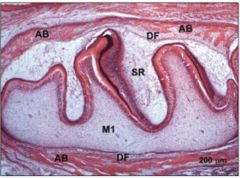
Follicle and tooth germ |
|
|
Rat alveolar bone showing the dental follicle (DF) surrounding an unerupted tooth (M1) encased in alveolar bone (AB) SR= stellate reticulum |
|
|
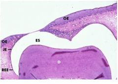
Reduced enamel epithelium |
Thin multilayered epithelium that covers the enamel of an unerupted tooth (naysmyths membrane) |
|
|
Primary enamel cuticle |
basal lamina lying between the reduced enamel epithelium and the enamel surface (naysmyths membrane) |
|
|
Nasmyth's membrane |
reduced enamel epithelium and primary enamel cuticle |
|
|
Junctional epithelium |
portion of gingival epithelium that is attached to the tooth enamel on one side and gingival connective tissue on the other. The coronal end lines the base of the gingival sulcus. The primary function is as a permeable barrier layer involved in limiting and controlling infection of periodontium |
|
|
Acquired enamel cuticle |
Protective, acellular protein layer covering the enamel surface coronal to the gingival margin derived |
|
|
Dental biofilm |
|
|
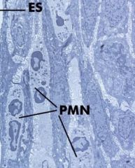
Inflamed junctional epithelium |
the EM of the junctional epithelium in inflamed gingiva Marked distention of intracellular spaces by PMNs) that are migrating from the connective tissue toward the gingival sulcus. Fluid exudate from the connective tissue also flows through the sulcus through the Intracel spaces. |
|
Healthy junctional epithelium |
orientated with long axis parallel to tooth surface Intracell spaces narrow |
|
|
Arrows show the junctional epithelia is the most permeable region of gingival epithelia |
|
|
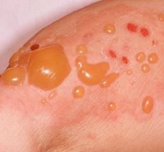
Bulls pemphigoid |
|
|
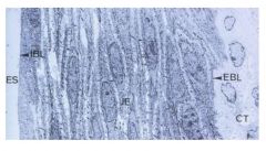
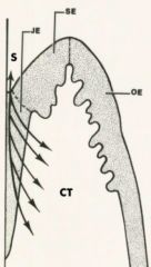
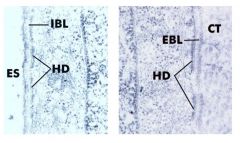
Secondary junctional epithelium IBL= internal basal lamina (faces tooth enamel) EBL= External basal lamina (faces CT) Attached to enamel surface via hemidesmosomes |
|
|
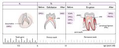
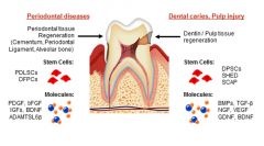
Regeneration of tooth tissues Periodontal diseases: stem cells (PDLSCs and DFPCs) and molecules ed PDGF BDNF Dental caries/pulp injury: SCs= DPSCs, SHED, SCAP and molecules= BMPS, GDNF, BDNF |
|
|
Bright et al 2015 |
irrespective of defect type of animal tissue used: PDL SC implantation can result in beneficial outcome for periodontal regeneration |
|
|
Key factors for stem cell transplantation |
Use of multipoptent adult stem cells Need singling molecules/inductive morphogenetic signals Need growth factors Need a scaffold: biodegradable material that can be a synthetic polymer or pressed bio product (e.g. collagen) |
|
|
applications of dental stem cells |
|
|
|
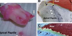
stem cells of apical papilla |
|
|
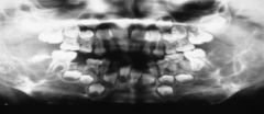
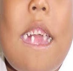
Anodontia Complete absence VERY RARE permanent dentition fail to form |
|
|
Supplemental Resembles normal tooth U2 more common than L5 more common that U5 |
|
|
Cherubim |
Familial multilocular cystic disease Genetic Autosomal dominant SH3BP2 gene Chromosome 4 Leads to abnormal bone due to the disruption of signalling pathways associated with the maintenance of bone |
|
|
Cherubim Multilocular cystic radiolucencies Abscent UEs |
|
|
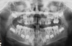
Supernumerary 1-3% of the population More females than males Mesiodens Paramolar (buccally) Distomolar (Behind 8s) |
|
|
who knows |
|
|
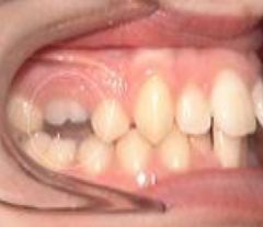
Submerged Es |
|
|
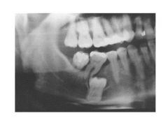
Dentigerous Cyst |
|
|
Cherubim |
|
|
|
|
|
|
|

|
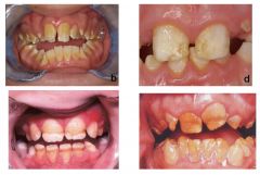
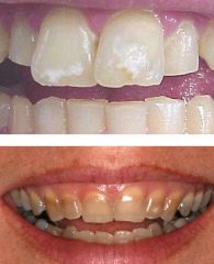
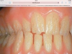
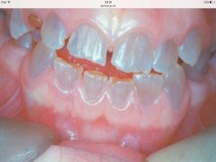
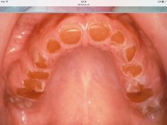
Tetracycline staining |
|
|
Amelgenesis imperfecta usually autosomal dominant and affects both dentitions Abrasion and attrition of enamel No increase risk to caries |
|
|
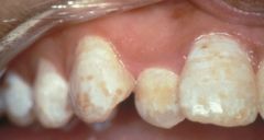
Fluorosis |
|
|
Mulberry molars |
|
|
Hutchinson's incisors |
|
|
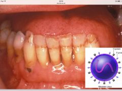
Effect of measles |
|
|
Turner teeth |
|
|
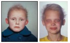
Hypohydrotic ectodermal dysplasia absence of ectodermal structures Usually X linked recessive (rare in females) Most cases caused by defects of EDA genes Fine/sparse hair Defective fingernails Absence of sweat glands and get hyperthermia |
|

|
Hypohydrotic ectodermal dysplasia Dental probs? Anodontia- due to failure of dental lamina to form Hypodontia- any teeth formed are deformed with conical crowns and delayed in eruption |
|
|
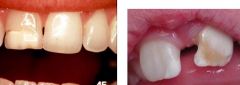
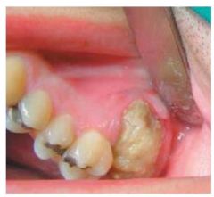
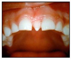
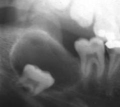
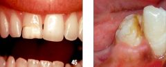
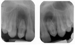
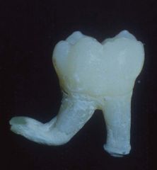
Delaceration |
Crown of tooth displaced from normal alignment Result of acute mechanical trauma |
|
|
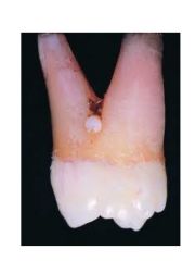
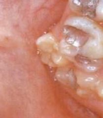
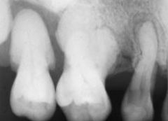
Enamel pearl |
Nodule of enamel at ADJ on maxillary molars HERS differentiate into ameloblasts |
|
|
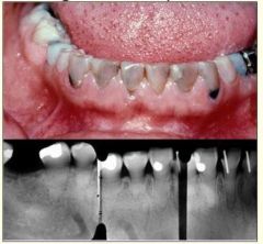
Dens in dente (dens invaginus) invagination of dental papilla may cause enamel linked cavity opening onto the surface of max incisors Increases caries risk |
|
|
Hypcementosis Cleidocranial dysplasia Hypophosphatasia (recessive autosomal) |
|
|
Ghost teeth |
|
|
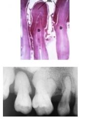
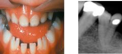
short, blunt roots, partial/total obliteration of the pulp chamber and root canal by dentine |
|
|
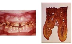
Double teeth |
|
|
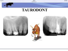
Taurodont |
|
|
Delaceration |
|
|
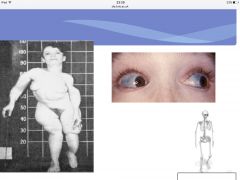
Scleroderma |
|
|
Scleroderma |