Reading...
![]()
Play button
![]()
Play button
![]()
Use LEFT and RIGHT arrow keys to navigate between flashcards;
Use UP and DOWN arrow keys to flip the card;
H to show hint;
A reads text to speech;
20 Cards in this Set
- Front
- Back
|
Onchocera Volvulus - Classification
|
Classification:
Onchocerca volvulus is a typical filarial nematode that is primarily a parasite of humans. Kingdom: Animalia Phylum: Nematoda Class: Secernentea Order: Spirurida Family: Onchocercidae Genus: Onchocerca Species: O. volvulu |
|
|
Onchocera Volvulus - Clinical Manifestations
|
Clinical Manifestations:
Patients present with subcutaneous nodules, dermatitis, and eye lesions. onchocerciasis or "river blindness" mostly in Africa. Long-term corneal inflammation, or keratitis, leads to thickening of the corneal stroma which ultimately leads to blindness. |
|
|
Onchocera Volvulus - Structure
|
Structure:
Long, thread-like adult worms live coiled in subcutaneous nodules and produce microfilariae that are slightly smaller than those of lymphatic filariae and are not sheathed. |
|
|
Onchocera Volvulus - Multiplication and Life Cycle
|
Multiplication and Life Cycle:
Adult worms live in subcutaneous nodules. Microfilariae wander through the skin, where they may be picked up by a feeding vector blackfly. In the blackfly they mature to infective larvae, which may enter a new host when the blackfly feeds. The larvae then move to the subcutaneous tissues, mature, mate, and produce microfilariae. |
|
|
Onchocera Volvulus - Pathogenesis
|
Pathogenesis:
Reaction to worms and microfilariae causes dermatitis with loss of skin elasticity and the formation of fibrotic subcutaneous nodules. Living and dead microfilariae in the eye cause trauma and reactions that can result in blindness. |
|
|
Onchocera Volvulus - Host Defenses
|
Host Defenses:
There is an inflammatory and immune response to living and dead parasites and to antigen-antibody complexes. Adult worms are localized in the subcutaneous tissues, surrounded by fibrotic nodules and ultimately calcify. |
|
|
Onchocera Volvulus - Epidemiology
|
Epidemiology:
Onchocerciasis is common in Africa; foci also occur in South and Central America and Mexico. The blackfly vectors breed in oxygen-rich water; thus, the disease characteristically is associated with fast-flowing streams. |
|
|
Onchocera Volvulus - Diagnosis
|
Diagnosis:
The clinical picture is indicative; final diagnosis is made by identifying microfilariae in skin snips. |
|
|
Onchocera Volvulus - Control
|
Control:
Individuals may be treated by removal of nodules and by administering microfilariacides that kill microfilariae. Control of vector blackflies minimizes reinfection. |
|
|
Onchocera Volvulus - General description
|
Onchocerca Volvulus
Onchocerca volvulus is a filarial worm that is transmitted to humans by blackflies (Simulium). Mature worms live in the subcutaneous tissues and produce microfilariae that migrate through the skin and connective tissues. |
|
|
Onchocera Volvulus - Clinical Manifestations
|
Clinical Manifestations:
Changes in skin pigmentation are often the first obvious signs of Onchocerca infection. Later stages of dermatitis present as atrophy and loss of skin elasticity. Although lymph nodes may become involved in onchocerciasis, involvement is not as prominent as with the lymphatic filariae. The subcutaneous nodules that harbor adult Onchocerca are usually firm and non-tender. They vary in size and location but usually are easily recognized when they occur in geographic areas where the disease is endemic. However, in some geographic regions, nodules may be in deeper tissue, and thus not easily palpable. The most serious clinical manifestation of onchocerciasis is blindness, caused by microfilariae that wander into the eye. In endemic areas, corneal opacities resulting from the reaction to dying microfilariae often suggest onchocercal infection. Alternatively, active living microfilariae may be seen when the eye is examined with a slit lamp. |
|
|
Onchocera Volvulus - Structure
|
Structure:
Adult Onchocerca may be up to 60 cm long, but are usually coiled in subcutaneous nodules. The microfilariae are slightly smaller than those of W bancrofti and B malayi and differ from them in lacking a sheath, having a different nuclear arrangement, and not usually being found in the blood. |
|
Onchocera Volvulus - Multiplication and Life Cycle
|
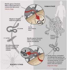
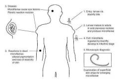
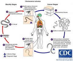
Multiplication and Life Cycle - Figure 92-3 shows the life cycle of Onchocerca. The microfilariae produced by adult female worms in subcutaneous nodules migrate into the skin and connective tissue; they do not generally enter the circulatory system. Microfilariae are ingested by vector blackflies, develop to the infective stage in the muscles of the flies, and then migrate to the mouth parts. When the infected flies feed on a new host, the larvae leave the mouth parts and enter the wound produced by the biting fly. Developing male and female worms congregate in subcutaneous tissue where they usually induce formation of a nodule
|
|
|
onchocera volvulus pathogenoses
|
|
Onchocera volvulus life cycle
|
Figure 92-3
Life cycle of Onchocerca. During a blood meal, an infected blackfly (genus Simulium) introduces third-stage filarial larvae onto the skin of the human host, where they penetrate into the bite wound . In subcutaneous tissues the larvae develop into adult filariae, which commonly reside in nodules in subcutaneous connective tissues . Adults can live in the nodules for approximately 15 years. Some nodules may contain numerous male and female worms. Females measure 33 to 50 cm in length and 270 to 400 μm in diameter, while males measure 19 to 42 mm by 130 to 210 μm. In the subcutaneous nodules, the female worms are capable of producing microfilariae for approximately 9 years. The microfilariae, measuring 220 to 360 µm by 5 to 9 µm and unsheathed, have a life span that may reach 2 years. They are occasionally found in peripheral blood, urine, and sputum but are typically found in the skin and in the lymphatics of connective tissues . A blackfly ingests the microfilariae during a blood meal . After ingestion, the microfilariae migrate from the blackfly's midgut through the hemocoel to the thoracic muscles . There the microfilariae develop into first-stage larvae and subsequently into third-stage infective larvae . The third-stage infective larvae migrate to the blackfly's proboscis and can infect another human when the fly takes a blood meal . |
|
|
Onchocera Volvulus - Pathogenosis
|
Pathogenesis:
Adult worms in the subcutaneous tissues cause varying degrees of inflammation and may induce subcutaneous nodules. Nodules appear 3 to 4 months after infection, but microfilariae are not generally detectable until I year after infection. Adult worms may be surrounded by an inflammatory response that progresses to granuloma formation and fibrosis or calcification, depending on the condition of the worm and age of the nodule (Fig. 92-3). Microfilariae appear to move upward, and in chronic heavy infections may be seen in the eye. Ocular damage is thought to be due both to the trauma caused by living microfilariae and to a hypersensitivity reaction to dead ones. A major problem in the management of onchocerciasis patients is the acute inflammatory response to dying microfilariae in the eye during treatment. Antigen-antibody complexes probably play a role in the development of eye lesions resulting from microfilariae. onchocerciasis or "river blindness" mostly in Africa. Long-term corneal inflammation, or keratitis, leads to thickening of the corneal stroma which ultimately leads to blindness. |
|
|
Onchocera Volvulus - Host Defenses
|
Host Defenses:
Nodules containing adult worms are surrounded by inflammatory cells that are replaced by collagen and fibrotic tissue, thereby localizing the worms. A study of nodules has shown that they are areas of high cellular activity. Some microfilariae appear to be killed before they ever leave the nodules where they are produced by the female worm. Obviously many others escape the chronic inflammatory responses and the granulomas commonly seen surrounding nematodes in tissue. The exact composition of the nodules varies, depending on the distance from the adult worms and the age of the nodule. In general, neutrophils are followed by eosinophils and macrophages. The involvement of eosinophils in killing microfilariae in untreated patients suggests specific immune reactions similar to those reported in other helminth infections. The fibrous outer layer of the nodule contains blood vessels, which may be surrounded in older nodules by cellular infiltrates that include plasma cells and eosinophils. |
|
|
Onchocera Volvulus - Epidemiology
|
Epidemiology:
Transmission occurs through bites of vector blackflies in the family Simuliidae. Although a few vector species breed in slow-moving streams, most require fast-flowing, highly oxygenated streams or rivers. For this reason, ocular onchocerciasis is often called “river blindness.” Onchocerciasis is prevalent in many parts of tropical Africa and has been reported in a few places in the Middle East. In the Western hemisphere, it is an important and widespread infection in Guatemala and the southern states of Mexico. It also appears in other areas of Central America, and a few foci have been found in Venezuela, Colombia, Surinam, Brazil, and Ecuador. |
|
|
Onchocera Volvulus - Diagnosis
|
Diagnosis:
Onchocerciasis is suggested by subcutaneous nodules or by the characteristic scaly dermatitis in individuals living in endemic areas. Positive diagnosis is usually made by identifying microfilariae in a superficial skin biopsy made with a scalpel or an appropriate punch. The skin samples, which are usually taken from the shoulder, are placed in a drop of saline or distilled water on a microscope slide, incubated for 30 minutes, and examined (Fig. 92-3). The diagnosis may also be made by finding adult worms in nodules or by observing microfilariae in the eye. Serologic tests are being developed to aid in diagnosis when microfilariae cannot be detected but onchocerciasis is suspected. |
|
|
Onchocera Volvulus - Control
|
Control:
Three main measures - vector control, nodule removal, and drug treatment - provide limited control of onchocerciasis. Control of blackfly vectors is difficult because most species breed in fast-flowing streams to which insecticides cannot easily be applied. Nodules harboring adult worms are usually removed, which presumably reduces the rate at which microfilariae are produced and thus the risk to the eyes (as well as the number of microfilariae available to vectors). Mass treatment poses difficulties, because suramin, the drug of choice for killing adult worms, must be given intravenously and is toxic. Diethylcarbamazine kills microfilariae but has limited efficacy against adult worms and has the disadvantage that some patients experience a severe reaction, which may exacerbate eye damage, to dying microfilariae. Ivermectin, a semisynthetic macrolide antibiotic, has recently been shown to be more effective and to produce fewer side effects than diethylcarbamazine. |