Reading...
![]()
Play button
![]()
Play button
![]()
Use LEFT and RIGHT arrow keys to navigate between flashcards;
Use UP and DOWN arrow keys to flip the card;
H to show hint;
A reads text to speech;
33 Cards in this Set
- Front
- Back
|
Polyhydramnios may result from impaired swallowing in a fetus with ______________.
|
- epignathis
- macroglossia - cleft lip and/or palate |
|
|
What is the best plane to visualize a cleft lip?
|
coronal
|
|
|
Is a cleft lip bilateral or unilateral?
|
can be either uni- or bilateral
|
|
|
What percentage of cleft palates is present with a unilateral and bilateral cleft lip, respectively?
|
unilateral - 70%
bilateral - 85% |
|
|
Which facial cleft is more associated with hypotelorism and holopronsencephaly?
|
medial cleft lip/palate
|
|
|
Describe the sonographic findings of bilateral cleft lip and a medial cleft.
|
- large gap in upper lip on modified coronal view
- nose is flattened and widened - premaxillary protrusion - tongue is visualized in a higher than normal position in the mouth |
|
|
A sonogram on a 28 week fetus reveals a mass protruding from the mouth. Some of the other ultrasound findings might be ______________ and _____________.
|
polyhydramnios; small stomach
|
|
|
What area of the body are most cystic hygromas seen?
|
posterior neck
|
|
|
A sonographic examination reveals a multi-loculated, cystic mass adjacent to the posterior aspect of the fetal head, edema in the soft tissues of the upper extremities, and ascites. This pattern most likely represents _________.
|
cystic hygroma
|
|
|
What other abnormalities or conditions can be associated with a cystic hygroma?
|
- hydrops
- Turner's syndrome: cardiac defects 50% associated with a chromosomal abnormality |
|
|
Gas or air in a fetus without cardiac activity indicates fetal maceration. What other ultrasound findings would also be seen?
|
- no cardiac activity
- spalding sign - exaggerated curvature of spine - anasarca - oliohydramnios |
|
|
What would situs inversus look like on ultrasound?
|
the heart and abdominal organs are completely reversed
|
|
|
How does normal fetal lung tissue appear on ultrasound?
|
normal fetal lung tissue appears homogeneous and more echogenic than the liver
|
|
|
List abnormalities of the chest that can be visualized on ultrasound.
|
- CCAM
- pulmonary sequestration - bronchogenic cyst - pleural effusion (hydrothorax) - diaphragmatic hernia |
|
|
Is CCAM unilateral or bilateral?
|
usually unilateral
|
|
|
CCAM Type II is associated with ___________.
|
- renal agenesis
- pulmonary anomalies - diaphragmatic hernia |
|
|
A sonographic examination of a 32 week fetus demonstrates a highly echogenic & homogeneous mass within the lower lobe of the left lung. Fetal hydrops is seen and a possible GI abnormality is suspected. What is the most likely diagnosis?
|
Extralobar pulmonary sequestration
|
|
|
True or False. A fetus diagnosed with extra lobar pulmonary sequestration most likely has a poor prognosis due to the associated abnormalities.
|
true
|
|
|
An obstetrics sonogram of a 30 week fetus demonstrates a single cystic mass adjacent to the heart of the left thoracic cavity. The hear tis deviated toward the right of midline. The abdominal circumference is small for gestational age and polyhydramnios is seen. What does this pattern most likely represent?
|
diaphragmatic hernia
|
|
|
Fetus' with diaphragmatic hernias have coexisting abnormalities ___________ of the time.
|
more than 50%
|
|
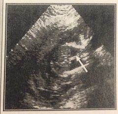
Identify the abnormality seen in this image.
|
pleural effusion (hydrothorax)
|
|

Identify the abnormality seen in this image.
|
unilateral cleft lip
|
|
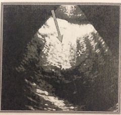
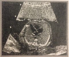
Identify the abnormality seen in this image.
|
CCAM
|
|
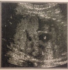
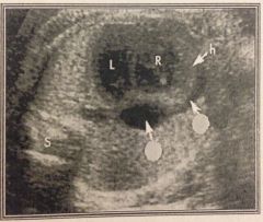
Identify the abnormality seen in this image.
|
left-sided diaphragmatic hernia
|
|
Identify the abnormality seen in this image.
|
VSD - ventricular septal defect
|
|
Identify the abnormality seen in this image.
|
rhabdomyoma
|
|
Identify the abnormality seen in this image.
|
pericardial effusion
|
|
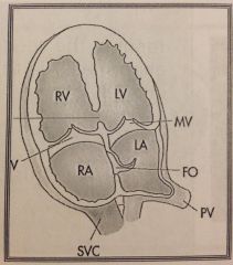
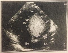
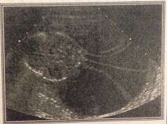
What does this ultrasound image demonstrate?
|
a normal 4 chamber heart
|
|
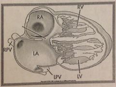
What is this heart schematic demonstrating?
|
left hypostatic heart
|
|
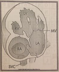
What is this heart schematic demonstrating?
|
atrial septal defect (ostium primum)
|
|
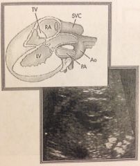
What is this heart schematic demonstrating?
|
normal LVOT & 5 chamber view
|
|
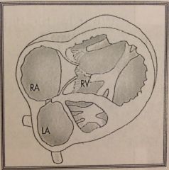
What is this heart schematic demonstrating?
|
right hypoplastic heart
|
|
Identify the abnormality seen in this image.
|
cystic hygroma
|