Reading...
![]()
Play button
![]()
Play button
![]()
Use LEFT and RIGHT arrow keys to navigate between flashcards;
Use UP and DOWN arrow keys to flip the card;
H to show hint;
A reads text to speech;
292 Cards in this Set
- Front
- Back
|
microcytic?
|
small RBC's (MCV < 80)
|
|
|
Microcytic anemias are?
|
iron deficiency, thalassemia, sideroblastic anemia
|
|
|
Macrocytic?
|
Large RBC
(MCV >100), |
|
|
Macrocytic anemias are?
|
B12 deficiency, folate deficiency, liver disease, acute hemolysis, severe hypothyroidism
|
|
|
Anemia is a sign of illness, not so much a?
|
diagnosis in itself.
|
|
|
Anemia refers to low?
|
Hg or Hct.
|
|
|
The most common cause of asymptomatic anemia?
|
is iron deficiency.
|
|
|
Daily iron requirements for men and post menopausal women is?
|
0.5-1.0 mg
|
|
|
Menstruating and pregnant women need how much iron?
|
2.0-2.5 mg.
|
|
|
Iron defic is more common in which gender?
|
women than men
|
|
|
When Fe defic is detected in men, the GI tract must be checked for?
|
occult bleed.
|
|
|
Pernicious anemia is more common in pts of what descent?
|
northern European descent, with a prevalence about 0.1%.
|
|
|
Folate defic is common in?
|
pregnancy, and in alcoholics, accompanied by sideroblastic anemia
|
|
|
Sickle cell is the most common?
|
hemoglobinopathy
|
|
|
Normal hemoglobin (Hg) for men is?
|
14-18
|
|
|
Normal Hg for women is?
|
12-16
|
|
|
Normal Hg in pregnancyis?
|
over 11, and in aging, slightly lower.
|
|
|
An acute blood loss induces signs of?
|
postural hypotension, increased heart rate, and shock if over one liter: it takes 72 hours to detect anemia since the loss decreases all parameters proportionally.
|
|
|
Anemia results from what 3 reasons?
|
blood loss, inadequate production or excessive red cell destruction.
|
|
|
Symptoms associated with anemia include?
|
fatigue, headache, irritability when mild and hyperkinetic symptoms appear when the hemoglobin drops below 7.5: such as elevated heart rate and palpitations.
|
|
|
Pica?
|
consumption of ice, starch, clay, or other substances
|
|
|
Craving ice is known as?
|
pagophagia
|
|
|
What sympts may occur at very low Hg levels?
|
|
|
|
Finger stick tests have a higher margin of error than?
|
venous blood work
|
|
|
Iron defic what kind of anemia?
|
microcytic, hypochromic
|
|
|
Iron defic anemia's due to?
|
blood loss or inadequate iron supply.
|
|
|
Nutritional sources influence absorption: iron in greens are?
|
poorly absorbed than animal sources (5% vs. >30%).
|
|
|
Of supplements, ferrous sulfate is more readily absorbed in the?
|
ferric form.
|
|
|
Most absorption of Fe occurs in the?
|
proximal small bowel.
|
|
|
Transferrin?
|
is a protein that transports iron in plasma
|
|
|
Transferrin can be measured by?
|
iron binding capacity or transferrin saturation.
|
|
|
With low plasma iron, transferrin transports iron to?
|
bone marrow from storage cells.
|
|
|
The hallmarks of iron defic are?
|
elevated total iron binding capacity (TIBC) and low transferrin saturation.
|
|
|
Red cell life span up to?
|
120 days.
|
|
|
Immature red cells stay in the?
|
marrow until fully developed and then are released into the circulation.
|
|
|
Ferritin?
|
is storage form of iron, stored in the liver.
|
|
|
Hemosiderin?
|
is the storage form stored in bone marrow.
|
|
|
Types of anemias, based on?
|
microcytic and macrocytic
|
|
|
How much iron is absorbed from the average adult dietary intake of ___?
|
Only 5-10% from the dietary intake of 10-20 mg daily.
|
|
|
Hematocrits (Hct) normally ___for men?
|
41-51
|
|
|
Hematocrits (Hct) normally ___for women?
|
37-47
|
|
|
Changes that can also occur at very low Hg levels are?
|
Pica, (consumption of ice, starch, clay, or other substances) blue sclera, nail changes and most commonly craving ice (pagophagia)
|
|
|
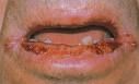
Cheilitis is a medical condition involving inflammation of the lip.
It is associated with many conditions, including megaloblastic anemia from Vitamin B12 deficiency, iron deficiency anemia (which in severe cases can lead to Plummer-Vinson syndrome and oral candidiasis. It can also be a symptom of allergies, such as allergy to Balsam of Peru. Cheilitis can also be caused by taking the (retinoid) drug Isotretinoin. |
|

What is this?
|
Koilonychia (also known as spoon nails[1]:782) is a nail disease that can be a sign of hypochromic anemia, especially iron-deficiency anemia.[2]:656[3] Koilonychia literally means "spoon nails." It refers to abnormally thin nails (usually of the hand) which have lost their convexity, becoming flat or even concave in shape. In a sense, koilonychia is the opposite of nail clubbing.
|
|
|
Iron is lost daily via?
|
stool, urine, skin, and nails.
|
|
|
Women lose about how much iron a day?
|
2 mg per day and men 1 mg daily.
|
|
|
History for Fe def should include questions regarding?
|
any note of change in bowel habits, abnormal blood loss, melena, increased use of aspirin or anti inflammatories, cancer, HIV, chronic disease symptoms, diet, lead exposure, pica, number of pregnancies, menstrual blood loss, gastric surgery, change in nails or sore tongue.
|
|
|
On PE with Fe def look for?
|
glossitis, chelitis, koilonychias, lymphadenopathy, hepatosplenomegaly, rectal exam for mass or occult blood. Check for pelvic masses.
|
|
|
How does one visualize red cell morphology, color and size?
|
Blood smear technique
|
|
|
With Fe def, target cells indicate?
|
hemoglobinopathy, such as thalassemia
|
|
|
Howell-Jolly bodies occur with?
|
DNA remnants post spleen removal
|
|
|
Heinz bodies are seen with?
|
glucose-6-phosphodehydrogenase defic or a decreased enzyme reducing power of the RBC
|
|
|
Neutrophils?
|
resolve bacterial infection. Toxic granulation in neutrophils indicates infection
|
|
|
Platelets are counted and should number?
|
140-400
|
|
|
Reticulcyte count?
|
is an estimation of new RBC production
How do immature RBCs compare in size to mature RBCs? |
|
|
RBCs increase in number after?
|
blood loss, with marked hemolysis, or in B12 or folate replacement
|
|
|
Red cell distribution width (RDW) assesses?
|
variation in the 20-80th percentile of the RBC population.
|
|
|
Increases in RDW indicate ?
|
many new and larger RBC most often following acute blood loss.
|
|
|
What parameter helps to distinguish anemia of chronic disease?
|
RDW
|
|
|
Normochromic-normocytic anemia is classified according to reticulcyte count as?
|
a manifestation of marrow response.
An increased retic count suggests? |
|
|
Ask about use of meds such as penicillins, cephalosporins, sulfa, quinidine, methyldopa since these may induce increase. With anemia, ask about history of ?
|
sickle cell disease or trait, family history of anemia, recent viral infection (especially with mono, CMV, or hepatitis), lupus, or a lymphoproliferative disease.
|
|
|
With anemia,on PE check for?
|
splenomegaly or signs of underlying connective tissue disease.
|
|
|
Check hemolytic indices?
|
for bilirubin, haptoglobin, and LDH: haptoglobin
|
|
|
Drug induced hemolysis can be detected via?
|
Coombs test, especially if pt is African American and recently took sulfa or antimalarial drugs, check for G6PD defic.
|
|
|
If suspect connective tissue disease or a lymphoproliferative disorder, check?
|
IgG autoantibodies.
|
|
|
Check cold m\hemagglutinin level for?
|
IgM based hemolysis with concurrent viral infection.
|
|
|
If the reticulocyte count is not appropriately increased, check for a?
|
metabolic cause for marrow suppression, such as renal failure, Addison’s disease, myxedema, or alcoholic liver disease.
|
|
|
A very low or absent reticulocyte count is suspicious for?
|
aplastic anemia, which will also include very low RBC, WBC, and platelet counts: this can occur secondary to toxin exposure via drugs or radiation and requires platelet transfusion.
|
|
|
Anemia workup should include measure of?
|
red cell indices and a peripheral blood smear. Determine morphological classification based on MCV as previously noted.
|
|
|
The test of choice for iron defic is?
|
the serum ferritin level, which will be low along with a low iron and elevated TIBC count.
|
|
|
Ferritin is likely to increase in pts with?
|
inflammatory disease or hepatocellular dysfunction.
|
|
|
Low levels of ferritin may be induced by?
|
GI bleed including peptic ulcer disease or colon CA
|
|
|
Anemia of chronic disease is a__________ state? Why?
|
hypoproliferative, iron metabolism is altered so insufficient RBC are produced. This type accounts for about 25% of anemias in primary care and is associated with conditions such as CA, inflammation, chronic liver disease, chronic infections, CHF, CAD, chronic renal failure, DM, TB, chronic fungal infection, and collagen vascular disease. See low iron, low TIBC, and increased serum ferritin with normal RDW, low retic, and Hct 27-35. Treatment is supportive.
|
|
|
Sideroblastic anemia increases?
|
iron level, increases TIBC, increases ferritin, then check the bone marrow for ringed sideroblasts.
|
|
|
Thalassemia?
|
ask patient about family origin/ethnicity since there is an assoc with those of Mediterranean ancestry. Do a peripheral blood smear looking for target cells, poikilocytosis.
|
|
|
Poikilocytosis refers to?
|
the presence in the blood of poikilocytes. Poikilocytes are abnormally shaped red blood cells as seen on a blood film in humans and many wild and domestic species of animals
|
|
|
Check a hemoglobin electrophoresis to assess?
|
hemoglobin A2.
|
|
|
Common condition affecting about 10-15% of premenopausal women, In men?
|
must identify the underlying cause, do not just treat.
|
|
|
Ferrous sulfate is better absorbed than?
|
ferric.
|
|
|
Iron is bound to?
|
phosphates and phytates in food.
|
|
|
Ferrous sulfate is least expensive. Dose often?
|
300 mg up to three times a day
|
|
|
When should Fe be taken in re to meals?
|
Take after a meal lessens GI discomfort but lessens absorption.
|
|
|
Taken with a __________ improves absorption?
|
vitamin C source
|
|
|
Advise, start Fe with?
|
once a day dosing and gradually increase to three times daily since nausea and constipation are common side effects.
|
|
|
Response to Fe should occur in?
|
10 days and be evidenced by an increase in reticulocyte count, then increase in Hg 0.1-0.2 g/100 cc/day.
|
|
|
For Fe, treatment is needed for several weeks to improve ?
|
Hg then for several months to improve iron stores.
|
|
|
Drug interactions are significant with substances such as ?
|
levodopa, methyldopa, tetracycline, or fluorquinolone antibiotics so pt should not take these simultaneously. Dose several hours apart.
|
|
|
Pts with IBS may need?
|
parenteral iron, though it is not absorbed any more effectively than PO
|
|
|
Iron should not be given IV secondary to possibility of?
|
sarcoma formation at the injection site. Parenteral iron has also induced anaphylaxis and asthma.
|
|
|
Indications for Fe replacements include?
|
a symptomatic pt with limited cardiac reserve
|
|
|
Condition is best prevented in target populations.Food sources high in iron include?
|
meat, fish, iron-enriched breads and cereals.
|
|
|
The iron in ______________ are bound to phosphates and phytates and is unavailable for absorption?
|
eggs and green vegetables
|
|
|
Excessive iron intake is assoc with?
|
increased malignancy risk and atherosclerotic disease.
|
|
|
Average American diet contains about?
|
12 mg iron per 2000 calories.
|
|
|
About 20% is absorbed by iron deficient patients and 5-10% in others. For most people, consuming?
|
a balanced diet suffices.
|
|
|
Macrocyctic anemia criteria include MCV > 95, with normal MCHC and macrocytes on peripheral smear. To distinguish megaloblastic from non-megaloblastic via peripheral smear, one would note that with megaloblastic you would see?
|
hypersegmented polymorphonuclear leukocytes (nuclei with over 5 lobes)
|
|
|
To differentiate folate vs. B12 anemia: B12 is?
|
more likely after gastric surgery, IBS, hypothyroid, or with condition of fish tapeworm (from consuming raw fish)
|
|
|
Folate defic can be assoc with?
|
poor nutrition, pregnancy, blood dyscrasias, alcoholism, sprue, severe psoriasis, anticonvulsant use, and methotrexate is a folate antagonist and can cause megaloblastic anemia with normal folate.
|
|
|
B12 assay is affected by?
|
recent antibiotic use.
|
|
|
B12 and folate must be measured?
|
together
|
|
|
Nonmegaloblastic macrocytic anemia: divide into subgroups secondary to marrow activity as increased, normal or decreased via reticulocyte count. Normal is?
|
0.8-2.5 % in men and 0.8-4.1 % in women. Multiply this by Hct and divide by 0.45
|
|
|
Increased retic count occur with?
|
hemorrhage and hemolysis.
|
|
|
Low normal or decreased retic occurs with?
|
myxedema or liver disease.
|
|
|
Very low levels fo retic count with?
|
sideroblastic anemia or myelophthisic processes.
|
|
|
In summary, do a peripheral smear to check for?
|
hypersegmented polymorphnuclear leukocytes and oval macrocytes:
|
|
|
if peripheral smear is +, check?
|
B12 and folate levels or try a small dose B12 or folate and monitor retic count. If B12 defic is +, do Schilling test to differentiate lack of intrinsic factor and malabsorption. Bone marrow biopsy if sideroblastic or myelophthisic process is suspected.
|
|
|
Folic acid Food sources are ?
|
green veget such as asparagus, lettuce, spinach, broccoli, liver, yeast, mushrooms. Increased alcohol intake is responsible for inadequate intake. Impaired absorption alos occurs with ilieal disease, phenytoin use. Increased demand occurs in pregnancy, malignancy, florid psoriasis, or severe hyperthyroidism. Pt having hemodialysis needs replacement therapy
. |
|
|
Anemia occurs after ?
|
3-4 months deficiency. Clinically with megaloblastic anemia may see glossitis, low serum folate (under 15 ng/ml), and marked reticulocytosis in response to physiologic doses (200).
|
|
|
Treat megaloblastic anemia with?
|
1-2 grams folic acid daily for 4-5 weeks. Pt with malabsorption will need long term replacement.
|
|
|
Nonspecific use of folic acid may mask?
|
B12 deficiency.
|
|
|
Management of B12 deficiency results from?
|
inadequate intake, faulty utilization (unusual and due to a genetic defect in transcobalamin), or an increased requirement.
|
|
|
Inadequate intake of B 12 is a problem usually for?
|
vegetarians who don’t eat eggs, dairy products, or meat.
|
|
|
Most B12 defic is due to?
|
pernicious anemia, the lack of intrinsic factor compromises absorption. Absorption can be impaired by disease in the terminal ileum.
|
|
|
A slowly developing megaloblastic anemia, glossitis, and neuropathy. Macrocytosis is the first hematologic manifestation and can precede anemia by ?
|
1-2 years, followed by hypersegmentation of neutrophils.
|
|
|
Neuropathy manifests itself by ?
|
symmetric paresthesias in hands and feet, progressing from ataxia to loss of vibratory and position sense.
|
|
|
Memory loss, disorientation, depression, hallucinations, agitation, personality changes, perversions in taste and smell, irritability, central vision scotomata can occur which if untreated, can become ?
|
permanent.
|
|
|
With pernicious anemia, there may be ?
|
thyroid disease, rheumatoid arthritis, vitiligo, or gastric CA.
|
|
|
Achlorhydria after histamine stimulation is characteristic. Diagnose via ?
|
Schilling test.
|
|
|
Treatment is cyanocobalamin, (or hydroxycobalamin which is excreted less rapidly) dose of 100-1000 micrograms daily for 1-2 weeks, then same dose twice a week for one month, then monthly for life. Response should be in?
|
72 hours with reticulocytosis and rapid improvement of any neurologic defects
|
|
|
Summary: check retic count: if increased, check for ?
|
recent hemorrhage or hemolysis: if hemolysis is confirmed, look for etiology via Coombs (drug induced) autoimmune (IgG or cold agglutinin) or hemoglobinopathy (SS, G6PD). If not increased, check for underlying renal, endocrine, or liver disease and check early iron defic or anemia of chronic disease.
|
|
|
If retic count is very low, and assoc with pancytopenia, or if peripheral smear shows many teardrop forms and fragmented cells, then,_____________is needed, especially if discontinuing offending drugs does not induce improvement.
|
bone marrow biopsy .
|
|
|
Treatment for low retic count?
|
first correctly diagnose, then treat appropriately. If pt is compromised, or elderly, transfuse.
|
|
|
Polycythemia ?
|
(erythrocytosis)
|
|
|
Polycythemia?
|
is an absolute increase in the red cell mass.
Red cell count increases, Upper limit normal Hct for men is? |
|
|
Polycythemia vera is a myeloproliferative disease which affects all marrow elements subsequent to an intrinsic celluar defect and not dependent on erythropoietin. It produces?
|
erythrocytosis, leukocytosis, thrombocytosis, splenomegaly, hyperuricemia. It is not a common condition (about 1000 annually diagnosed in the US.
|
|
|
Polycythemia onset age is?
|
50-60.
|
|
|
Polycythemia can have either a?
|
benign or malignant course. Symptoms develop gradually and are vague, nonspecific.
|
|
|
What happens with polycythemia?
|
First RBS increase, later WBC and platelets increase. If HCT > 55, usual symptoms include headache, vertigo, tinnitus, a sense of a full head, blurred vision, angina or claudication with coexisting vascular disease.
|
|
|
With polycythemia, pt may also have?
|
malaise, sweating, GI symptoms like belching, and most of all, itching (pruritis after bathing).
|
|
|
Joint complaints are common with elevated ?
|
uric acid level.
|
|
|
LUQ discomfort is expected in cases of ?
|
splenomegaly.
Bleeding may occur as a manifestation of ? |
|
|
Pt often looks ruddy but with peripheral cyanosis and ecchymosis. ?
|
BP normal. Hepatomegaly 40%, splenomegaly 70%. WBC over 12000, platelets up in half of pts. ESR low. No increase in erythropoietin level.
|
|
|
Erythropoietin is formed via?
|
the action of erythrogenin released form the kidney and acts on plasma protein substrate to form erythropoietin.
|
|
|
Physiologic or pathologic secondary erythrocytosis results from?
|
inappropriate erythropoietin production without tissue hypoxia, such as that due to renal disease, extrarenal CA, or rarely due to uterine myomas or hemangiomas.
|
|
|
Relative spurious erythrocytosis?
|
(Gaisbock’s syndrome) induces Hct elevation without an increase in red cell mass. Occurs primarily in middle aged, obese, hypertensive men.
|
|
|
Work up for erythrocytosis (polycythemia)?
|
careful history, including check for use of diuretic, vomiting and diarrhea, hypertension, stress.
|
|
|
Secondary causes of erythocytosis include?
|
living at high altitude, known congenital heart disease, murmur and cyanosis, smoking over 2 PPD, chronic lung disease, positive family history, history of renal disease.
|
|
|
On PE for polycythemia, check?
|
BP, look for cyanosis, clubbing, ecchymosis, chronic lung disease, HSM, abd or pelvic masses of murmur with right to left shunt.
|
|
|
Work up for erythrocytosis: check?
|
CBC, platelets, smear. Two thirds of pts have very elevated WBC, half with thrombocytosis. Check ABGs if significant hypoxia.
|
|
|
Renal sono should be ordered
|
IVP or CT abdomen.
|
|
|
When Hct is > 55, hyperviscosity, thrombosis and impaired hemostasis are risks. Treatment is?
|
via phlebotomy often enough to drop the Hct to low to mid 40s: via up to 250-500 cc every 2-3 days.
|
|
|
Treatment for polycythemia vera is also?
|
phlebotomy: goal is Hct 50. Monitor monthly. Manage pruritis with H1 blockers (such as astemizole 10 mg daily)
|
|
|
Anemia secondary to hemolysis occurs due to?
|
RBC membrane alteration. Usually RBC has diminished life span. Destruction exceeds production, see anemia with jaundice, maybe hemoglobinuria, and elevated LFTs. This is an inherited disease.
|
|
|
Hemolytic anemia secondary to metabolic defects:?
|
|
|
|
G6PD pts should not be given what drugs?
|
sulfa drugs.
|
|
|
Sickle cell affects what ethicities?
|
African Americans almost exclusively
|
|
|
Adults need screening to identify ?
|
carriers (AS = heterozygous). Prevalence about 0.15% disease (SS), and trait about 7%.
|
|
|
Pt in SS crisis presents?
|
dehydrated, infections, hypoxia.
Complications of SS chronic disease include? |
|
|
AS (AS = heterozygous) clinical manifestations?
|
only painless hematuria.
|
|
|
AS has a small risk of?
|
sudden death with extremely vigorous exercise especially at high altitude.
|
|
|
AS Diagnosed prenatally via ?
|
chorionic villus sampling at 6-8 weeks gestation. Later sickle-dex screening with hemoglobin electrophoresis form peripheral smear.
|
|
|
Treatment is to keep?
|
Hg and Hct levels relatively low.
|
|
|
Thalassemia is usually seen among?
|
African Americans. Alpha and beta chains may be reduced in amount and establish labeling of type.
|
|
|
Beta thal ?
|
is more common and = insufficient beta chain production. It is assoc with increase in hemoglobin A2 (this type affects those of Mediterranean ancestry). There are no assoc PE findings.
|
|
|
On peripheral smear beta cells present?
|
note target cells, basophilic stipling, polychromatophilia, poikilocytosis, anisocytosis.
|
|
|
Lymphadenopathy significance depends ?
|
on age of pt (<50 usually benign), location of involved nodes (characteristic of infection vs malignancy), characteristics of nodes (small, nontender, mobile vs hard, irreg and fixed)
|
|
|
Approach for lymphadenopathy?
|
watch and wait if little suggestion of malignancy: treat underlying infection, biopsy as needed.
|
|
|
More commonly due to reactive or secondary_______?
|
erythrocytosis a physiologic tissue response with a chronically low PaO2 (<55) or arterial oxygen saturation under 92%.
|
|
|
Hemoglobin electrophoresis needed. Recommend that pt stop ?
|
smoking: level starts to drop in one week and normalizes 3-4 months.
|
|
|
: Glucose 6 phosphate dehydrogenase (G6PD) affects ?
|
10% of African American in the US, some pts of Mediterranean descent. Gene is on X chromosome.
|
|
|
When sickle cell becomes deoxygenated or dehydrated, it?
|
sickles. Transfusions are often required with surgery.
|
|
|
Thrombocytopenia occurs via?
|
decreased production (cytotoxic drugs, genetic inability, or radiation therapy)
|
|
|
Clinical manifestations of thrombocytopenia?
|
petechiae in lower extremity, buccal mucosa, sock tops, area with belt friction. With lower counts, ecchymosis, mucosal bleeds, bruising.
|
|
|
Work up for thrombocytopenia?
|
CBC, platelet and retic counts
|
|
|
Idiopathic thrombocytopenic purpura (ITP) affects?
|
young women, HIV + pts, those with mono, Graves disease and Hashimotos (both are immune-mediated thyroid conditions). Can affect children after a viral illness and resolves in about 3 months. In adults, tends to be more chronic.
|
|
|
Treatment for ITP?
|
avoid aspirin if symptom range includes petechiae or occas bruising. If mucosal bleed develops, refer for treatment (usually prednisone). Pt needs pneumovax.
|
|
|
A complication of ITP is?
|
intracranial bleed. ITP in pregnancy need to check baby since baby’s platelets are reduced by prednisone. C-section may be needed.
|
|
|
For ITP, avoid any drugs which?
|
caution against use in aspirin sensitive people.
|
|
|
What can produce thrombocytopenia?
|
Lupus, Hodgkins, and non-Hodgkins’ lymphoma
|
|
|
Thrombocytosis?
|
platelet count over 500,000. Occurs as a response to hemorrhage, cessation of high alcohol consumption (which suppresses production), or in response to cancer, osteomyelitis, TB, IBS, splenectomy. Characterized by normal platelet function, just increased numbers.
|
|
|
Vastly elevated platelet count occurs with?
|
thrombocythemia with abnormal function and risks of hemorrhage or thromboembolism. Aspirin is helpful at low levels.
|
|
|
Malignant cells arise in?
|
marrow, proliferate, more into the circulation and overrun it. Eventually the pt becomes functionally aplastic and decrease in space for normal WBC and RBC.
|
|
|
Diagnose thrombocythemia?
|
via peripheral smear: + abnormal and immature cells.
|
|
|
Chronic myelogenous leukemia starts with?
|
malignancy, stem cell, chromosome 9 and 22. First chronic phase over several years, but pt experiences weight loss, + HSM, and very elevated WBC. Bone marrow transplant recommended. First need to drop WBC count.
|
|
|
Acute myeloblastic leukemia (AML) ?
|
malignancy of clone of stem cells, rapidly fatal without treatment. Occurs secondary to treatment for ovarian CA, Hodgkins, or as primary disease.
On PE for AML note? |
|
|
Chronic lymphocytic leukemia is diagnosed with?
|
peripheral smear. Demonstrating marked lymphocytosis.
|
|
|
Red cells are created by?
|
intact marrow micro environment and functional erythropoietin mechanism (sources = kidney secondary to respiratory oxygen level)
|
|
|
DNA synthesis forms?
|
all cells in the body
|
|
|
Life cycle of the RBC?
|
daily 0.8% are replaced
|
|
|
Reticulocytes are?
|
young RBCs which mature in 24-48 hours
|
|
|
Morphology of reticulocytes are?
|
large cells (i.e. macrocytic with MCV around 140) and hyperchromic
|
|
|
What happens with the reticulocytes?
|
after 120 days, about 80% are recycled 99% of the iron in heme molecule is recycled, so old RBCs are a rich source of iron which is lost with a bleed
|
|
|
With chronic illness, RBC span may drop to about?
|
60 days
|
|
|
Causes of diminished RBCs?
|
poor nutrition, bone marrow suppression, chronic disease, blood loss or excessive hemolysis
|
|
|
Check for proportional reduction of Hg, Hct, RBC count: when Hct and Hg are low with normal RBC, suggests?
|
alpha or beta thalassemia trait
|
|
|
Check cell size, MCV normally 80-100. When MCV is normal but Hg and Hct are low, implication is?
|
normal marrow and ingredients for RBCs-with lower amount Hg, cell size smaller
|
|
|
Cell should also be normal color =?
|
normochromic
|
|
|
MCHC ?
|
normal, mean corpuscular hemoglobin concentration.
|
|
|
Decreased MCHC?
|
Not normally indicative of a nutritional deficiency, but more like due to chronic disease or increased loss
|
|
|
When MCV is low, small cells are produced =?
|
microcytosis.
|
|
|
Increased MCV =?
|
macrocytosis. The Hg amount is normal, and what is disturbed is usually DNA synthesis usually due to decreased folate, B12 ingestion of absorption, or due to meds like phenytoin, estrogen in high doses, anticonvulsants (Tegretol or Depakote) or AZT
|
|
|
Elevated MCV without anemia is usually?
|
a problem is with folic acid uptake
|
|
|
False + macrocytosis can occur with?
|
poorly controlled DM since hyperglycemia causes red cells to swell slightly
|
|
|
Reticulocytosis occurs with?
|
recovery from acute anemia or blood loss, retic count is normal
|
|
|
RDW = red cell distribution width quantifies?
|
anisocytosis
|
|
|
If RDW's elevated over 15%?
|
cells are made secondary to differing conditions and become smaller if formed with less iron
|
|
|
What's an excellent marker of early anemia?
|
RDW
|
|
|
When does a flow murmur occur?
|
with rapid blood passage through the heart with other PE signs when Hg < 8 (e.g. pale conjunctiva, tachycardia, murmur): order CBC with diff and iron studies.
|
|
|
Example of results: MCV 71, Hg 6.8, Hct 22.4, MCHC 30.4, RDW 17.4, serum iron 13, TIBC 479 (indicates available iron platelets 420 (which commonly increase with?
|
iron deficiency without comorbid clotting disorders), and 3+ hypochromasia, 1+ anisocytosis (backs up the increased RDW), and poikolocytosis (=cells or different shape).
|
|
|
Ferritin drops to 2 (excellent marker of iron stores
|
if ferritin is < 12 there are no marrow iron stores. If iron stores are < 200, exhausts supply early
. |
|
|
Average menses exhausts?
|
0.8 ml packed RBC
|
|
|
Replacement iron therapy is most effective if taken when?
|
HS
|
|
|
Monitor after several days, and check reticulocyte count to document recovery. If retic. Count triples with appropriate treatment, assume?
|
normal renal function
|
|
|
The lower the Hct, the _______ retic cells circulate?
|
longer (e.g. with Hct 45%, one day for retic, but if Hct 15%, 2.5 days as retic)
|
|
|
Order of failure for anemia?
|
first the ferritin drops, then serum iron, then RDW increases, TIBC increases, Hg falls, and indices last
|
|
|
Case: a 65 year old male with a poor diet (few fruits and veget) c/o fatigue. Labs: Hg 11.2, Hct 34%, + microcytosis, MCV 70, platelets increased, retic 1.2% and stool guaiac + 3/3.What type of anemia is it?
|
Even though he has a poor diet (low in folate sources), the cause of his anemia can’t be B12 or folate deficiency since he’s not macrocytic. He has iron defic due to likely colon CA
|
|
|
Case: 28 yo female, of Medit ancestry. Hg 11.9, Hct 35%, RBC 5.9 (increased), MCV 67, MCHC 30% (slightly low). No complaints. H/O chronic anemia. Hg electrophoresis is next and identifies?
|
beta-thalassemia trait. Normal iron studies.
|
|
|
2 parents with beta thal trait have ____% of producing offspring with thalassemia per Mendelian genetics?
|
25%
|
|
|
If MCV and RBC are low, do work up for?
|
iron defic anemia.
|
|
|
Case: 16 yo female c/o arthralgia, rash, wt loss, fatigue. Hg 10, Hct 28%, MCV 84, MCHC 35.?
|
Normocytic, normochromic anemia (indicates an inadequate number but normally made cells). Often due to chronic disease. Consider likely causes. Lupus in this case per symptoms. Follow up testing includes retic 0.3%, ESR 96, LDH 102, ANA 1:640 with homogeneous pattern.
|
|
|
Anemia of chronic disease occurs with?
|
malignancy, infection, and inflammation.
|
|
|
Check for B12, normal in this case and folate, 1 (normal 1.5-20.6)?
|
It takes about 20 week for folate defic to appear clinically. Treat underlying cause, in this case poor nutrition. Often also note low WBC in such cases, especially neutrophils, which are rich with folic acid. Treatment is folic acid supplement. Retic count should rise within 5-10 days therapy.
|
|
|
Case: 66 yo female European ancestry c/o fatigue, chest pain, murmur, jaundice. Hg 5, Hct 17%., MCV 132, WBC 2.9, platelets 76, retic 3%. Patients develop chest pain when Hg <?
|
7 since can’t get enough oxygen to myocardium secondary to inability to deliver oxygen to myocardium. With lower Hg, it’s easier to pull oxygen off Hg molecule.
|
|
|
Pernicious anemia: risk includes older age. This is an autoimmune problem due to lack of intrinsic factor often due to gastric surgery, malabsorption, or strict vegan diet. Usually this anemia presents with?
|
neuro signs often paresthesias, especially around the mouth or in the fingers.
|
|
|
Rapid improvement of pernicious anemia follows?
|
treatment with B12 injection (IM). During recovery, see markedly elevated retic levels.
|
|
|
With pernicious anemia, monitor?
|
anginal symptoms since it is easy for these pts to develop volume overload. Since RBCs have elevated potassium levels, also monitor for hyperkalemia.
|
|
|
The normal Hg:Hct ratio is?
|
1:3.
|
|
|
Cause is?
|
deficiency of G6PD, a X chromosome linked hereditary condition inducing painless jaundice and hemolytic anemia when pt takes sulfa drug. Also occurs in those of Mediterranean descent.
|
|
|
Total LDH and isoenzymes (LDH 1 & 2 will be markedly increased?
|
retic willl be up during hemolysis and indirect bilirubin will also be elevated with jaundice.
|
|
|
Haptoglobin needs?
|
free Hg in circulation, so with decreased haptoglobin occurs with hemolytic anemia.
|
|
|
Abnormal cell forms are identifiable including abnormal inclusion bodies. Pt with G6PD should not be exposed to?
|
Dapsone, pyridium, ASA, chloramphenicol, Vit C over 250 mg, benzene, mothballs, and for some, beta-lactams, quinidones, Aldomet, and streptomycin.Need to monitor these pts closely during recovery.
|
|
|
Leukocytes?
|
are heterogeneous cell groups that arise from single stem cells with differentiation during stem cell maturation. Part of the reticuloendothelial system.
|
|
|
Forms include?
|
Granulocytes
|
|
|
Granulocytes?
|
include neutrophils, eosinophils, and basophils.
|
|
|
Life span of granulocytes is?
|
10 hours in circulation, 4-5 days in tissue
|
|
|
Granulocytes are effective in?
|
bacterial infections
|
|
|
Chemotaxis?
|
attracts neutrophils via substances produced by microbes, cell injury, or plasma proteins.
|
|
|
MOA of Granulocytes?
|
they degrade pathogens, generate oxidants, and degrade tissue.
|
|
|
Neutrophils?
|
(polys or segs) are mature granulocytes
|
|
|
Bands?
|
are a neutral form, more immature with granules, but nucleus is immature (i.e. contains bands or lobulated). Usually there are a small number in circulation. Similar to the reticulocytes of RBCs. They replace older neutrophils.
|
|
|
Cell differentiation of neutophils takes?
|
7-10 days. With increased demand, e.g. due to infection, inflammation, injury, the process speeds up to 48-72 hours.
|
|
|
Granulopoiesis?
|
in the marrow, there’s a maturing pool or “storage pool” waiting for a need to be released. About 50% grans are in circulation at any one time the “circulating pool”
A “marginating pool”? |
|
|
Most WBCs are?
|
neutrophils. When WBCs are increased, secondary to increased % neutrophils.
|
|
|
Neutrophilia = ?
|
about 7000/mm cubic. If total WBC is 10,000, about 68% are neutrophils, 5% bands and 73% are therefore neutrophil forms
|
|
|
Causes of neutophilia include?
|
infection such as abscess, pneumonia, rheumatic fever, septicemia.
|
|
|
Tissue injury from?
|
tumor necrosis, MI, or burns with infection, see marked increase with MI with high % neut. (which is tissue injury without infection)
|
|
|
Physiologic causes of neutophilia occur, unrelated to any of the above causes, including as part of ?
|
a normal stress response, menses, exercise which increases body temp, labor, or steroid use (around dose 40 mg) about 4-6 hours after the dose which is gone after 24 hours.
|
|
|
Prednisones effects on WBCs?
|
increases the absolute neutrophil count 9ANC) to 1700 to 7500, decreases the monocytes by 90%, and lymphs by 70%.
|
|
|
If band level increases with prednisone too, check the?
|
absolute band count (usually < 500 cells per cubic mm in health).
|
|
|
WBC 7200, 50% neutrophils (ANC 3600: above upper normal limit) 3% bands (ABC 216: at upper normal limit), may be caused by?
|
The above may be noted in OM in adults or in minor UTI
|
|
|
Granulocytes # in infection?
|
see no > 10,000-30,000
|
|
|
Granulocytes # in malignancy, increase to?
|
> 50,000
|
|
|
Case: 42 yo male c/o fever, ache in right side of chest, cough, T 101 F, HR 110, R 24 with bronchial breath sounds. Diagnosis is?
|
pneumococcal pneumonia which is more common in people > 40, caused by Gram + diplococcus.
|
|
|
His Hg 15.5, Hct 47, WBC 13,500 with neut (ANC 10,800: over upper normal limit)
|
bands 4% (ABC: 542)
|
|
|
Left shift?
|
pulls up less mature granulocytes from from various pools. Increased band and younger forms like metamylocytes.
|
|
|
Treatment for thrombocythemia?
|
is chemo or bone marrow transplant.
|
|
|
RBC life span?
|
120 day life span
|
|
|
Every day about 20-30 mg iron is recovered with about ____ lost to hemolysis?
|
15%
|
|
|
How may bone marrow disorders & inflammation affect the count?
|
can diminish the number of new RBCs
|
|
|
Hemoglobin takes up a large part of the?
|
red cell
|
|
|
When WBC count is high?
|
(leukocytosis) may be misread with automated system as RBCs
|
|
|
Thalassemia trait?
|
2/4 genes responsible for production Hg A 95%, Hg A2 5% (abnormally high). Affects those of Asian ancestry too.
|
|
|
Case: 38 yo alcoholic. RBC 3.9, Hct 36%, MCV 102 ( mild macrocytosis) MCHC 34, retic 0.8%, AST 95, ALT 45, LDH 44 HDL 61. Common def in alcohol abusers?
|
due to B12 defic, Folic acid is lost well due to alcoholism
|
|
|
: Case: 12 yo old African American male recently has taken Bactrim. C/O dark urine, yellow eyes, fatigue and dizziness. On PE, note pale conjunctiva, tachycardia, and low BP. Hg 6, Hct 18 MCV 93, retic 18%, platelets 200. UA contains blood.?
|
|
|
|
Case: 56 yo female s/p TBI from MVA with decreased LOC, dysphagia, fever, vomiting: check for aspiration pneumonia. Her WBC 22,100, neut 80% (ANC 17, 680), bands 15% (ABC 3,315), toxic granulation, platelets 892,000 (very high) BUN 45 Cr 1.9 this indicates?
|
severe infection with marked dehydration
|
|
|
myeloblastic anemia?
|
mature neut with > 5 distinct lobes
|
|
|
Doehle bodies ?
|
small blue cytoplasmic inclusions
|
|
|
see toxic granulation?
|
coarse black or purple granules in cytoplasm
|
|
|
Check WBC morphology. With infection, or inflammation, see?
|
toxic or Doehle bodies .
|
|
|
Hypersegmented neut seen with?
|
iron defic and myeloblastic anemia .
|
|
|
Degenerative left shift occurs when?
|
need to access less mature forms secondary to exhausting the supply of bands. Total WBC down with less supply.
|
|
|
E.g. 8 month old female with 10 day URI and temp 103, RR 40 not making eye contact, fussy. Hg 11, Hct 33, WBC 5.8, but neut 22% (ANC 1232), bands 42% (ABC 2436, very elevated), lymphs 34%, monos 2%. Neut and bands together 64%, but predominance of immature forms. Possible Diagnosis?
|
septicemia, pneumococcal.
|
|
|
Case: 72 yo female, with Gram neg sepsis due to UTI. Hct 38%, Hg 11. Normal Hg: Hct ratio is 1:3 so this indicates relative dehydration, and as such is a late marker of dehydration. Also note WBC 2.6, neut 35% (ANC 910), bands 48% (ABC 1248 indicates exhausted bands calling up metas which occurs with severe infection), metas 2%. This is a regenerative left shift: rise in total WBC, with decrease in immature forms, rise in monos predicts recovery. After hydration, and antibiotic therapy,?
|
see regenerative left shift: WBC 6200, neut 57% and bands 12% (together sum of 69 is getting better), monos 9%.
|
|
|
Neutropenia ?
|
ANC < 2000. Increased WBC 5000 with or without neutrophils induce ANC 2000. Mild ANC 1000-2000. Moderate ANC 500-1000.
|
|
|
Severe neutropenia =?
|
agranulocytosis, ANC < 500. Risk with agranulocytosis is increased susceptibility to serious bacterial infection, with organsisms such as Klebsiella, Escherichia, Pseudomonas, Staph.
|
|
|
Drug induced neutropenia, due to what meds?
|
PTU, Phenytoin, Carbamazepine, Chloramphenical, CA chemo agents (methotrexate, plaquenil).
|
|
|
Neutropenia has nutritional causes, such as?
|
deficiency of B12, folate, copper. May occur as response to select infections like TB, hepatitis, mono, aplastic anemia. In serious illness neut supply gets exhausted with thyrotoxicosis, Addison’s disease, acromegaly.
|
|
|
Case: 34 yo female with 6 month h/o nervousness, involuntary weight loss, and lump at the base of her neck. Diagnosis: Graves’ disease. Her WBC 3200, neut 30%, bands 1%, ANC 992. Mild neutropenia. Need to monitor CBC and WBC on antithyroid drugs. She will improve on?
|
Tapazole and neut count will go up.
|
|
|
This also occurs in 30% of the African _______in health. ANC usually 1000-2000, with no increased risk of serious infection?
|
population of those of Middle Eastern ancestry
|
|
|
Lymphocytes arise from?
|
stem cells, are the second most numerous leukocyte from in the periphery. Mature into T and B cells in other lymphoid tissue after arise in the marrow.
|
|
|
T cell: found in?
|
lymph nodes and spleen -80% of all lymphocytes -life span is months to years -includes surface markers CD4 and CD8 -originally arise in marrow -in thymus, develop into cells with immunologic properties (T cells develop sense of “self” vs. “nonself”)
|
|
|
T cells functions?
|
in cell mediated immunity -aids in recovery from infection and assists in production of future resistance to infection
|
|
|
CD4 accounts for 35-45% of all lymphocytes in health-acts as____, assists the ___, are ____ help moderate_____?
|
CD4 act as helper cells, -assist beta cells develop into antibody producing plasma cells, -assist CD8 cells to mature with cytolytic action, -active macrophages, contain intracellular bacteria and allow destruction of organisms, -help moderate delayed hypersensitivity reaction
|
|
|
CD8 account for 30-49% of lymphocytes in health. What are they?
|
helper “killer” cells, cytotoxic cells, at are capable of killing viral infected and tumor cells, suppresses cells that inhibit antibody production and reactions of delayed hypersensitivity
|
|
|
B cell arises from?
|
marrow,-mature in lymph tissue,-about 20% of all lymphocytes,-found in lymph nodes, spleen, gut, respiratory tract,-active against select bacterial infections, via humeral or antibody mediated immunity,-especially important in toxin-induced disorders with or without infection by encapsulated bacteria (like pneumonia)
|
|
|
Lymphopenia: normal lymph =?
|
20-40% WBC-normal ALC 1000-4000 / mm cubic
|
|
|
lymphopenia =?
|
ALC < 1000 in adults or 2500 in children
|
|
|
Critical Lymphocyte value?
|
< 500
|
|
|
Causes of Critical Lymphocyte values?
|
immunosuppressive interventions (chemo or radiation), steroid use, Hodgkin’s, HIV or any debilitating infection.
|
|
|
Case: ?
|
32 yo male HIV+ on no meds. WBC 4600, neut 79%, ANC 3200, lymphs 18%, ALC 828
|
|
|
Lymphocytosis = ?
|
> 4000 adult, > 7200 child. Lymphocyte is usually the fist cell to enter virally infected tissue. Increases commonly in viral infections. Occurs with leukocytosis, normal counts, or leukopenia.
|
|
|
Lymphocytosis seen in?
|
URI, Varicella, hepatitis, measles, mumps, hepatitis.
|
|
|
Case: ?
|
28 yo male CC: Headache x 3 days, stiff neck, x 2 days, + photophobia, intermittent fever. PE: + nuchal rigidity. Dx: viral meningitis. His WBC 5.2 (4-10.8), neut 25% (40-70%), bands 7% (2-6%), lymphs 54% (20-42%), atypical lymphs 14%.
|
|
|
Eosinophils are a form of granulocyte similar in size and shape to neutrophils with a bi-lobed nucleus?
|
|
|
|
-antiparasitic action capability?
|
when a parasite is too large for phagocytosis, eos can release substances from granules directly onto the surface of the parasite to ingest.-when excessively stimulated, release granule, tissue injury, eosinophilic bronchitis in asthma
|
|
|
Eosinophilia?
|
AEC > 250
|
|
|
Eosinophilia seen in?
|
the “3 Ws”: worms, wheezes, weird diseases-especially with allergic or seasonal rhinitis, parasitic infection, Addison’s disease, Cancer, pernicious anemia, drug reaction. May be seen during acute infection, with increase in number and likely represents healing process.
|
|
|
Case: 37?
|
yo male with asthma, c/o URI with asthma exacerbation. WBC 6000, neut 33%, lymph 55% (viral shift), eos 7%
|
|
|
Eosinopenia is uncommon, but can occur in?
|
glucocorticoid use and pancytopenia (e.g. aplastic anemia)
|
|
|
Monocytes are?
|
the largest of the leukocytes, normally 2-5% WBC, migrate to wounded area, attracted via chemotaxis, phagocytosis, production macrophages,-once in place, develop organelles and enzymes for phagocytosis,-cytolytic action,-remain in healthy tissue months to years,-seen in TB, brucellosis, bacterial endocarditis, typhus, RA< lupus,-recovery from acute infection,-500 mm cubic, > 10% differential
|
|
|
Basophils?
|
mediate allergic and inflammatory disease-cytoplasm contains basophilic granules, deep purple color,-slightly smaller that neutrophil-few in circulation-contain heparin and histamine-when basophils migrate to tissue they become mast cells, mast cells have IgE attachment sites, release histamine with degranulation
|
|
|
What illness induces lymphocytosis with characteristic changes including appearance of atypical lymphocytes or Downy cells (large cells with deep blue cytoplasm).?
|
Mononucleosis
|