![]()
![]()
![]()
Use LEFT and RIGHT arrow keys to navigate between flashcards;
Use UP and DOWN arrow keys to flip the card;
H to show hint;
A reads text to speech;
349 Cards in this Set
- Front
- Back
|
What are the main carriers of N2. |
Alanine, Glutamine |
|
|
Method protein digestion (Locations, enzymes, types AA) |
Stomach: Pepsin: Activation: Pepsinogen -- HCl --> Pepsin SI: Trypsin: + AA (Lysine, Arginine) Chymotrypsin: Nonpolar bulky aromatic AA Elastase: Small nonpolar AA Activation: Enteropeptidase most critical for activating all Trypsin -- Enteropeptidase --> Trypsin Chymotrypsinogen --Trypsin --> Chymotrypsin Proelastase -- trypsin --> Elastase |
|
|
Where does Purine and pyrimidine synthesis take place? a. Mitochondria b. Rough ER c. Cytosol d. Nucleus e. Ribosome |
c. Cytosol |
|
|
What is the important precursor for purine and pyrimidine biosynthesis? How is it produced? Importance o the precursor. |
PRPP R5P + ATP -ribose phosphate pyrophosphokinase---> PRPP + AMP > or in other words, adding PPi to R5P Importance: Providing phosphoribose unit for both purine and pyrimidine nucleotides |
|
|
Requirements for purine biosynthesis. |
Requirements: PRPP, IMP, ATP (for GMP prod.), GTP (for AMP prod.) R5P (from pentose phosphate pathway) + ATP --> PRPP + AMP > PPi added to R5P PRPP --> IMP Producing AMP: IMP -- (GTP > GDP + PPi) --> AMP Producing GMP: IMP -- (ATP > AMP + PPi) --> GMP |
|
|
Regulation purine biosynthesis (Reason, method) |
Reason:
> Avoid wasting N2 and energy (ATP, GTP) > Avoid purine accumulation (toxic), eventually leading to Gout (uric acid deposition in joints > pain) |
|
|
Precursors purines, pyrimidines |
Purines: (all purine components have A, G first letters) Aspartate Glutamine Glycine Pyrimidines: Aspartate Carbamoyl phosphatae |
|
|
Breakdown proteins |
Stomach: into peptides: Via Pepsin SI (1st part into di and tripeptides): Trypsin (+ Lysine, Arginine) Chymotrypsin (Large bulky nonpolar AA) Elastase (Small nonpolar AA) Carboxypeptidase SI (2nd part into AA and absorbed): Dipeptidase Tripeptidase |
|
|
Cystic fibrosis (patho, result, tx) |
Thickening of exocrine secretions Result: Decreased ability to secrete pancreatic enzymes Tx: Give pancreatic enzyme supplements |
|
|
Tx. cystinuria |
H2O Restrict methionine (Methionine > cysteine) |
|
|
Classes proteins |
Primary structure: Linear Secondary: Folding or coiling of linear AA (stabilized by H2 bonding) Tertiary (globular AA): Overall 3D structure protein > Micelle like folding (stabilized by disulfide, hydrophobic, electrostatic interactions) Quaternary: >1 tertiary subunits (hydrophobic interior, hydrophilic exterior) |
|
|
Types H2 bonding |
In secondary structures Intramolecular H2 bonding: alpha helices (corkscrew like) Intermolecualr H2 bonding: B pleated sheets > also intramolecular H2 bonding in B pleated sheet |
|
|
Properties collagen |
Pro, HYP confer rigidity in collagen (Vitamin C required to hydroxylate proline) Tropocollagen: fundamental unit collagen |
|
|
Nutritional collagen disorders |
Scurvy |
|
|
Essential AA in growing people? |
Arginine, Histidine for children and pregnant women |
|
|
Patient has dark urine. What does he have? Reason. |
Alcaptonuria Mechanism: Accumulation of homogentisic acid b/c defect in homogentisate oxidase. |
|
|
Patient has defect in homogentisate oxidase. What does he have? |
Accumulate of homogentisic acid and thus, alcaptonuria. |
|
|
A patient has CNS problems. NH4+ is elevated and glutamine is elevated. What is the patient expected to have? a. Cystinuria b. Hemocystinuria c. Maple syrup urine disease d. Marasmus e. Kwashiorkor f. OTC deficiency g. Deficiency in Urea cycle after arginosuccinate h. Carbamoyl phosphate synthetase 1 i. Alcaptonuria j. Phenylketonuria |
Since NH4+ and glutamine elevated (AKG expected low), there is a problem in the Urea cycle. To diff, between the 2 possible conditions: Carbamoyl phosphate I synthetase and OTC deficiencies check for orotic acid: > no orotic acid = CP1 deficiency > orotic acid present = OTC deficiency No orotic evident thus, g. CPS 1 deficiency |
|
|
What is the typical kind of AA found in humans? |
L AA (NH3 on left) |
|
|
A patient is being administered arginine therapeutically. What does the patient have? a. Cystinuria b. Hemocystinuria c. Maple syrup urine disease d. Marasmus e. Kwashiorkor f. OTC deficiency g. Deficiency in Urea cycle after arginosuccinate h.. Carbamoyl phosphate synthetase 1 i. Alcaptonuria j. Phenylketonuria |
Arginine generallly given when defect in Urea cycle after arginosuccinate step. Thus g. Deficiency in Urea cycle after arginosuccinate |
|
|
A patient is suspected of having a deficiency in the formation of citrulline. Which of the following best confirms this? a. There is an elevation in Glutamine b. The patient exhibits mental retardation and irreversible brain damage. c. There is an elevation of Orotate d. There is decreased urea production e. There is an elevation in carbamoyl phosphatase f. There is an elevation in ornithine |
This patient has a defect in ornithine transacarbamoylase (conversion O + > Cittruline)This, Carbamoyl phosphatase is elevated and there are the following resultsIncreased GltuamineDecreased AKG (resulting in decreased holding of NH3, increasing NH3)Increased Carbamoyl phosphatase is elevated Increased CP conversion to OrotateThe increased Orotate is most specific for ornithine transcarbamoylase deficiency (differentiates from CP1 deficiency, no orotate increase). c. There is an elevation of Orotate |
|
|
Which of the following patients required immediate attention? a. Patient with Urea = 1 g/day,1.5 g/day non-urea nitrogenous waste b. Patient with Urea = 3 g/day, 1 g/day non-urea nitrogenous waste c. Patient with Urea = 3g/day, 2/day non-urea nitrogenous waste d. Patient with Urea = 10 g/day, 2g/day non-urea nitrogenous waste e. Patient with Urea = 12 g/day, 2g/day non-urea nitrogenous waste |
In order from fed to most starved is below: Fed: 1g/day urea, 2 g/day other N2 waste Less fed: 2g/day urea, 1g/day other N2 wasteFasting 12 hrs: 12g/day urea, 2g/day other N2 waste Fasting 3 days: 10g/day urea, 2g/day other N2 waste Fasting 5-6 weeks: 3g/day urea, 2g/day other N2 wasteThus the patient fasting 5-6 weeks, 3 g/day urea, 3 g/day other N2 waste, c is starved the most and is of highest priority. c. Patient with Urea = 3g/day, 2/day non-urea nitrogenous waste |
|
|
A patient has a decreased amount of cysteine. What does the patient have? What are other expected symptoms? a. Cystinuria b. Hemocystinuria c. Maple syrup urine disease d. Marasmus e. Kwashiorkor f. OTC deficiency g. Deficiency in Urea cycle after arginosuccinate |
b. hemocysturia Other expected symptoms? decreased cysteine, elevated methione |
|
|
A patient has a deficiency in cystathione synthase and cystathionase. What is expected? a. Decreased cysteine reabsorption in the kidneys b. Dementia and diarrhea c. Increased methionine d. Increased valine and isoleucine e. Increased orotate |
c. Increased methionine Other facts: Patient has homocysteinuria |
|
|
A patient has a deficiency in the breakdown of arginosuccinate into its products. What is expected in this patient? a. Decreased NAD+/NADP+ b. Decreased cysteine c. Increased cysteine d. Increased branched chain amino acids e. Increased orotate |
e. Increased orotate Mechanism: Accumulation of carbamoyl phosphatase, increased conversion into orotate. Tx: Patient would be given arginine |
|
|
Method of purine degradation (Relevant pharmacology) |
AMP --> hypoxanthine GMP --> guanine hypoxanthine + guanine --> xanthine xanthine -- xanthine oxidase(- Allopurinol for gout) --> uric acid (poorly water soluble) Uric acid > builds up in joints Result allopurinol: Bulidup of xanthine and hypoxanthine + guanine (H2O soluble products) |
|
|
Common form DNA Its properties? |
B-DNA right handed orientation |
|
|
How is DNA compacted? |
DNA compacted by combining with histone proteins to form nucleosomes Histone proteins: 2 H2A, 2 H2B, 2 H3, 2 H4 (all attached to nucleosome) But 2 H1 in linker DNA Multiple nucleosomes and linker DNA (w/ H1) form the chromatin |
|
|
Types chromatin |
Euchromatin: light on microscrope in nucleus Mechanism acquiring: acetylation of histone proteins Heterochromatin: darker on microscope in nucleus Mechanism acquiring: methylatino of histone proteins |
|
|
Proteins abundant in histone proteins. |
Lysine Arginine x histidine |
|
|
Diff. between euchromatin and heterochromatin |
Euchromatin: DNA transcriptionally active >Method: less bound to histone proteins (more acetylation less methylation) Heterochromatin: DNA not transcriptionally active > Method: more bound to histone proteins (more methylation, less acetylation) |
|
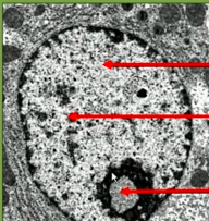
Label each arrow 1. top arrow 2. middle arrow 3. bottom arrow Significance of some of them if applicable. |
1. Euchromatin (Lighter nucleolus) > Method: More transcriptionally active nucleolus (or less bound to histone proteins via more acetylation or less methylation) 2. Heterochromatin (Dark nucleolus) > Method: Less transcriptionally active nucleolus (or more bound to histone proteins via less acetylation or more methylation) 3. Nucleolus |
|
|
What is produced in the nucleolus? a. mRNA b. rRNA c. tRNA |
Ribosomal RNA (rRNA) is produced in the nucleolus b. rRNA |
|
|
What will occur in the following reaction. Phenylalanine + Alpha keto glutarate -- Transaminase, PLP --> ? a. Formation of the alpha keto acid of phenylalanine and aspartate b. Formation of the alpha keto acid of phenylalanine and oxaloacetate c. Formation of the alpha keto acid of phenylalanine and glutamate d. Formation of glutamate and aspartate e. No reaction |
c. Formation of the alpha keto acid of phenylalanine and glutamate Note: only Lysine and threonine do not undergo transamination reactions |
|
|
What will occur in the following reaction. Lysine + Alpha keto glutarate -- Transaminase, PLP --> ? a. Formation of the alpha keto acid of lysine and aspartate b. Formation of the alpha keto acid of lysine and oxaloacetate c. Formation of the alpha keto acid of lysine and glutamate d. Formation of glutamate and aspartate e. No reaction |
e. No reaction. Reason: Lysine and threonine do not undergo transamination reactions |
|
|
What will occur in the following reaction. Threonine + Alpha keto glutarate -- Transaminase, PLP --> ? a. Formation of the alpha keto acid of threonine and aspartate b. Formation of the alpha keto acid of threonine and oxaloacetate c. Formation of the alpha keto acid of threonine and glutamate d. Formation of glutamate and aspartate e. No reaction |
e. No reaction. Reason: Lysine and threonine do not undergo transamination reactions |
|
|
What will occur in the following reaction. Glycine + Oxaloacetate -- Transaminase, PLP --> ?a. Formation of the alpha keto acid of glycine and aspartate b. Formation of the alpha keto acid of glycine and glutamate c. Formation of the alpha keto acid of glycine and glutamine d. Formation of glutamate and aspartate e. No reaction |
a. Formation of the alpha keto acid of glycine and aspartate Note: only Lysine and threonine do not undergo transamination reactions |
|
|
Vitamin B6 is deficient in a patient. What reaction is affected? a. Oxidation and reduction reactions b. Transamination reactions c. Phosphorylation reactions d. Xenobiotic transformation reactions e. Acetylation reactions |
b. Transamination reactions |
|
|
Glutamate is added to a beaker along with glutamate dehydrogenase and NAD+. The student discovers that PLP is not present. What will occur? a. Glutamate is converted into oxaloacetate b. Glutamate is converted into glutamine c. Glutamate is converted into alpha keto glutarate d. No reaction |
c. Glutamate is converted into alpha keto glutarate Rationale: This is the reaction: Glutamate -- GH, NAD+>NADH --> Alpha keto glutarate +NH3+ PLP is NOT required, so reaction occurs normally. |
|
|
Aspartate is added to a beaker along with its appropraite transaminase and a alpha keto acid. The student discovers that PLP is not present. What will occur? a. Aspartate is converted into oxaloacetate and an AA is formed b. Aspartate is converted into alpha keto glutarate and an AA is formed c. Aspartate is converted into glutamine and an AA is formed d. No reaction |
d. No reaction This is the reaction: aspartate + alpha keto acid -- Transaminase, PLP --> Oxaloacetate + AA Without PLP (necessary cofactor for Transaminase), reaction WILL NOT Occur. |
|
|
Which amino acid provides an additional component of urea in the urea cycle? a. alanine b. aspartate c. alanine and glutamine d. alpha keto glutarate e. none of the above |
b. aspartate Aspartate and NH3 give NH3 groups to form urea |
|
|
Arginase is defect. What will happen? |
Accumulation of arginine and NH3+ buildup (CNS problems) Tx: Arginine |
|
|
Arginosuccinate lyase defect. what will happen? |
Arginosuccinate accumulation and NH3+ buildup (CNS problems) |
|
|
Arginosuccinate synthetase defective. What will happen? |
Citruline accumulation and nH3+ buildup (CNS problems) |
|
|
Ornithine transcarbamoylase defective. What will happen? |
Accumulation of Carbamoyl phosphate, NH3 elevated (CNS problems) , increase glutamine, decreased AKG Increased Carbamoyl phosphate > increased orotate |
|
|
What reaction is responsible for combining carbamoyl phosphate and ornithine? a. carbamoyl phosphate synthetase I b. cittruline synthase c. ornithine transcarbamoylase d. arginase e. carbamoyl phosphate transcarbamoylase |
c. ornithine transcarbamoylase |
|
|
A patient has an accumulation of homogentisicacid. What condition does he most likely have? a. Phenylketonuria b. Homocystinuria c. Alcaptonuria d. Maple syrup urine disease e. Hartnup's disease |
c. Alcaptonuria |
|
|
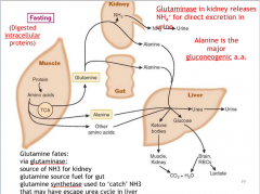
A patient is in the fasting state. Which of the following amino acids are mobilized in teh fasting state? Select all that apply. a. alanine b. aspartate c. glutamate d. glutamine e. oxaloacetate |
a. alanine d. glutamine Alanine and glutamine are both mobilized, however, glutamine is eventually deaminated by the kidney and lvier into alanine Alanine is then transported as a potential source of glucose for gluconeogenesis (NOT GLUTAMINE) |
|
|
Which of the following amino acids are used to produce glucose for gluconeogenesis in the fasting state? Select all that apply. a. alanine b. aspartate c. glutamate d. glutamine e. oxaloacetate |
a. alanine While Alanine and glutamine are both mobilized, glutamine is eventually deaminated by the kidney and gut into alanine Alanine is then transported as a potential source of glucose for gluconeogenesis (NOT GLUTAMINE) |
|
|
What is involved after mobilize of the AA without eating for a couple hours? |
Mobilize of glutamine and alanine Glutamine deamination to alanine via kidney and gut. Alanine transported to liver eventually for gluconeogenesis |
|
|
Purine breakdown steps (from notes) |
GMP > Guanosine > Guanine AMP > Hypoxanthine Both --> Xanthine > Guanine --> Xanthine > Hypoxanthine --(-Allopurinol)> Xanthine Xanthine -- (-Allopurinol)-->Uric acid |
|
|
Pyrimidine synthesis steps |
glutamine + CO2 + 2 ATP --(+PRPP)> Carbamoyl phosphate Carbamoyl phosphate --(+Aspartate)--> Orotate Orotate + PRPP --> UMP |
|
|
Regulators of pyrimidine synthesis? |
-: UMP +: PRPP |
|
|
Regulators of purine synthesis? |
-: GMP/GDP/GTP or AMP/ADP/ATP > inhibiting R-5-P, PRPP, IMP converison steps |
|
|
Which of the following is not a regulated step in purine synthesis pathway? a. IMP > b. R5P > c. 5-Phosphoribosyl 1-amine > d. PRPP > |
c. 5-phosphoribosyl 1 amine |
|
|
Which of the following is a negative regulatory of purine synthesis? a. PRPP b. GMP c. IMP d. UMP e. none of the above |
b. GMP - regulators purine: GMP, GDP, GTP / AMP, ADP, ATP no + regulators in purine - regulators pyrimidine synth: UMP + regulators pyrimidine synth: PRPP |
|
|
Which of the following is a negative regulatory of pyrimidine synthesis? a. PRPP b. GMP c. IMP d. UMP e. none of the above |
d. UMP - regulators purine:GMP, GDP, GTP / AMP, ADP, ATP no + regulators in purine - regulators pyrimidine synth: UMP + regulators pyrimidine synth: PRPP |
|
|
Which of the following is a positive regulatory of pyrimidine synthesis? a. PRPP b. GMP c. IMP d. UMP e. none of the above |
a. PRPP |
|
|
Precursors purine and pyrimidine |
Pyrimidine: Glutamine Aspartaet Purine: Glutamine Glycine Aspartate |
|
|
Which of the following converts protein into peptides? a. HCl b. trypsin b. pepsin d. elastase e. chymotrypsin f. carboxypeptidase |
b. pepsin |
|
|
Carboxypeptidse is secreted by which organ? a. salivary glands b. stomach c. brush border of SI d. pancreas |
d. pancreas |
|
|
Describe AA digestion |
Pepsin: Protein --pepsin +HCl> peptides Trypsin: break + charged AA (Lysine, Arginine) Chymotrypsin: break large bulky aromatic (tryptophan, tyrosine, phenylalanine) Elastase: break small nonpolar ---> broken into di and tripeptides Di,tripeptides --di + tripeptides --> Amino acids |
|
|
Di and tripeptides are produced by enzymes from what organ? a. saliva b. stomach c. pancreas d. brush border SI |
c. pancreas Trypsin, chymotrypsin, elastase convert peptides into tri and di peptides |
|
|
What ion is required for AA absorption? a. K+ b. Na+ c. Ca2+ d. Cl- e. Mg2+ f. PO43- |
b. Na+ |
|
|
Dysfunction of the Na+/k+ ATPase will most likely affect the reabsorption of what ion? |
AA Glucose as well |
|
|
What side is the AA/Na+ symport and Na/k atpase on? |
AA/Na+ symport: apical Na/k atpase: basolateral |
|
|
A patient has the symptoms of diarrhea, dementia, and neck redness. What does he most likely have? a. Cystinuria b. Maple syrup urine disease c. Alcaptonuria d. Hartnups disease e. Marasmus f. Von Gierkes |
d. Hartnup Deficiency of lack tryptophan reabsorption |
|
|
A patient has a lack of tryptophan reabsorption. What does he most likely have? a. Cystinuria b. Maple syrup urine disease c. Alcaptonuria d. Hartnups disease e. Marasmus f. Von Gierkes g. Kwashiorkor h. Deficiency essential AA i. sickle cell anemia |
d. Hartnups disease Lack tryptophan reabsorption > Hartnups (pellagra like symptoms: 4 Ds) |
|
|
A patient is experiencing anemia and ischemia during a low O2 state . What does he most likely have? a. Cystinuria b. Maple syrup urine disease c. Alcaptonuria d. Hartnups disease e. Marasmus f. Von Gierkes g. Kwashiorkor h. Deficiency essential AA i. sickle cell anemia |
i. sickle cell anemia Steps: Genetic mutation glutamic acid > valine on AA 6 on B globin chains of hemoglobin During deO2, B chains of Hg aggregate (b/c hydrophobic form good bonds together) forming insolubule fibers (rigid sickle shaped cells) > anemia when broken > ischemia, necrosis (pain) when sickle cells block blodo flow in capillaries |
|
|
What parpticipates in intracellular protein digestion? a. nucleus b. rough eR c. smooth eR d. golgi apparatus e. lysosome f. peroxisome |
e. lysosome Intracellular protein digestion: - lysosomes (proteases) - ubiquitin proteasome sysmptom |
|
|
What contributes to AA pool? |
Dietary and intracellular proteins |
|
|
What are blood AA used for? list all functions |
> Protein production > glucose formation = gluconeogenesis (alanine): fasting state = glycogenesis: fed state > TG with excess > form N2 molecules (purine, pyrimidine, NTs, hormones, heme, other functional N2 products) |
|
|
A child has normal appearances but emaciated. What does he most likely have a. Cystinuria b. Maple syrup urine disease c. Alcaptonuria d. Hartnups disease e. Marasmus f. Von Gierkes g. Kwashiorkor h. Deficiency essential AA i. sickle cell anemia |
e. Marasmus |
|
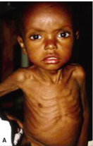
What does this child most likely have? Labs? |
Marasmus Normal albumin, but calorie deficient |
|
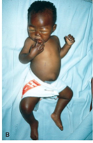
What does this child most likely have? Labs? |
Kwashiorkor Labs: > normal calorie, but Negative n2 balance (low albumin) > fatty liver (lack AA to transport Fats throughout: low VLDL) |
|
|
A patient has Kwashiorkor. Which of the following is the reason for a fatty liver? a. low albumin levels b. low chylomicrons c. low VLDL d. high LDLs |
c. low VLDL |
|
|
Fate AA intake. Describe. |
all sent to liver first via hepatic portal vein via digestion > AA converted into proteins > AA (alanine) converted into glucose = fasting state: gluconeogenesis = fed state: glycogenesis > fed state: AA converted into TG during fed state and sent via VLDL throughout body > Converted into N2 containing molecules (NT, hormones, heme) |
|
|
A patient has consumed an excessive amount of protein. Which of the following is most likely being produced at the highest rate? a. protein b. glycogen c. fat d. glucose |
c. fat |
|
|
What happens when gluatmine is released into the blood stream during the fasted state? a. glutamine is converted via glutaminase in the lvier to glutamate b. glutamine is converted via glutaminase in the kidney to glutamine c. glutamine is converted via glutamine dehydrogenase into glutamate |
b. |
|
|
Where is glutaminase located? |
kidney: glutamine enters --glutaminase--> NH3+ into urea and alanine also produced |
|
|
How is glutamine produced degraded? |
Glutamate -- glutamine synthetase (ATP>ADP+Pi) + NH3 --> Glutamien Glutamine -- glutaminase --> glutamate + NH3 |
|
|
Use glutamine Reactions. |
Catches NH3 that may have escaped urea cycle Via glutamate -- glutamine syntehtase --> glutamine Kidney: Glutamine -- glutaminase --> Glutamate + NH3 (urinated) Intestine: Glutamine --glutaminase--> glutamate + NH3 Rationale: glutamine used as energy by intestine Liver: NH3+ used as urea |
|
|
Describe protein use in fasting state. |
|
|
|
Formation of what molecules takes place in the mitochondria? a. ornithine b. arginosuccinate c. carbamoyl phosphate d. aspartate e. cittruline f. urea |
c. carbamoyl phosphate e. cittruline |
|
|
Urea is produced after what molecule is broekn down? a. carbamoyl phosphate b. arginosuccinate c. arginine d. fumarate e. ornithine f. aspartate g. citruline |
c. arginine |
|
|
Aspartate is added to what molecule in the urea cycle? a. carbamoyl phosphate b. arginosuccinate c. arginine d. fumarate e. ornithine f. aspartate g. citrulline |
g. citrulline |
|
|
Fumarate is removed from what molecule? a. carbamoyl phosphate b. arginosuccinate c. arginine d. fumarate e. ornithine f. aspartate g. citrulline |
b. arginosuccinate |
|
|
Ornithine is added to what molecule? a. carbamoyl phosphate b. arginosuccinate c. arginine d. fumarate e. ornithine f. aspartate g. citrulline |
a. carbamoyl phosphate |
|
|
How is carbamoyl phosphate synthesized in the urea cycle? |
NH3 + HCO3 + 2ATP --> 2 ADP + Carbamoyl phosphate +(Acetyl glutamate) acetyl-coa + glutamate -- arginine --> acetylglutamate |
|
|
How is the urea cycle regulated? a. positive feedback b. negative feedback c. feedbackward d. feedforward |
d. feedward |
|
|
How is the urea cycle promoted? |
High AA whether by: AA intake AA breakdown in fasting state (Gluconeogenessi) |
|
|
It is noticed that a person looks tired and says he has no eaten for over a week. Which of the following is expected? a. increased breakdown of glycogen to form glucose b. increased formation of triglycerides to be broken down into FA c. decreased breakdown of proteins d. increased PFK-1 activation e. increased glycogen phosphorylation activation |
The patient is in the prolonged fasting state with following occuring: > lipogenesis (FA used by majority organs except brain, RBC): skeletal muscles, x brain, x RBC > gluconeogenesis (little protein, glycerol, lactate): mainly RBC (only source is glucose) , some brain > decreased proteolysis to conserve protein for body and vital functions c. decreased breakdown of proteins > ketogenesis (used by brain) |
|
|
It is noticed that a person looks tired and says he has no eaten for over a week. Which of the following is expected? a. Urea levels of 12 g/day b. increased formation of triglycerides to be broken down into FA c. increased PFK-1 activation d. Urea levels of 2 g/day e. increased glycogen phosphorylation activation f. increased breakdown of glycogen to form glucose |
The patient is in the prolonged fasting state with following occuring: > lipogenesis (FA used by majority organs except brain, RBC): skeletal muscles, x brain, x RBC > gluconeogenesis (little protein, glycerol, lactate): mainly RBC (only source is glucose) , some brain > decreased proteolysis to conserve protein for body and vital functions d. Urea levels of 3 g/day stages urea: key: urea = N2 excretion in the form of urea non urea = other nitrogenous waste products 700 g intake: 1g urea, 2 g non urea N2 150 g intake: 2 g urea, 1 g non 12 hr fasting: 12 g urea 2 g non urea n2 3 day fasting (almost end brief): 10 g urea (beginning to decrease protein use) 2 g non urea n2 5 weeks: 2 g urea 2 g non urea N2 |
|
|
What is the most common urea cycle enzyme defect? a. carbamoyl phosphate synthase deficiency b. arginase deficiency c. arginosuccinate synthetase deficiency d. ornithine transcarbamoylase deficiency e. arginosuccinate lyase deficiency |
d. ornithine transcarbamoylase deficiency |
|
|
There is a deficiency in the production of fumarate in urea cycle. What is expected. |
High glutamine, Low akg, high NH3 Tx: Since block after arginosuccinate, provide arginine If block not after arginosuccinate, provide n removing and low protein intake |
|
|
A patient has a defect in the urea cycle. The patient is experiencing central nervous system problems. Which of the following is the best treatment for this patient? a. provide a low phenylalanine diet b. provide a low branched amino acid diet c. provide a low protein diet d. eliminate protein |
The patient has problem in urea cycle Tx: > lOW Protein diet > N2 reducing substances d. is too extreme, need some protein, but low protein a: refers to phenylketonuria, not correct as all AA must be reduced here not just phenylalanine b: is relevant to maple syrup urine disease, not relevant |
|
|
Which of the following must be elevated in a deficiency in the urea cycle? a. carbamoyl phosphate b. urea c. glutamate d.glutamine e. alpha keto glutatarate f. orotate |
a. carbamoyl phosphate d. glutamine UC def: elevated carbamoyl phosphate, elevated glutamine, low AKG (High AKG), low urea, elevated orotate (OTC deficiency only) ELimination: f: not necessarily elevated, only elevated in OTC deficiency |
|
|
A patient has an elevation in Thymine and Cytosine. Which of the following is most likely defective in this patient? a. carbamoyl phosphate synthase deficiency b. arginase deficiency c. arginosuccinate synthetase deficiency d. ornithine transcarbamoylase deficiency e. arginosuccinate lyase deficiency |
d. ornithine transcarbamoylase deficiency OTC: Elevated Carbamoyl phosphate, glutaminate, low AKG (Elevated AKG) Elevated orotate --> Pyrimidine (TC) |
|
|
Describe AA in anabolic and catabolic functions. Location describe |
In liver Anabolic (insulin, fed state, dephosphorylation) > glycogen formation (glycogen phosphatase) > TG formation (acetyl coa carboxylase) Catabolic (fasting, 12 hrs-3 days mainly, less catabolic beyond 3 days) > glucose formation (gluconeogenesis, mainly via alanine) > ketone body formation = via acetyl coa formation |
|
|
What molecules are responsible for the production of certain non essential AA? |
Pyruvate Oxaloacetate AKG 3-PG Pyruvate (+NH3)--> Serine Oxaloacetate (+NH3) --> Aspartate AKG (+NH3) --> Glutamate 3 Phosphoglycerate |
|
|
Which of the following is not responsible for the production of non essential AA according to slide 10? Select all that apply. a. 1,3-BPG b. 3-PG c. 2-PG d. Pyruvate e. oxaloacetate f. PEP g. alpha keto glutarate |
b. 3-PG d. pyruvate e. oxaloacetate g. alpha ketoglutarate |
|
|
A patient is lacking in methionine. Which of the following is considered essential? |
cysteine |
|
|
A patient is lacking in phenylalanine. Which of the following is considered essential? |
tyrosine |
|
|
A patient is now pregnant. Which of the following are essential? |
arginine, histidine = essential in people with + N2 balance arginine histidine |
|
|
Name the essential AA |
PVT TIM HALL Phenylalanien Valine Threonine Tryptophan Isoleucine Methionine Histidine Arginine (considered b/c most consumed in urea cycle) Lysine Leucine Branched AA: (Essential b/c body not capable of synthesizing branched) Aromatic: Body not capable of synthesizing aromatic (except tyrosine from phenylalanine) Incorporate sulfur into compounds: Methionine VIL Valine Isoleucine Leucine Conditionally: Arginine, histidine (+ N2 balance) Tyrosine (phenylalanine depletion) Cysteine (methionine depletion) |
|
|
which AA is considered essential b/c of its role in the urea cycle? |
Arginine although produced by the body, consumed in urea cycle. |
|
|
Which amino acid is produced by the body? a. phenylanine b. valine c. arginine d. histidine e. leucine f. lysine |
c. arginine although all considered essential, arginine is produced by the body, but still considered essential, b/c mainly consumed in urea cycle |
|
|
Describe AA conversions |
Alanine <-- transamination --> pyruvate AKG > Glutamate -- glutamine synthetase --> Glutamine Oxaloacetate --TA --> Aspartate --> Asparagine Methionine > Cysteine Phenylalanine > Tyrosine |
|
|
Oxaloacete can be converted to which of the following? Select all that apply a. Glutamate b. Aspartate c. Pyruvate d. Alanine e. Asparagine f. Tyrosine f. methionine |
OA > Aspartate > Asparagine b. aspartate e. asparagine |
|
|
The body is unable to add sulfur to amino acids. Which of the following is an application of this? Name AA |
Methionine |
|
|
Which of the following is not a category of essential amino acids in the body? a. branched b. aromatic c. polar d. sulfur containing |
c. polar Essential AA: branched aromatic sulfur containing |
|
|
Metabolic AA Forming AA, forming glucose/KB |
AA forming precursors: Oxaloacetate Alpha ketoglutarate Pyruvate 3-Phosphoglycerate Glucogenic AA: There are AA capable of forming glucose (Alanine) Ketogenic AA: There are AA capable of forming KB |
|
|
1. A Patient has a deficiency in Vitamin B12. Which of the following can be a result of this? a. marasmus b. alcaptonuria c. homocysteinuria d. maple syrup urine disease e. tay sachs disease f. X-linekd adreno... 2. What are possible lab findings? |
1. c. homocysteinuria 2. elevated: methionine homocysteine decreased: cysteine Exact deficiency: defective cystathione synthase defective cystathionase |
|
|
What is the allosteric inhibitor of cystathione? a. methionine b. homocysteine c. PRPP d. cysteine e. cysteine sulfinic acid f. serine |
d. Cysteine |
|
|
What are the cells in NS? |
CNS: Neuroglia neurons PNS: Schwann cells neurons |
|
|
Collagen, elastin, chondroblasts, osteoblasts, osteocytes, osteoblasts are found in what type of tissue? a. epithelial b. connective c. muscle d. nervous |
b. connective |
|
|
Which of the following detects changes in the internal and external environment and coordinates the appropriate response? a. epithelial b. connective c. muscle d. nervous |
d. nervous |
|
|
Which of the following is responsible for contraction resulting from rearrangement of internal bonds between proteins? a. epithelial b. connective c. muscle d. nervous |
c. muscle |
|
|
What are the types of muscles? |
Skeletal Cardiac smooth (visceral) |
|
|
Which of the following tissues are continuous sheets of lining bound together by tight junctions? a. epithelial b. connective c. muscle d. nervous |
a. epithelial |
|
|
Which of the following line inner and outer surfaces of the body to form a barrier? a. epithelial b. connective c. muscle d. nervous |
a. epithelial |
|
|
Which of the following function in absorption, substance movement, and secretion? a. epithelial b. connective c. muscle d. nervous |
a. epithelial |
|
|
A disorder in which of the following can lead to chronic diarrhea, metabolic acidosis, and severe dehydration? a. cilia b. flagella c. rough er d. lysosome e. microvilli f. peroxisome |
e. microvilli > composed of actin this describes microvillus inclusion disorder > Born without microvillie S&S: > chronic diarrhea > metabollic acidosis > severe dehydration > potentially fatal |
|
|
A patient has celiac sprue and bacterial infections. which is most likely affected? a. cilia b. flagella c. rough er d. lysosome e. microvilli f. peroxisome |
e. microvilli > composed of actin |
|
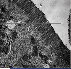
A slide is shown below. 1. What does this refer to? a. cilia b. flagella c. rough er d. lysosome e. microvilli f. peroxisome 2. What are the components of this? a. actin b. intermediate filaments c. tubulin 3. What is a function of this? a. motility b. protection against phagocytosis c. increase surface area d. catching prey |
1. e. microvilli 2. a. actin Since actin made of MF and MF comprise microvillie 3. c. increase surface area |
|
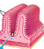
A following diagram is shown. Describe. (structure) (function) |
Microvilli in GI structure: actin (MF) function: increase S/A |
|
A diagram shown below.
1. Which of the following is an example of a pathology of this cell? a. Gauch disease b. Tay sachs c. Sinus intertisus d. Celiac sprue e. Mitochondrial disorder |
d. Celiac sprue This is a microvillie. Disorder of microvilli is celiac sprue |
|
|
Which of the following form centrioles? a. microfilaments b. intermediate filaments c. microtubules |
c. microtubules |
|
|
Which of the following form hollow tubes? a. microfilaments b. intermediate filaments c. microtubules |
c. microtubules |
|
|
Which of the following are found only in eukaryotes? a. cell membrane b. centrioles c. cytoplasm d. DNA e. plasmid f. outer membrane |
b. centrioles Centrioles found only in eukaryotes |
|
|
A 9 group trimer arrangement is see arranged around a central cavity. What does this refer to? a. centrosome b. microfilaments c. cilia d. centrioles e. intermediate filaments f. integrin proteins g. laminin proteins |
d. centrioles |
|
|
A 9 + 2 group arrangement is see arranged around a central cavity. What does this refer to?a. centrosome b. microfilaments c. cilia d. centrioles e. intermediate filaments f. integrin proteins g. laminin proteins |
c. cilia |
|
|
A patient has a congenital myopathy. Which of the following is most likely affected? a. keratin b. nuclear lamina c. actin d. centrioles e. cilia |
c. actin slide 67 |
|
What does this refer to? a. keratin b. laminin c. actin d. fibronectin e. cilia f. flagella g. microvilli |
c. actin |
|
|
A patient has an accumulation of Hydrogen peroxide 1. What is most likely affected? a. lysosome b. peroxisome c. cilia f. nucleus 2. What disorder is this most likely? a. Hartnups disease b. Microvilli inclusion disease c. Tay sachs disease d. X linked adrenoleukodystrophy e. Pellagra f. Zellweger syndrome g. Maple syrup urine disease h. Marasmus i. Kwashiorkor j. cystic fibrosis k. Pellagra l. Zellweger syndrome m. sinus inversus |
1. b. peroxisome 2. disorders peroxisomes (accumulation H2O2 b/c unable to create enzymes to break down hydrogen peroxide) > x linked adrenleukodystrophy > zellweger syndrome d. x linked adrenoleukodystrophy l. zellweger syndrome |
|
|
1. Lysosomal storage diseases are usually inheritted through what mode? a. heterosomal recessive b. heterosomal dominant c. autosomal recessive d. autosomal dominant 2. These disorders are usually of what type? a. digestive disorders b. absorptive disorders c. transfer disorder d. phagocytic disorder |
1. c. autosomal recessive (cell bio 1, slide 64) 2. c. transfer disorder |
|
|
A patient has a defect in hexosaminidase A alpha chain. Which of the following does he most likely have? a. X-linked adrenoleukodystrophy b. Pemiphigus c. Tay sachs disease d. Ornithine transcarbamoylase deficiency e. Gaucher disease f. Mitochondrial storage disease |
c. Tay sachs |
|
The following is shown. 1. What does this patient have? a. X-linked adrenoleukodystrophy b. Tay sachs c. Pemphigoid d. Pemphigus vulgaris e. Kwashiorkor 2. What is the pathophysiological basis for this disease? a. Antibodies towards laminin proteins b. Antibodies towards cadherin of hemidesmosomes c. Antibodies towards cadherin of tight junctions d. Antibodies towards cadherin of desmosomes e. Antibodies towards integrin of hemidesmosomes f. Antibodies towards integrin of desmosomes 3. What is another possible result of this? a. looseining of adhesion between hemidesmosomes and basal lamine b. loosening of adhesion between adjacent cells c. Lack of tryptophan d. Lack of desmosome e. Lack of hemidesmosome |
1. d. pemphigus vulgaris 2. d. antibodies towards cadherin of desmosomes 3. b. loosening of adhesion between adjacent cells |
|
|
Which disorder is autosomal recessive? a. Pemphigus b. X-linked adrenoleukodystrophy c. Maple syrup urine disease d. Osteoporosis e. Gaucher disease f. Tay sachs g. Alcaptonuria h. hOMOcysteinuria |
e. Gaucher disease f. Tay sachs |
|
|
Defects of which of the following (discussed in class), are autosomal recessive? a. tryptophan reabsorption b. desmosomes c. microvilli d. lysosome e. peroxisome f. cilia g. flagella h. pili i. branched amino acid breakdown |
d. lysosome (Gaucher disease, Tay sachs) |
|
|
Function Golgi apparatus |
> Glycosylation of proteins > Packaging, sorting, modifying proteins for secretion > Lysosomal production |
|
|
There is a defect in the production of lysosomes. Which of the following is most likely responsible? a. Tay sachs disease b. X-linked adrenoleukodystrophy c. Defect RER production productino of lysosomes d. Defect SER production production of lysosomes e. Defect Golgi apparatus production of lysosomes f. Gaucher disease g. Zellweger syndrome h. Marasmus |
e. Defective Golgi apparatus production |
|
A defect of the following will cause what? |
Defects in: glycosylation of protiens modifying, preparing for secretino of substances |
|
|
1. Which cells are most affected by mitochondrial diseases? a. epithelial tissue b. muscle tissue c. connective tissue d. nervous tissue 2. What S&S are expected from mitochondrial diseases? a. bloating abdomin b. myopathy c. diarrhea, dementia, pallor of wrists and hands d. sinus intervtis e. ptosis f. exercise intolerance |
1. b. muscle tissue d. nervous tissue 2. S&S mitochondrial storage disease: myopathy ptosis exercise intolerance b. e. f. |
|
Describe. What type? Structure. How to distinguish |
Gram + Structure: C/M CW: Thick peptidoglycan with teichoic acid (stains purple with gram stain) No outer membrane May have capsule (generally polysaccharide, except bacillis anthracis): evade phagocytosis, nutrients, prevent dehydration |
|
Describe. What type?Structure. How to distinguish |
Gram - C/M Thin peptidoglycan cell wall without teichoic acid Outer membrane (lipopolysaccharide, endotoxin) May have capsule (generally polysaccharide, except bacillis anthracis): evade phagocytosis, nutrients, prevent dehydration |
|
|
A certain organ lacks polysaccharides in its capsule. what is it and whats its shape? |
Bacillus anthracis Rod like shape. |
|
|
Which of the follwoing do not include polymers? a. carbs b. lipids c. nucleic acids d. proteins |
b. lipids |
|
|
Describes lipids Structure Classes |
Hydrophobic tail 3 classes: fats (triglycerides: 1 glycerol esterified to 3 FA via dehydration synthesis) > ester has C=O bonded to O steroids (4 rings: 3 hexose w/ OH group, 1 pentose) phospholipids (amphipathic) |
|
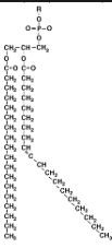
What does this indicate. |
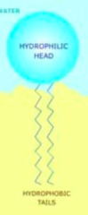
Phospholipid Nonpolar region: hydrocarbons Polar: Phosphate (PO43-) Properties: amphipathic move mainly laterally, rotation, limited flip flop (only flip flop with proteins using ATPase) |
|
|
A certain molecule has 16 or 18 C atoms. What is it? a. phospholipid b. steroid c. fatty acid d. sphingosine |
c. fatty acid |
|
|
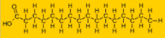
Fats -structure -properties |
1 glycerol 3 fa > FA repelled from H2O |
|
|
What is a FA molecule composed of? |
1 glycerol esterified to 3 FA (3 H2O dehydration synthesis) Glycerol FA > Long linear unbranched carbon skeleton (16-18C) > Carboxyl group 1 one end (head) > long hydrocarbon tail attached to carboxyl group |
|
|
Components of TG (Fat) |
1 glycerol 3 FA > Carboxyl group (Head) |
|
|
What is the head of FA? |
Carboxyl group |
|
|
What is the tail of FA? |
Hydrocarbon |
|
|
Properties FA |
Mainly hydrocarbon (little to no affinity for water) |
|
|
Main component of biological membranes? |
Phospholipid bilayer main other: steroids proteins carbs glycolipids glycoproteins |
|
Describe. |
Saturated FA |
|
Describe. |
Unsaturated FA |
|
Describe. |
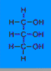
Glycerol |
|
Describe. |
1 glycerol esterified to 3 FA Ester bond ebtween glycerol and FA: C=O bonded to O |
|
|
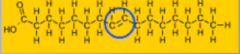
Unsaturated vs. saturation -Criteria (requirements) |
Requirement: presence and # double bonds in hydrocarbon tails. Saturated fat Structure: no double bond (more packing, stronger intermolecular forces) properties: Increase saturation increasing melting point Unsaturated fat Structure: Increase double bonds (less closely packed) properties: lower meltingi point |
|
|
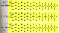
Saturated fat properties |
No double bonds (fully saturated carbons with H) High melting point Hydrocarbon tails pack tightly together Types: animal fat, butter |
|
|
Unsaturated fat properties |
1-3 double bonds (not fully saturated carbons with H) > causing kinks Low melting point Hydrocarbon tails pack tightly together Types: oils *Important influence on membrane fluidity in different temperatures |
|
|
whICH OF THe following has an important influence on membrane fluidity at different temperatures? a. saturated fats b. unsaturated fats c. phospholipids d. cholesterol |
b. unsaturated fats (cell bio 2, slide 10) |
|
|
What causes kinks? |
Double bonds in unsaturated fats |
|
|
Which types fats are saturated |
Animal fat, butter |
|
|
Which types fats are unsaturated |
plant oils |
|
|
How many double bonds in unsaturated fats? a. 0 b. 1-2 c. 1-3 d. 1-4 e. 1-7 f. 1-19 |
c. 1-3 (slide 10) |
|
|
How are fats formed? a. phosphorylation reactions b. hydrolysis reactions c. glycosylation reactions d. dehydration reactions |
d. dehydration reactions |
|
|
Are fats large molecules? T/F |
Yes. |
|
|
Components fats |
Glycerol, FA |
|
|
Another name fats |
Triglycerides |
|
|
What determine physical properties of FA |
# C Sites and # double bonds |
|
Describe. Location Properties |
Saturated fat Animal products, butter High melting point Closing packing, no double bonds (no kinks) : strong intermolecular forces |
|
Describe. Location Properties |
Unsaturated fat: Vegetable oils Low melting point Weaker packing, 1-3 double bonds (kinks) : weaker intermolecular forces Unsaturated fats play important role in regulation of membrane fluidity |
|
|
Function fats |
- Energy storage (9 cal/g) (vs. 4 cal/g for carbs) -Cushion for vital organs -Insulation |
|
|
What organs contains cushion fat? |
Kidneys |
|
Describe.
|
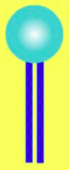
phospholipid symbol |
|
Describe. |
space filling model of phospholipids |
|
|
Describe phospholipid |
Esterification of 2 FA and 1 phosphate to glycerol Nonpolar: 2 FA Polar: 1 Phosphate (PO43-) Application: basic component of cell membrane |
|
|
How to form different types phospholipids |
Link a variety of small polar molecules to the phospholipid to form various types phospholipids |
|
|
Which of the following are amphipathic? a. fats b. steroids c. phospholipids b. hydrocarbon tails |
c. phospholipids B/c contain hydrophobic and hydrophilic regions |
|
|
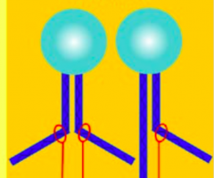
What happens when adding water to phospholipids |
Form aggregates PO43- binds to water Hydrocarbon tails away water > form phospholipid or micelles |
|
|
What happens when water added to phospholipids? a. interaction of polar heads with hydrocarbon tails b. interactions of water with polar head c. intearctions of water with hydrocarbon tails d. no formation of micelles or lipid bilayers |
b. interactions of water with polar head formation of micelles or lipid bilayers |
|
|
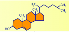
Steroids Structure Function Properties |
Structure: 3 Hexose sugars, 1 pentose sugar (reach across half of the bilayer) (same length at C16 FA) Function: Component of animal cell membrane Stabilization the membrane and protection against extreme conditions > at warm temperatures, reduce fluidity > at low temperature, increase fluidity Precursor to steroid hormones Precursor to bile acids Properties: Contributes to atherosclerosis Amphipathic(along with phospholipids) Increases with: increase food intake increase synthesis by body genetic defects (i.e: faulty LDL) Interspected in cell membrane -OH heads point toward hydrophilic polar head |
|
|
Which of the following is responsible for stabilization of the membrane? a. saturated FA b. unsaturated FA c. phospholipids d. cholesterol |
d. cholesterol |
|
|
Which of the following are amphipathic? a. saturated FA b. unsaturated FA c. phospholipids d. cholesterol |
c. phospholipids d. cholesterol (-OH on first hexose) |
|
|
Which of the following form aggregates when added to water? a. saturated FA b. unsaturated FA c. phospholipids d. cholesterol |
c. phospholipids |
|
|
Which of the following can form micelles and phospholipid bilayers? a. saturated FA b. unsaturated FA c. phospholipids d. cholesterol |
c. phospholipids |
|
|
Which of the following contributes to atherosclerosis in high levels? a. saturated FA b. unsaturated FA c. phospholipids d. cholesterol |
d. cholesterol |
|
|
Estrogen and testosterone are example of what type of molecule? a. saturated FA b. unsaturated FA c. phospholipids d. cholesterol |
d. cholesterol |
|
|
Which of the following reaches across half the bilayer? a. saturated FA b. unsaturated FA c. phospholipids d. cholesterol |
d. cholesterol |
|
|
Which of the following is a precursor for steroid hormone synthesis? a. saturated FA b. unsaturated FA c. phospholipids d. cholesterol |
d. cholesterol |
|
|
Which of the following is a precursor for bile acids?? a. saturated FA b. unsaturated FA c. phospholipids d. cholesterol |
d. cholesterol |
|
|
What are bile acids? derivation function |
Derived from cholesterol 'Biological detergents' that solubilize fats in the sI |
|
|
Cholesterol increase with what? |
Increase synthesis in mammals High intake with food Genetic deficiencies (i.e: LDL carrier protein deficeincy) |
|
Describe. |
Cholesterol 4 fused rings > 3 6 C (hexose) rings = 1 -OH in the first hexose > 1 5C (pentose) rings -amphipathic |
|
|
Describe cholesterol in the c/M |
Interspersed -oH Polar head Of cholesterol always faces hydrophilic heads of phospholipids |
|
|
Are cholesterol amphipathic? |
Yes. along with phospholipids |
|
|
What describes C/M Structure Functino |
Fluid musaic model of: Phospholipids Lipids Proteins Carbohydrates -Semipermeable barriers |
|
|
C/M is a phospholipid bilayer composed of what? |
Phospholipid Lipid Carbs Proteins |
|
|
What does the biological illustrate? |
Relatinoship between structure and function |
|
|
What describes cell membranes? a. completely permeable barriers b. semi permeable barriers c. super permeable abrriers d. impermeable barriers |
b. semi permeable barriers |
|
|
Describe the movement of phospholipid molecules in cell membrane |
Random Types movement: lateral (22 um/sec) rotation fast flip-flop : rarely switch from 1 Phospholipid layer to the other [but can with ATPase] reason: hydrophilic head of molecule must cross the hydrophobic core of the molecule |
|
|
Explain flip flopping of phospholipid bilayer molecules in cell membrane? |
Rarely phospholipid bilayer molecules flip flop Reason: Hydrophilic head must cross thh hydrophobic core of the membrane |
|
|
Why is it difficult for flip flopping of phospholipid molecules in cell membrane? a. hydrophobic tail of molecule must cross hydrophilic head b. hydrophobic head must cross water molecules c. hydrophilic head must cross hydrophobic tail |
c. hydrophilic head must cross hydrophobic tail |
|
|
Effect of unsaturations? |
Lower melting temperature Reason: Lower packing because of kinks |
|
|
Which of the following is responsible for protection against extreme conditions? a. saturated FA b. unsaturated FA c. phospholipids d. cholesterol |
d. cholesterol |
|
|
Which of the following is responsible for maintaining fluidity despite changes in temperature? a. saturated FA b. unsaturated FA c. phospholipids d. cholesterol |
d. cholesterol |
|
|
Membrane proteins -derivation -structure -types -properties |
Derivation: derived from the 20 AA Types -Integral membrane: Hydrophobic interior Hydrophilic exterior -Peripheral membrane: Hydrophilic exterior Loosely bound to the surface of the membrane Not embedded in the lipid bilayer Properties: Mobility proteins: Much slower movement Most membrane proteins immobile by virtue of their attachment to the cytoskeleton But some move in directional manner via shuttle or transport Few proteins drift largely Function: Receptors Membrane transport Enzymes (transferases, hydrolases, oxidoreductases) Cell adhesion molecules: selectins, integrins |
|
|
Which of the following accounts for more than 50% of the cell dry weight? a. carbohydrates b. lipids c. proteins d. phospholipids e. cholesterol |
c. proteins |
|
|
Which of the following contains hydrophobic and hydrophilic regions? a. triglycerides b. phospholipids c. cholesterol d. peripheral proteins e. integral proteins |
b. phospholipids
c. cholesterol e. integral proteins |
|
|
Which of the following is interspersed throughout the cell membrane? a. triglycerides b. phospholipids c. cholesterol d. peripheral proteins e. integral proteins |
c. cholesterol |
|
|
Which of the following is loosely bound to the surface of the membrane a. triglycerides b. phospholipids c. cholesterol d. peripheral proteins e. integral proteins |
d. peripheral proteins |
|
|
Which of the following is not embedded in the lipid bilayer? a. triglycerides b. phospholipids c. cholesterol d. peripheral proteins e. integral proteins |
d. peripheral proteins |
|
|
Which of the following are structurally sophisicated molecules according to Dr. H? a. carbohydrates b. proteins c. lipids d. DNA |
b. proteins |
|
|
Which of the following are immobile by virtue of their attachment to the cytoskeleton? a. phospholipid b. cholesterol c. protein d. glucose residues |
c. protein |
|
|
Compare size protein and phospholipid |
Proteins --size --> >> phospholipid But thus move slower |
|
|
Which of the following is true regarding proteins and phospholipids in C/M? a. proteins are smaller and move faster b. proteins are smaller and move slower c. proteins are larger and move faster d. proteins are larger and move slower |
d. proteins are larger and move slower |
|
|
Which of the following is true regarding molecules in cell membranes? a. peripheral proteins can sometimes adhere to the interior of cell membranes b. phospholipid molecules move at a rate of about 44 um / s in the cell membrane c. most membrane proteins are immobile d. phospholipid rotation occurs rarely e. many proteins drift largely |
c. most membrane proteins are immobile Explain: a. peripheral proteins adhere to the exterior of the cell membrane, sometimse adhering to hydrophilic area of integral membrane protiens b. phospholipid molecules move at rate of 22 um/s d. phospholipid rotate occurs rapid (its flip flopping that rarely occurs) e: few molecules drift largely |
|
|
Which of the following is true regarding molecules in cell membranes? a. cholesterol is only hydrophobic b. most membrane proteins are mobile c. At low temperature, cholesterol makes membrane less fluid d. few proteins drift largely e. rotation of phospholipid occurs slowly |
d. few proteins drift largely elimination: a. cholesterol is amphipathic (-OH on 1st hexose ring) b. most membrane proteins are immotile c. at low temperature, cholesterol makes membrane more fluid (resist tendency to be more solid) d. correct e. rotation of phospholipid occurs rapidly almost all of the time. |
|
|
What are the primary forces holding cell membranes together? a. electrostatic b. hydrophobic c. hydrophilic d. H2 bonding e. covalent |
b. hydrophobic Hydrophobic interacitons are the primary forces that hold membrane components together. |
|
|
Which is the following is true regarding cell membranes? a. phospholipid molecules flip flop rapidly b. phospholipid molecules move at a rate of 100 um/s laterally c. cholesterol spans less than 10% of the cell membrane length d. phospholipid molecules move randomly e. A membrane is a rigid sheet of molecules |
d. phospholipid molecules move randomly Explanation: a: flip flop occurs rarely b: move at rate of 22 um/s c: cholesterol spans about half of the C/M length d. Correct phospholipid molecules do move randomly (Cell Bio 2, Slide 23) e: a membrane is not a rigdi sheet of molecules, they move quite a lot. |
|
|
Function of membrane proteins |
Receptors > GPCR (Gs, Gi) > Tyrosine kinase Membrane enzymes > Transferase > Hydrolases > Oxidoreductases Cell adhesion molecules Cell adhesion molecules > Cell-cell identification and interaction >>selectins, integrins |
|
|
Example functions of membrane enzymes |
Hydrolase Transferase Oxidoreductease |
|
|
Which of the following is not an example of a function of membrane enzymes? a. hydrolase b. transferase c. oxidoreductase d. acetylase |
d. acetylase |
|
|
ER Function Mechanism |
Function: P/M built by ER ER determines asymmetric distribution of proteins, lipids, carbohydrates Mechanism: ER delivers PM in reversed orientation > molecules start out in inside face of ER and up on outside face of PM |
|
|
The plasma membrane it build by which of the following? a. nucleus b. endoplasmic reticulum d. GA e. centriole f. centrosome g. lysosome h. peroxisome |
b. ER |
|
|
Explain membrane asymmetry |
Inside face distinct outside face Reason asymmetry: proteins diff (some inside, some outside) carbohydrates are restricted to exterior surface of P/M |
|
|
Describe membrane carbohydrates |
Branched oligosaccharides usually with < 15 monosaccharides Oligosaccharides: form glycoproteins form glycoproteins Function: adhesion cell-cell interaction |
|
|
Which of the following is the component of membrane carbohydrates? a. unbranched monosaccharides b. branched monosaccharides c. unbranched oligosaccharides d. branched oligosaccharides e. unbranched polysaccharides f. branched polysaccharides |
d. branched oligosaccharides < 15 monosaccharides |
|
|
Which of the following is correct regarding cell membranes? a. cholesterol span less than half of the width of the cell membrane b. unbranched oligosaccharides are part of the cell membrane c. membrane proteins are small and move slow as compared to phospholipid molecules d. membrane carbohydrates have less than 20 monosaccharides e. carbohydrates are restricted to the exterior surface of the plasma membrane |
e. carbohydrates are restricted to the exterior surface of the plasma membrane explan: a: cholesterol spans 1/2 the width of C/M b: branched oliosaccharides are part of the C/M c: membrane proteins are large and move slow compared to phospholipid molecules d: membrane carbs have less than 15 monosaccharides, not less than 20 e: correct |
|
|
Properties carbohydrates in C/M |
Branched oligosasccharides < 15 monosaccharides in the oliosascchardes Restricted to exterior of P/M Function: Adhesion Cell-cell interaction form: glycoproteins glycolipids properties: outer membrane surface oligosaccharide vary among species, individuals and from cell type to another |
|
|
What is a nucleoside |
Sugar + N2 base |
|
|
What is a nucleotide |
Nucleoside + PO43- = Sugar + N2 base + PO43- |
|
|
Differentiate between deoxyribsoe and ribose |
H on C-2 of deoxyribose OH on C-2 of ribose |
|
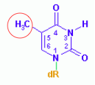
Describe. a. Adenine b. Guanine c. Thymine d. Cytosine e. Uracil |
Thymine c. thymine > pyrimidine b/c 1 ring only > T or U because no NH2 at 12 o clock position > methylated form of uracil |
|
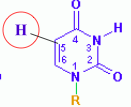
Describe. a. Adenine b. Guanine c. Thymine d. Cytosine e. Uracil |
> Pyrimidine b/c 1 ring only > T or U because no NH2 at 12 o clock position > unmethylated form of thymine: uracil e. Uracil |
|
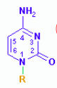
Describe. a. Adenine b. Guanine c. Thymine d. Cytosine e. Uracil |
> Pyrimidine b/c 1 ring only > C because NH2 at 12 o clock position d. cytosine |
|
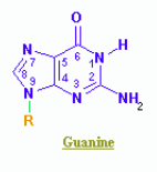
Describe. a. Adenine b. Guanine c. Thymine d. Cytosine e. Uracil |
> Purine b/c 2 rings (A or G) > No -NH2 on 12 o clock, so guanine b. Guanine |
|
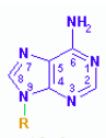
Describe. a. Adenine b. Guanine c. Thymine d. Cytosine e. Uracil |
> Purine b/c 2 rings (A or G) > -NH2 on 12 o clock, so adenine a. Adenine |
|
|
1. Describe the orientation of DNA. a. 3'>5' WITH PO43- at 5' and OH at 3' b. 3'>5' with OH at 5' and PO43- at 3' c. 5'>3' with PO43- at 5' and OH at 3- d. 5'>3' with OH at 5' and PO43- at 3' 2. What is the name of the bonds between 5' and 3'? a. phosphoanhydride b. ester c. diester d. phosphodihydride e. phosphodiester |
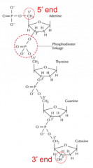
1. c. 5'>3' with PO43- at 5' and OH at 3- 2. e. phosphodiester |
|
|
1. The OH is located at what carbon of sugar in DNA? 2. The PO43- is located at what carbon of sugar in DNA? a. 1' C b. 2' C c. 3' C d. 4' C e. 5' C. |
1. c. 3' C 2. e. 5' C |
|
|
What are the Watson Crick discoveries? |
>double stranded >antiparallel >sugar phsophate backbone >base pairs inside >complementary base pairing >> 2 H between A-T >> 3 H between G-C |
|
|
Which of the following is not a correct Watson Crick discovery in 1953? Select all that apply a. double stranded helical structure b. parallel strands c. G-C base pairs have 2 H2 bonds d. A-C base pairs have 2 H2 bonds e. sugar phosphate backbone on outside |
a. e. Explan: a: correct b: they are antiparallel strands c: G-C base pairs have 3 H2 bonds not 2 d: A-C does not even base pair e: correct,sugar phosphate bakcbone on outside |
|
|
Describe the DNA replication process. a. OH attacks at 5' PO43- b. OH attacks at 3' PO43- c. PO43- attacks at 5' OH d. PO43- attacks at 3' OH |
d. PO43- attacks at 3' OH |
|
|
What describes the DNA replication process? a. non conservative b. semi conservative c. completely conservative d. moderately conservative e. mildly conservative f. semi republican |
b. semi conservative |
|
|
Describe the central dogma of molecular genetics |
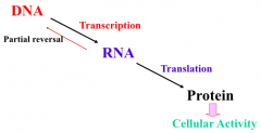
DNA --transcription --> RNA DNA <--reverse transcriptase-- RNA (usually retroviruses such as HIV RNA -- translation --> protein --> cellular effects |
|
|
Describe RNA polymerase transcription compelx |
TFIID first binds recruiting other TF (TFIIA, TFIIB, TFIIF) that also binds to promoter (TATA box), recruit RNA polymerase II |
|
|
What is the first TF binding to promoter during transcription? a. TFIIB b. TFIIC c. TFIID d. TFIIF e. TFIIL |
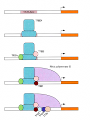
c. TFIID |
|
|
What RNA polymerase is promoters via TF IID? a. 1 b. 2 c. 3 |
b. 2 RNA polymerase II via TF IID binding |
|
|
The coding strand is 5'TCAGCGACC3'. What is the corresponding mRNA? |
5'UCAGCGACC3' Since coding, only exchange T with u, keep rest same. |
|
|
Important codons and applicable AA to know |
Start (AUG) codon > methionine AA for eukaryotes > met-formyl start AA for prokaryotes Phenylalanine codon: UUU, UUC (phenylalanine AA) End codon: UAA, UAG, UGA (no AA) |
|
|
Tempalte DNA is 3' AGTCGCTGG 5' What is the mRNA? |
5'UCAGCGACC 3' |
|
|
Tempalte DNA is 5'GGTCGCTGA3'. What is the mRNA? |
5' UCAGCGACC 3' |
|
|
Polyadenylation for the 10 As or so depends on what? a. CstF b. CPSF c. PAP d. PAB e. 5'cap f. AAUAAA sequence |
b. CPSF f. AAUAAA sequence Steps in polyadenylation: Polyadenylation depends on CPSF, AAUAAA sequence for the first 10 As or so. After that, depends on existing poly A tail. CPSF and CstF multi protein complexes bind to rear of advancing > CPSF and CstF transfer to the new pre-mRNA > CPSF bind to THE aauaaa SEQUENCE > CsTF bind to the GU or U rich sequence following it RNA cleavage at apoint 35 nucleotides after end of AAUAAA sequence > PAP starts synthesis of poly A tail > PAB protien immediately binds to new polyadenosine sequence |
|
|
What complexes bind to the rear of advancing RNA polymerase II during polyadenylation process? a. CstF b. CPSF c. PAP d. PAB e. 5'cap f. AAUAAA sequence |
a. CstF b. CPSF |
|
|
What binds to the AAUAAA sequence in polyadenylation process? a. CstF b. CPSF c. PAP d. PAB e. 5'cap f. AAUAAA sequence |
b. CPSF |
|
|
What binds to the GU or U rich sequence in polyadenylation process? a. CstF b. CPSF c. PAP d. PAB e. 5'cap f. AAUAAA sequence |
a. CstF |
|
|
When does RNA cleavage occur in polyadenylation process? |
At a point 35 nucleotides after end of AAUAAA sequence. > PAP starts poly A tail synthesis and continues polyadenylation > PAB immediately binds to new polyadenosine sequence, but acts as a molecular ruler, specifying adenylation stop point |
|
|
What starts the synthesis poly A tail? a. CstF b. CPSF c. PAP d. PAB e. 5'cap f. AAUAAA sequence |
c. PAP |
|
|
What immediately binds to the new polyadenosine sequence during polyadenylation process? a. CstF b. CPSF c. PAP d. PAB e. 5'cap f. AAUAAA sequence |
d. PAB |
|
|
What continues polyadenylation on the A tail, about 50-250 nucleotides, depending on organism? a. CstF b. CPSF c. PAP d. PAB e. 5'cap f. AAUAAA sequence |
c. PAP |
|
|
What acts as a molecular ruler, specifying adenylation stop point? a. CstF b. CPSF c. PAP d. PAB e. 5'cap f. AAUAAA sequence |
d. PAB |
|
|
What determines mRNA export from nucleus? a. CstF b. CPSF c. PAP d. PAB e. 5'cap f. AAUAAA sequence |
d. PAB e. 5'cap |
|
|
Location of start and stop codons? |
AUG (Methionine) at 5' NH2 (Amino terminus) UAA/UAG/UGA (stop) at 3' COOH (Carboxy terminus) |
|
|
Which of the following is incorrect? a. Phenylalanine's codon is UUU. b. The start codon for prokaryotes is formyl-methionine c. The stop codon for eukaryotes is UAA/UAG/UGA d. The start codon is located at the 5' N terminus |
b. The start codon for prokaryotes is formyl-methionine > The start codon is AUG, the AA is formyl-methionine (methionine for eukaryotes) |
|
|
Which of the following is true? Select all that apply. a. phenylalanine's codon is UAG b. phenylalanine's codon is UUC c. Eukaryote's start codon is methionine d. Prokaryote's starting amino acid is formyl-methionine e. Stop codon is UAA f. The stop codon located at 5' NH2 terminus |
b. phenylalanine: UUU, UUC, UCU d. e. Explanation: a. phenl: UUU, UUC, UCU b: correct c: eukaryote's start codon: AUG not methionine AA d: correct e: correct f: stop codon located at 3' COOH terminus |
|
|
Describe translation steps |
Step 1: Bind to AUG methionine codon on small unit ribosome, bringing methionine (formyl-methionine in prokaryotes) > Recruitment of large subunit ribosome Formation of initiation complex Step 2: Elongation steps: Methionine at E, New AA at P Peptide bond between E and P, E displaced and new Peptide at P New AA at A Peptide bond between PA, P released and new peptide at A Peptide moves back to P in order to bind with future A Step 3: When Stop codon at A, TF recruited at A (No AA) formed And end of translation |
|
|
Describe translation. |
When UAG/UAA/UGA codon at A site, termination factor binds to UAG. |
|
|
What best explains the termination process in translation? a. the stop codon binds to the Amino acid representing the stop function b. the stop codon binds to an anticodon, which then binds to the transcription factor, ending transcription c. the stop codon binds to methionine, ending transcription d. the stop codon binds to termination factor, ending transcription |
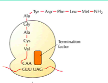
d. the stop codon binds to termination factor, ending transcription |
|
|
Translocaiton occurs in what step of translation? a. initiation b. elongation c. termination |
b. elongation |
|
|
Describe gene regulation in bacteria |
Bacteria have several genes If needed: gene transcribed If not needed: gene not transcribed Rationale: Avoid wasting energy, nutrients Types genes: house keeping genes 'environmental genes' |
|
|
Describe operon in abcteria |
Operator around -10 - +1 -10: TATAAT (Pribnow box) -35: TTGACA Promoter and operator located to the left (upstream of the start site) |
|
|
Location of promoter Location of operator |
Upstream (left) to the start site (+1) |
|
|
1. Which of the following refers to the location of the Pribnow box? 2. TTGACA location? a. -55 b. -35 c. -25 d. -15 e. -10 f. -5 g. 0 h. +1 |
TATAAT (Pribnow box): -10 TTGACA: -35 1. e. -10 2. b. -35 |
|
|
Describe positive and negative control in the context of the bacterial operon |
Positive control: protein required to initiate transcription Negative control: protein requried to block transcription > Lac operon |
|
|
Describe induction of lac operon |
Normally Repressor protein (Lac I gene product) binds to operon, inhibiting transcription of lac operon relevant genes But when lactose present, converted to allolactose, and binds to the repressor protein (Lac I gene product), and now transcription can occur > in this function, lactose serves as the inducer RNA polymerase transcribed following genes: Lac z: B galactosidase * Lac y: B-galactoside permease Lac a: B-galactoside transacetylase Lactose --B-galactosidase --> glucose + galactose |
|
|
Transcription of which of the following allows for cleavage of lactose ? a. lac a gene b. lac b gene c. lac x gene d. lac y gene e. lac z gene |
RNA polymerase transcribed following genes: Lac z: B galactosidase Lac y: B-galactoside permease Lac a: B-galactoside transacetylase Lactose --B-galactosidase --> glucose + galactose e. lac z gene |
|
|
Describe Trp operon. |
Normally without tryptophan, RNA polymerase present to transcribe tryptophan However if tryptophan present, it acts as a corepressor binding to the actual repressor. The corepressor (tryptophan)-repressor form a complex and bind to promoter, inhibiting transcription of tryptophan related genes |
|
|
Describe positive control. |
Normally without effector (activator bind to effector creating inactive compelx: Act-E complex, normal transcription With activator, more transcription |
|
|
What are the eukaryotic transcription regulation factors? |
Transcription factors: > Basal TF > Specific TF (coner specifity of expression) Activators: enhance expression Repressors: reduce expression |
|
|
Which of the following codes for essentially all the genes? a. basal TF b. activator c. specific TF d. repressor |
a. basal TF |
|
|
Which of the following confers specificity of expression? a. basal TF b. activator c. specific TF d. repressor |
c. specific TF |
|
|
Which of the following enhances expression? a. basal TF b. activator c. specific TF d. repressor |
b. activator |
|
|
Which of the following reduces expression? a. basal TF b. activator c. specific TF d. repressor |
d. repressor |
|
|
Describe different layers of tissue, formation, tumors |
Epithelial: Endoderm: Forms vascular endothelium Benign: angioma Metastatic: sarcoma Ectoderm: Epithelium Benign: adenoma Metastatic: sarcoma Mesoderm: GI, Airways Benign: adenoma Metastatic: sarcoma Connective: Mesoderm (torso, limbs) : Benign: chondroma/osteoma/lipoma Metastatistc: sarcoma Neuroectoderm (head/neural crest) Muscle: Mesoderm Benign: myoma Metastatic: myosarcoma Nervous: Neuroectoderm |
|
|
Describe teratoma. |
A tumor composed of cells from more than 1 tissue type. Can differentiate into virtually any tissue type and often contains muscle, hair, nerve, teeth |
|
|
Derivations of epithelial tissue. |
Mesoderm (blood vessels) benign tumor: angioma metastatic tumor: sarcoma Ectoderm (epidermis) benign tumor: adenoma metastatic tumor: carcinoma Endoderm (gut respiratory) b tumor: adenoma mestatic tumor: carcinoma |
|
|
Derivations of connective tissue |
Mesoderm (limbs, torso) benign tumor: osteoma, chondroma, lipoma mestatisc; sarcoma Neuroectoderm (head, neural crest) |
|
|
Derivations of muscle tissue |
Mesoderm Benign: myoma Carcinoma: myosarcoma |
|
|
Derivations of nervous tissue |
Neuroectoderm |
|
|
Which of the following tissue types can develop from any of the 3 embryonic layers? a. epithelial tissue b. connective tissue c. muscle tissue d. nervous tissue |
a. epithelial Epithelial: mesoderm (vascular endothelium) ectoderm (epidermis, ependyma) endoderm (gut, respiratory) |
|
|
1. The vascular endothelium is formed from what tissue layers? a. ectoderm b. endoderm c. mesoderm d. neuroectoderm 2. What is a benign tumor of the vascular endothelium? a. adenoma b. angioma c. carcinoma d. fibroma e. myoma f. myosarcoma g. sarcoma 3. What is a metastatic tumor of the vascular endothelium? a. adenoma b. angioma c. carcinoma d. fibroma e. myoma f. myosarcoma g. sarcoma |
1. epithelium: mesoderm c. mesoderm 2. Benign tumor: angioma 3. Mestatic tumor: sarcoma |
|
|
1. The epidermis is formed from what tissue layers? a. ectoderm b. endoderm c. mesoderm d. neuroectoderm 2. What is a benign tumor of the epidermis? a. adenoma b. angioma c. carcinoma d. fibroma e. myoma f. myosarcoma g. sarcoma 3. What is a metastatic tumor of the epidermis?a. adenomab. angiomac. carcinomad. fibromae. myomaf. myosarcomag. sarcoma |
Epidermis Ectoderm Benign tumor: adenoma/papilloma Metastatic: carcinoma 1. a. ectoderm 2. a. adenoma 3. c. carcinoma |
|
|
1. The gut and respiratory tracts iare formed from what tissue layers?a. ectodermb. endodermc. mesodermd. neuroectoderm2. What is a benign tumor of the vascular endothelium?a. adenomab. angiomac. carcinomad. fibromae. myomaf. myosarcomag. sarcoma3. What is a metastatic tumor of the vascular endothelium?a. adenomab. angiomac. carcinomad. fibromae. myomaf. myosarcomag. sarcoma |
gut and respiratory derivation: endoderm benign: papilloma/adenoma metastatic: carcinoma 1. b. endoderm 2. a. adenoma 3. c. carcinoma |
|
|
1. The limbs and torso formed from what tissue layers?a. ectodermb. endodermc. mesodermd. neuroectoderm2. What is a benign tumor of the limbs and torso?a. adenomab. angiomac. carcinomad. fibromae. myomaf. myosarcomag. sarcoma3. What is a metastatic tumor of the limbs and torso?a. adenomab. angiomac. carcinomad. fibromae. myomaf. myosarcomag. sarcoma |
limbs and torso C/T mesoderm: limbs and torso >benign tumor: lipoma/fibroma/osteoma/chondroma >metastatic tumor: sarcoma neuroectoderm: head (neural crest) 1. c. mesoderm 2. benign: osteoma/chondroma/fibroma/lipoma 3. metastatic: sarcoma |
|
|
1. The head (neural crest) formed from what tissue layers?a. ectodermb. endodermc. mesodermd. neuroectoderm 2. What tissue forms the head? a. epithelial b. connective c. muscle d. nervous |
1. d. neuroectoderm 2. b. connective |
|
|
1. The muscles formed from what tissue layers?a. ectodermb. endodermc. mesodermd. neuroectoderm2. What is a benign tumor of the limbs and torso?a. adenomab. angiomac. carcinomad. fibromae. myomaf. myosarcomag. sarcoma3. What is a metastatic tumor of the limbs and torso?a. adenomab. angiomac. carcinomad. fibromae. myomaf. myosarcomag. sarcoma |
1. c. mesoderm 2. e. myoma 3. f. myosarcoma |
|
|
Nervous tissue is formed from what embryonic layer? a. ectoderm b. endoderm c. mesoderm d.neuroectoderm |
d. neuroectoderm |
|
|
which tissue covers surfaces, lines cavities, and forms glands? a. e/t b. c/t c. m/t d. n/t |
a. e/t |
|
|
Which tissue haa a free surface, basal lamina, and specialized cell to cell junctions? |
E/T |
|
Describe. |
cartialge (hyaline) |
|

|
dense regular c/t |
|
|
dense irregular |
|
|
Cytoskeleton made up of what protein filamenets |
Actin (microfilaments) Intermediate filaments Microtubules |
|
|
Microfilaments -Structure -Function |
Structure: Actin 8 nm Function: Anchor centrosomes Provide mechanical strength Cytokinesis Cytoplasmic streaming (amoeboidal like movement) Locomotion in WBC and amebas |
|
|
Whihc of the following is found in epithelial cells and also form hair and nails? a. nuclear laminins b. keratins c. neurofilaments d. vimentins |
b. keratins |
|
|
Whihc of the following forms a meshwork that stabilizes the inner membrane of the nuclear envelope? a. nuclear laminins b. keratins c. neurofilaments d. vimentins |
a. nuclear laminins |
|
|
Whihc of the following strengthens the long axons of neurons? a. nuclear laminins b. keratins c. neurofilaments d. vimentins |
c. neurofilaments |
|
|
Whihc of the following provides mechanical strength to muscle cells? a. nuclear laminins b. keratins c. neurofilaments d. vimentins |
d. vimentins |
|
|
What required for microtubule growth and decrease? |
Growth: GTP (expanding microtubule) Decrease: GDP (shrinking microtubule) |
|
|
Colchicine binds what and prevents its polymerization? a. actin b. intermediate filaments c. tubulin d. keratin e. laminin f. fibronectin |
c. tubulin |
|
|
Which of the following move vesicles, granules, organelles like mitochondria and choromosomes? a. actin b. nuclear laminins c. keratins d. neurofilaments e. vimentins f. microtubules |
f. microtubules |
|
|
Which of the following forms a heterodimer of alpha and beta segments? a. actin b. nuclear laminins c. keratins d. neurofilaments e. vimentins f. microtubules |
f. microtubules |
|
|
Microtubules -Structure -Function -Constituents |
Structure: Function: Move vesicles, granules, organelles Cytoskeleton role Constituents: Cilia, Flagella (9+2 arrangement) Centrioles (9 triplets surrounding core) |
|
|
Desribe ciliary and flagellar axonome |
9 (Doublets) + 2 (singlets) 2 singlets > C1: fibers bound > C2: no fibers bound A and B on each of 9 doubles On A: inner and outer dynein arms radial spokes towards middle radial head on radial spokes |
|
|
Aspects C1 and 2 singlets in axoneme of cilia and flagella |
C1 fibers C2 no fibers |
|
|
Explain Kartagener syndrome Patho Result |
Patho: Autosomal recessive Lack dynein arms in A of 9 doublets Result (Immotile cilia) Bronchiectasis Sinusitis Infertility Sinus invertisus |
|
|
What diseases discussed are considered autosomal recessive? Select all that apply. a. X linekd adrenoleukodystrophy b. Cystic fibroiss c. Kartagener syndrome d. Zellweger e. Gaucher f. Tay Sachs |
Lysosomal storage diseases > Gaucher > Tay Sachs Kartagener syndrome c. e. f. |
|
|
Describe Cadherin |
Homophilic binding > Involve His-Ala-Val Ca2+ requiring Structure: single pass transmembrane glycoprotein |
|
|
Which cell adhesion involves His-Ala-Val? a. cadherin b. selectin c. integrin d. Ig |
a. cadherin |
|
|
Which CAMs are glycoprotein membrane ? a. cadherin b. selectin c. integrin d. Ig |
a. cadherin c. integrin |
|
|
Types cadherins? |
E-cadherin, P-Cadherin (N-Cadherin) Tissue: Epithelial (Nerve, muscle, lens cells) Structure: Adhesion belts Cytoplasmic filaments: actin Desmosomal cadherins Tissue: Epidermis, Placenta Structure: Desmosomes Cytoplasmic filaments: actin |
|
|
Which of the following are found in the placenta? a. E-cadherin b. P-cadherin c. desmosomal cadherin d. N-cadherin |
c. desmosomal cadherin |
|
|
Which of the following are found in epithelial tissue? a. E-cadherin b. P-cadherin c. desmosomal cadherin d. N-cadherin |
a. E-cadherin b. P-cadherin |
|
|
What signals promote cell survival in mammary gland? |
B1 integrin focal adhesion binase (FAK) integrin linekd binase (ILK) |
|
|
Steps collagen formation |
Fibers assembeld after N and C termini propeptides cleaved The triple helical domain with nonhelical peptides are both ends Regular assembly at monomers into banded fibrils |
|
|
What involves the first step in collagen formation? a. Formation of triple helical domains with nonhelical peptides at both ends (monomers) b. Fibers assembeld after N and C terimini propeptides cleaved c. regular assembly of monomers into banded fibrils |
b. Fibers assembeld after N and C terimini propeptides cleaved |
|
|
What involves the last step in collagen formation? a. Formation of triple helical domains with nonhelical peptides at both ends (monomers) b. Fibers assembeld after N and C terimini propeptides cleaved c. regular assembly of monomers into banded fibrils |
c. regular assembly of monomers into banded fibrils |
|
|
Elastic fiber structure |
Hydrophilic (purple) domains Hydrophobic (pink) domains |
|
|
Integrin Structure, Properties |
Structure (Heterodimer): alpha beta (alpha beta 2) msot common may involve actin filaments Transmembrane binding glycoproteins Properties: Ca2+ dependent Types: LFA1 (WBC) Mac-1Macropahges Patho: Leukocyte adhesion deficiency (unable to synthesize Beta 2 subunit, resulting in WBC lacking entire Beta 2 subunit to bind to endothelial cells) |
|
|
Types Non Ca2+ CAM |
ICAM: (Heterophilic) Expressed endothelial cells Ligand for WBC N-CAM: (Homophilic) Expressed variety cell types, incudling nerve cells L1: Neural cell to cell adhesion Important in developing nerve processes |
|
|
Which of the following is important in developing nerve processes? a. LFA-1 b. L1 c. N-CAM d. ICAM |
b. L1 |
|
|
Which of the following is epxressed by a variety of cell types, including nerve cells? a. LFA-1 b. L1 c. N-CAM d. ICAM |
c. N-CAM |
|
|
Which of the following is expressed on activated endothelial cells? a. LFA-1 b. L1 c. N-CAM d. ICAM |
d. ICAM |
|
|
The Arg Gly Asp is a part of what? a. cadherin b. selectin c. integrin d. laminin e. fibronectin |
e. fibronectin Arg-Gly-ASP (RGD) |
|
|
The His Ala Val is a part of what? a. cadherin b. selectin c. integrin d. laminin e. fibronectin |
a. cadherin |
|
|
Integrin -Structure -Properties -Function |
Structure: alpha beta heterodimer (typical alpha beta 2) Binding: External: Fn-RGD Intracellular: Talin Alpha actinin Intermediate filaments (hemidesmosomes bind to IF ) |
|
|
Describe fibronectin |
RGD sequence (Arg-Gly-Asp) Binds to: collagen heparin specific integrins |
|
|
Describe laminins |
3 subunits (Alpha, 2 beta) linked via disulphide bonds Function: promote differentiation neurite extension cell adhesion |
|
|
Different types integrins |
alpha 1 beta 2: WBC bind to endothelial cells alpha m beta m2: macrophage bind to endothelial b3: bind to fibrinogen > dysfunction Glanzzman (bleeding) alpha 6 ebta 4: integrin on hemidesmosomes bind to laminin in basal lamina alpha 5 beta 1: integrin bidn to fibronectin |
|
|
What does fibrinogen bind to? |
integrin collagen heparin |
|
|
Which contains 2 subunits bound by disulfide bonds? a. cadherin b. integrin c. laminin d. fibronectin |
d. fibronectin |
|
|
Describe laminin structure function |
1 alpha 2 beta > alpha chai contains the cell surface receptor binding domains at N and C termini Functions: promotion differentiation cell adhesion neurite extension Binds collagen IV/ Heparin sulfate |
|
|
Which of the following is correct regarding laminin? a. binds to heparin, collagen, integrin b. contains 2 subunit bonded via disulfide bonds c. alpha chain contains the cell-surface receptor binding domains at N-, and C-terimini d. contains the sequence Gly-Ala-Val |
c. alpha chain contains the cell-surface receptor binding domains at N-, and C-terimini Explan a: refers fibronectin b. refers fibronectin c: correct d: refers cadherin |
|
|
Which of the following is correct regarding fibronectin? a. contains 3 subunits: 1 alpha, 2 beta b. binds to alpha 6 beta 4 integrin c. contains gly-ala-val sequence d. contain arg-gly-asp sequence |
d. contain arg-gly-asp sequence |