![]()
![]()
![]()
Use LEFT and RIGHT arrow keys to navigate between flashcards;
Use UP and DOWN arrow keys to flip the card;
H to show hint;
A reads text to speech;
59 Cards in this Set
- Front
- Back
|
Except for collar bones, all bones of the body inferior to the skull form by the process of |
Endochondral ossification |
|
|
___ ___ invade the___ covering the hyaline cartilage model and convert it to___. |
Blood vessels; perichondrium; periosteum. |
|
|
___ at the___ surface of the periosteum secrete___ ___ around the hyaline cartilage model, forming a___ ___. |
Osteoblasts; inner; bone matrix; bone collar. |
|
|
___ in the shaft center___ and then hollows out, forming an internal cavity. What ossification process? |
Cartilage ; calcifies. Endochondral ossification. |
|
|
___ bone forms. It is then removed by___ to form the___ cavity. What ossification process? |
Spongy; osteoclasts; medullary. Endochondral ossification |
|
|
First major step of Endochondral ossification |
Chondrocytes enlarge, die, and calcify |
|
|
Second major step of Endochondral ossification |
Blood vessels invade the perichondrium of the cartilage model |
|
|
Third major step of Endochondral ossification |
Perichondrium is converted to periosteum so that the inner layer produces bone |
|
|
Fourth major step of Endochondral ossification |
Osteoblasts replace calcified cartilage with spongy bone |
|
|
Fifth major step of Endochondral ossification |
Osteoclasts create a marrow cavity |
|
|
First major step of intramembranous ossification |
Osteoblasts differentiate within a connective tissue at the site of arterioles |
|
|
Second major step of intramembranous ossification |
Osteoblasts cluster together to form ossification center |
|
|
Third major step of intramembranous ossification |
Clusters of osteoblasts form osteoid that become calcified |
|
|
Fourth step in intramembranous ossification |
Trabeculae radiate outward from ossification center (spongy bone) to join with neighboring trabeculae |
|
|
Osteocytes: 1) mature bone cells 2) repair damaged bone 3)dissolve bone matrix 4) are in lacunae 5) secrete collagen fibers 6) found on inner layer of periosteum |
1, 2, 4 |
|
|
Osteocytes 1) found in periosteum 2) divide 3) most abundant cell in bone 4) differentiate to osteocytes 5) maintain protein and minerals 6) located in endosteum |
3 and 5 |
|
|
Osteoclasts 1) divide 2) multinucleated to dissolve bone matrix 3) maintain population of osteoblasts 4) in exposed matrix of endosteum 5) derive from stem cells 6) in lacunae |
2, 4, 5 |
|
|
Osteogenic 1) divide 2) mature bone cells 3) most abundant bone cell 4) located inner layer of periosteum 5) Stem cells 6) secrete collagen fibers |
1, 4, 5 |
|
|
Osteogenic 1) differentiate to osteocytes 2) differentiate to osteoblasts 3) dissolve bone matrix 4) derive from stem cells 5) mature bone cells 6) important in repair of fracture |
2, 6 |
|
|
Osteoblasts 1) mature bone cell 2) dissolve bone matrix 3) secrete collagen fibers 4) differentiate to osteocytes 5) differentiate to osteoclasts 6) create bone matrix |
3, 4, 6 |
|
|
Osteogenesis |
Formation of bone |
|
|
Osteolysis |
Bone matrix is dissolved |
|
|
Osteolysis releases what minerals to where |
Calcium and phosphorus to ECF or blood |
|
|
Ossification |
Existing tissue is replaced with bone tissue |
|
|
Calcification |
Depositing calcium salts within a tissue |
|
|
Osteoid |
Organic component of bone |
|
|
Medullary cavity contains |
Marrow |
|
|
Calcitonin |
Decrease the level of calcium in blood. (hypocalcemia) |
|
|
Hypocalcemia |
Low calcium in bloood |
|
|
Parathyroid hormones (PTH) |
Increase level of calcium in blood (hypercalcemia) |
|
|
Hypercalcemia |
High level of calcium in blood |
|
|
Cause of hypercalcemia |
Osteoclasts breaking down bone |
|
|
Result of hypocalcemia |
Calcium from blood is released in urine |
|
|
Calcitonin and PTH are both___ that affect ___ ___ level? |
Hormones. Blood calcium |
|
|
Vitamins necessary for bone formation |
A, B12, C, D, K |
|
|
Vitamins not necessary for bone formation: 1) A 2) E 3) C 4) K 5) D 6) B6 |
2 and 6 are not necessary |
|

A |
Osteogenic/osteoprogenitor |
|

B |
Osteoblast |
|
C |
Osteocyte |
|

D |
Osteoclast |
|

Zone of calcified cartilage |
A |
|

Zone of hypertrophic cartilage |
B |
|

Zone of proliferating cartilage |
C |
|
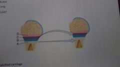
Zone of resting cartilage |
D |
|

Lacuna |
A |
|

Lamella |
B |
|

Canaliculi |
C |
|

Osteoclasts |
D |
|
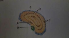
Osteocyte |
E |
|
|
Osteomalacia |
Softening of bones due to lack of minerals |
|
|
Most common cause of osteomalacia |
Vitamin D deficiency |
|
|
Osteomalacia in children |
Ricketts. Bowed legs |
|
|
Osteoporosis |
Reduction in bone mass |
|
|
Correct order of bone repair 1) formation of bony callus 2) formation of hematoma 3) formation of fibrocartilaginous callus 4) remodeling the callus |
2, 3, 1, 4 |
|
|
Pott fracture |
Fracture of the distal end of lateral fibula |
|
|
Colles fracture |
Distal end of lateral radius |
|
|
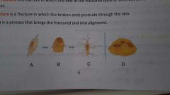
Comminuted fracture |
Bone is crushed and small bone fragments lie between two main fragments |
|
|
Impacted fracture |
One end of the fractured bone is forcefully driven into another |
|
|
Reduction |
Process that brings the fractured end into alignment |