Reading...
![]()
Play button
![]()
Play button
![]()
Use LEFT and RIGHT arrow keys to navigate between flashcards;
Use UP and DOWN arrow keys to flip the card;
H to show hint;
A reads text to speech;
19 Cards in this Set
- Front
- Back
|
What types of movements does the nervous system control?
|
❤Voluntary (purposeful, goal directed), usually learned
❤Reflex responses (involuntary, automatic, rapid responses to external stimuli e.g. coughing, the knee jerk, withdrawing away from a hot stimulus) ❤Rhythmic movements combine features of voluntary and reflex movements -initiation and termination are voluntary, while other aspects are almost automatic |
|
|
Describe the pyramidal system
|
-the development of the pyramidal system is directly related to the capacity of the animal to perform skilled movements
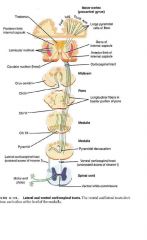
-well developed in primates with digits and opposable thumbs -poorly developed in domestic animals, especially in the horse, ox and sheep ❤NEURON 1 -the cell body of the pyramidal system are located in the cerebral cortex in the motor area which is in the precruciate area -it is arranged somatotopically -regions invovled with more highly skilled functions have a larger representation -muscles with small motor units also have a larger area of representation -the primary motor area of one cerebrum serves the musculature on the contralateral side of the body -from the cell body the axon of neuron 1 passes through the internal capsule into the crus cerebri (still maintaining somatotopic arrangement) on the ventral surface of the midbrain -the axons of the pyramidal system are divided into the corticonuclear axons (controlling muscles of the head) and corticospinal axons (controlling skeletal muscles of the body) -the corticonuclear fibers decussate near the nuclei of the cranial nerves that innervate striated muscle of the head (III, IV, V, VI, VII, IX) before entering those nuclei to synapse within them -striated muscle of the head includes the extraocular muscles (CN III, IV, VI), the muscles of mastication (CN V), the muscles of facial expression (CN VII) and the tongue (IX) -the remaining axons (the corticospinal axons) continue in the pyramid (somatotropic arrangement) after passing underneath the transverse fibers of the pons, then decussate at the level of the medulla oblongata and cross to the opposite side of the spinal cord to form the lateral corticospinal tract -a few axons don't decussate but stay ipsilateral within the ventral corticospinal tract before decussating within the spinal cord ❤NEURON 2 -the crossed pyramidal system of axons form the lateral corticospinal tract medial to the cranial projecting spinocerebellar tracts in the dosolateral funiculus -most synapse on interneurons in the ventral gray column that influence LMNs located there ❤NEURON 3 -Cell body in cranial nerve (III, IV, V, VI, VII, or IX) or in the ventral horn gray matter of the spinal cord -this cell is the gamma motor neuron but sometimes can be the alpha motor neuron |
|
|
What feedback pathways exist for the pyramidal system?
|
FEEDBACK PATHWAYS
-feedback pathways take a copy of information from the motor cortex to the cerebellum to inform about the action that is about to take place -the cerebellum gets info from the unconcious proprioception pathways, and sends feedback projection to the motor centers and regulates the subsequent actions as they take place -the cerebral cortex of one side is regulated by the contralateral cerebellar cortex |
|
|
What is the reticular formation? What does it do? What tracts arise from it?
|
Anatomy: network (mixture) of gray & white matter, found throughout the brainstem
-gets synaptic output from collateral branches of ascending tracts (e.g. spinothalamic tract) PHYSIOLOGY -spontaneously active neuronal circuits; perform 3 major functions: 1) Ascending system to alert the cerebral cortex (via non-specific thalamic nuclei) vs. coma 2) Vegetative centers: regulate heart rate, respiration, digestion, micturition etc. 3) Standing posture and muscle tone via two pathways to alpha and gamma neurons ●pontine reticulospinal tract – arises from neurons located laterally in pons -dominant and spontaneously active; runs ipsilateral in the ventral funiculus -important in the involuntary control of axial and proximal extensor musculature -the reticulospinal tract is particularly important in controlling the magnitude of muscle tone of these antigravity muscles -activates alpha & gamma motor neurons to extensor muscles of proximal joints -tend to have an excitatory effect on lower motor neurons to the antigravity muscles ●medullary reticulospinal tract -arises from neurons located in the medially in the medulla -inhibits neurons to extensor muscles and excites neurons to flexor muscles -not spontaneously active -driven by cerebral cortex to present movement of posture -descends bilaterally in lateral funiculus FUNCTIONS 1) the first function of these tracts is related to skilled voluntary movement requiring a stable postural background -just before the execution of such a voluntary movement, the reticulospinal tract subconsciously activates the appropriate axial and proximal musculature that will compensate for the postural destabilization that will be produced by the intended voluntary movement (usually the distal musculature) 2) it also plays a role in the voluntary execution of crude (non-skilled) stereotypical movements of the proximal musculature, such as those involved in simple pointing or locomotion |
|
|
What are the two descending tracts from the vestibular nuclei?
|

VESTIBULAR NUCLEI
2 descending tracts: ●lateral vestibulospinal tract – which also drive standing posture & ●medial vestibulospinal tract – (mlf) which controls neck muscles -plays a major role in the involuntary control of axial and proximal extensor musculature that prevents the animal from falling to the ground -vestibulospinal tract originates from cell bodies in the vestibular nuclear complex, which lies primarily in the medulla, just ventral to the 4th ventricle 0this complex consists of several subnuclei that receive their principal synaptic input from 8th cranial nerve fibers carrying sensory input from the vestibular apparatus of the inner ear -the vestibular apparatus provides sensory information about the position of the head with respect to gravity and about acceleration of the head through space, thus indicating body position and disturbances of balance -the vestibular nuclear complex also receives significant input from the cerebellum, but not from the forebrain levels of motor system hierarchy -axons synapse within medial regions of the spinal cord gray matter that primarily control extensor musculature -a disturbance of balance of balance detected by the vestibular apparatus results in excitation of the antigravity musculature in an attempt to counteract the disturbance -it seems to make some contribution to control of antigravity muscle tone as well |
|
|
How do posture and movement occur?
|
-posture and movement result from excited alpha and gamma motor neurons
-a motor unit is one alpha motor neuron plus all of the muscle fiber it innervates -interneurons hardwire motor neurons so that logical movement patterns are produced -thus nearly all descending pathways synapse directly on motor neurons -a minority synapse directly on motor neurons e.g. vestibulospinal & a minority of pyramidal neuons -thus both voluntary and reflex pathways compete for control of the same interneurons and motor neurons -to gain control of motor units, voluntary neurons must suppress reflex neurons and vice versa |
|
|
What is the red nucleus?
|

-large neurons of the of the red nucleus give rise to the rubrospinal tract - the principle descending tract for voluntary movement in domestic animals (rubrobulbar fibers go to cranial motor nerve nuclei. Other small neurons project to the olivary nucleus)
-the red nucleus is merely a collection of projection neurons -axons from the motor area of the cerebral cortex synapse on large projection neurons in the red nucleus and control their activity -the rubrospinal tract decussates in the midbrain and descends in the dorsal half of the lateral funiculus -rubrospinal fibers synapse on spinal interneurons and produce independent movements of shoulder/hip; elbow/stifle; and carpus/hock (not digits) |
|
|
What tectum? What tracts arise from it?
|

tectum = roof of midbrain
-2 tracts arise from deep neurons of the rostral colliculus 1. tectospinal fibers - descend to the cervical spinal cord (for head turning) -synapse within medial regions of the spinal cord gray matter that primarily controls the axial and proximal musculature -only project as far as the upper cervical regions of the cord 2. tectobulbar fibers - to cranial nerve nuclei that control ear & eye movement. Via horizontal (pons) and vertical (midbrain) gaze centers within the reticular formation, the tectum reflexly shifts the eyes (saccadic movements) to focus on novel stimuli within the visual field -note saccade = quick eye movement used to shift focus to new visual feature; perception occurs at stops between saccades -the tectospinal tract is involved in producing a movement of the head toward the stimulus that corresponds with the rapid eye movement so that the animal's gaze is fixated directly on the stimulus |
|
|
What is the substantia nigra?
|
-a dorsal region (pars compacta) contains dopaminergic neurons that project to the striatum (caudate nucleus & putamen) to facilitate movement initiation
-a ventral region (pars reticulata) functions like the globus pallidus. It tonically inhibits brainstem motor nuclei such as those in the tectum -this inhibition is lifted by descending input from the striatum (disinhibition) |
|
|
What is the subthalamus?
|
-the subthalamic nuclei participates in basal nuclei circuits
-the nucleus contains glutaminergic output neurons that excite the globus pallidus which tonically suppresses movement activity |
|
|
What makes up the basal nuclei or ganglia?
|
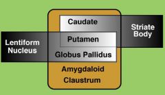
-referes to non-cortical gray matter of the telencephalon
-5 major nuclei: -the major nuclei may be anatomically grouped in different ways 1) striatum = caudate & putamen 2) lentiform nucleus = putamen and globus pallidus; together they have the shape of a lens ***Physiologically, only caudate, putamen, and globus pallidus play a motor role |
|
|
What are the basal ganglia? What do they do? Why is the globus pallidus so important?
|
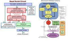
❤Although they do not give rise to descending tracts, basal nuclei regulate voluntary movement onset and cessation
-they facilitate initiation of selected movements and suppression of non-selected movements -they participate in a circuit interposed between non-motor cortex precipitating the movement and motor cortex executing the movement. ❤The non-motor areas of cerebral cortex do not communicate their movement needs directly to the motor cortex -instead the cortices are linked by a circuit involving basal nuclei and thalamus, including also subthalamus and substantia nigra ❤In general the globus pallidus is tonically active in suppressing movement -excitation of the globus pallidus by the subthalamus facilitates movement suppression -when non-motor cerebral cortex excites the striate body, the caudate and putamen specifically inhibit neurons in the globus pallidus (and subthalamus) -this specific disinhibition enables movement initiation, by releasing excitatory thalamic neurons NOTES: — the non-motor cerebral cortex communicates movement needs to the motor cortex via basal nuclei - thalamic circuits — tonic activity of the internal globus pallidus suppresses movements in general; re-inforced by the subthalamus, this inhibition suppresses non-selected movements — selected movements are allowed (initiated) by a process termed selective disinhibition, as cerebral cortex selectively excites inhibitory neurons of the striatum that selectively inhibit tonic neurons of the globus pallidus. — inhibitory neurons (-) are GABAergic; excitatory neurons (+) are glutamatergic; substantia nigra dopaminergic neurons are excitatory via D1 receptors |
|
|
What are the areas of the cerebral cortex that are important in motion?
|
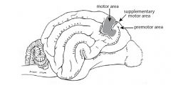
The motor area of the cerebral cortex executes voluntary movement; other motor-related areas of cortex are involved in planing and selecting voluntary movements in response to cues:
• motor area - located around the cruciate sulcus; principal source of output for all voluntary movement; it is the main origin of two descending pathway systems: a direct pyramidal & an indirect extrapyramidal (see below). Each output neuron of the motor cortex innervated multiple muscles (movement organization). • premotor area—located in frontal lobe rostral to the motor area; required for generating learned, rapid-sequence movements (motor pattern generator circuits). supplementary motor area—located medial to premotor area; active when thinking about a proposed movement; projects to the motor area |
|
|
Describe in general the difference between the pyramidal & extrapyramidal pathway
|
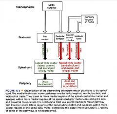
1] Pyramidal tract = a direct connection from motor and premotor areas of cerebral cortex to efferent
neurons, generally via local interneurons -axons travel in pyramids of the medulla oblongata. -most axons (85% in human) decussate at the medullary-spinal junction and form the lateral corticospinal tract which terminates in lateral motor nuclei of ventral horn enlargements -the remaining axons form the ventral corticospinal tract which crosses at the level of termination in the spinal cord and innervates medial motor nuclei (postural muscles) -thus, the pyramidal tract controls particularly musculature of the manus and pes; it is especially concerned with precise movements of individual digits (also lips and tongue) 2] Extrapyramidal tracts = the term applied to all other (non-pyramidal) voluntary movement tracts -under the direction of cerebral cortex (motor area), these tracts control proximal musculature and thus generate relatively coarse components of posture/movement locomotion -naturally, this system is most important in domestic animals The principal extrapyramidal tracts are: rubrospinal tract, pontine reticulospinal tract, and medullary reticulospinal tract. Note: a specific voluntary movement, e.g., a feline paw swipe involving rubrospinal or pyramidal tracts, requires an associated postural adjustment (involving reticulospinal or rubrospinal tracts) that often must precede the specific action. Thus voluntary movements require multiple tracts. |
|
|
What lesions can occur in the motor system?
|
-lesions in the primary motor cortex in the dog often result in relatively mild gait deficits contralateral to the side of the lesion, which may only become apparent on proprioceptive function testing
-the animal will often be able to walk normally (although it may do so in circles or compulsively) -however lesions in the midbrain or caudal will often produce severe ataxia or hemi/tetraparesis with significant obvious gait deficits including knuckling etc. -this is true for cervical (C1-C5) lesions - with ataxia either in all 4 limbs or in 2 limbs ipsilateral to the side of the lesion In all cases, the lesion can be described as being ‘upper motor neuron’ in nature – i.e. the upper motor neurons are the neurons that are selectively knocked out, leaving hyper-functioning lower motor neurons. Since the upper motor neuron pathways are extensively inhibitory to the lower motor neurons, the effect of an upper motor neuron is to reveal overzealous functioning of the lower motor neurons: hypertonia (increased muscle tone) and hyperreflexia (increased strength of reflexes). In particular, the extensor (antigravity) muscle groups become more active. |
|
|
What is spinal shock?
|
An exception to this is the condition known as spinal shock. This is a state of transient total voluntary
movement paralysis and areflexia (a lack of spinal reflexes) that results immediately after the severing or severe lesions of the spinal cord. It is due to loss (paradoxically) of descending influence on spinal cord neurons and can last from a minimum of 2 weeks to several months in man. The length of time for which spinal shock occurs is species dependent and also depends on the degree to which descending signals control movement e.g. Lasts for about an hour in the frog and up to several days in the cat. After the period of spinal shock, the reflexes reappear and become exaggerated i.e. a state of hyperreflexia exists. After spinal shock is over, tendon jerks and flexion reflexes are hyperactive and a small stimulus can cause prolonged withdrawal of a limb. |
|
|
What is hemiparesis or hemiplegia?
|
If descending influences from the cerebral cortex are lost as in human stroke victims (e.g. Damage to fibres
in the internal capsule), voluntary movement of opposite side of the body is reduced or abolished – Hemiparese or Hemiplegia. Limbs are flaccid for a while and reflexes are lost (as in spinal shock). However reflexes return within a few hours to a few days and again they are exaggerated. Muscle Tone increases due to a loss of inhibitory control over extensor stretch reflexes from the cerebral cortex, allowing the excitatory action of the vestibulospinal and pontine reticulospinal tracts to continue unopposed on the extensor |
|
|
What are the motor centers the comprise the extrapyramidal system?
|
FOREBRAIN:
1) cerebral cortex 2) basal nuclei MIDBRAIN: 3) descending reticular formation 4) red nucleus 5) tectum (dorsal midbrain) HINDBRAIN 6) pontine motor reticular centers 7) lateral medullary motor reticular centers 8) medial medullary motor redicular centers 9) vestibular nuclei |
|
|
Which pathways are facilitatory? inhibitory?
|
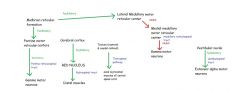
The midbrain reticular formation sits in the central region of the midbrain. It is facilitated by input from the globus pallidus and sends (faciliatory) projections to the pontine motor reticular centre, the lateral medullary reticular centre and the medial medullary reticular centre.
The tectum of the midbrain is composed of the rostral and caudal colliculi. They are joined to the lateral and medial geniculate nuclei (respectively) in the diencephalon and form the start of the tectospinal pathway. The pontine motor reticular centres exert a mainly facilitatory drive over gamma neurons in the spinal cord via the pontine reticulospinal tract. They are controlled by the midbrain reticular formation (via facilitation). The lateral medullary motor reticular centres have no direct pathway to the reticulospinal tracts but work to inhibit the medial medullary motor reticular centres (and are therefore facilitatory through disinhibition of the medullary raticulospinal pathway). They, in turn are facilitated by the midbrain reticular formation, The medial medullary motor reticular centres control the medullary reticulospinal tract, exerting a massive inhibitory influence on gamma neurons in the spinal cord. They are in turn controlled by the lateral medullary motor reticular centres (which inhibit them) and the midbrain reticular formation (facilitatory). The red nucleus lies in the midbrain (buried in the reticular formation) underneath the colliculi. It receives facilitatory input from the cerebral cortex and is the start of the rubrospinal pathway. The vestibular nuclei are strongly facilitatory via the vestibulospinal pathways, and act mainly on extensor alpha neurons in the spinal cord. They are in turn inhibited by the cerebral cortex. The tectospinal pathway mediates rapid (reflex) turning of the head and neck in response to noise or visual stimuli (eg the reflex head jerk towards the direction of a firework that you don’t expect to have gone off just behind you). |