![]()
![]()
![]()
Use LEFT and RIGHT arrow keys to navigate between flashcards;
Use UP and DOWN arrow keys to flip the card;
H to show hint;
A reads text to speech;
57 Cards in this Set
- Front
- Back
|
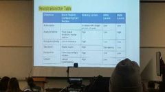
Indolamines of monoamines |
Seratonin(5ht)
Made from tryptophan
Tryptophan plus tyrosine makes 5htp 5htp plus decarbo makes 5ht
9 confirmed receptors most metabotropic
Cell bodies that start Seratonin are in raphe nuclei
Drugs that affect are Ssris Block reuptake Agonist Ex; prozan zoloft
Mdma(ectasy) Makes transporters run in reverse Agonist Hallucinations
Maois
Termination is Seratonin transporter |
|
|
Ablation |
Cut out n test it out |
|
|
Surgical method |
Experimental ablation Removing brain tissue n see its affects Oldest method Lesson studies |
|
|
Brain lesions |
Radio frequency lesions Insert wire radiofrequency n heat kills cells touching wire
Excitotoxic lessioms Selective Excitatory amino acid injected thru cannula (lil funnel) into specific rgion Like kainic acid Glutamate agonist Gets so excited it kills cell bodies not axons
Both permanent |
|
|
Rebersible lesions |
Gaba agonist (muscimol) Temporal disruption of brain activity Blocks aps in axons in area |
|
|
Sham lessioms |
Cut thru tissue Causes unintended damage Animals undergo surgery for electeode/cannula But recieve no treatment cux u need a control group
Used to see effects of treatments |
|
|
Sterotaxic surgery |
How they know where to put electeode or cannula Put animal im stereotaxic apparatus
This way u get a 3d ish look
Bregma is closed junction of skill bones
Refrence point Rostral n caudul to bregnas |
|
|
Histological methods (study of tissue) |
Tissue placed in fixative (formalin) Prevents post mortem decay Stengthens brain tissue Kills baceria or mold Then froze (dry ice) so u can cut Cut into thin sectioms (10 to 80 um thick) Microtome Cryostat Put on slide n stain |
|
|
Stains |
Cresyl violet Cell bodies Attaches to Rna and dna n nucleus stuff Golgi stain Whole cell |
|
|
Anterograde labelinh |
PHA L into the VMH travels into dwn axons to terminal buttons See what a neuron projects to |
|
|
Retrograde labelinh |
Fluorogold into vmh Gets taken up from terminal buttons n goes backwads to soma See what projects to a neueon |
|
|
Immunocytochemiccal |
Labels Use antibodies tha attaxj to different protoens Attach labels to specific tissue |
|
|
Imaging methods |
CT Computerised tomogrtaphy 1st method Xray beam
MRI Magmettic resonance imaging haad Passes magnetic field thru haadBrtter reolutiom than CT scans Brtter reolutiom than CT scans
diffuser tensor imaging Based on movement of water molecules in white matter Visualize bumdle of axons Colors myliens Mainly white matter cuz it has fat |
|
|
Vmh amd female copulatiom |
Medial amygdala goes to vmh To pag |
|
|
Electrophsdiological recordings |
Electrode of mesaure neurtal activity (Recordings/numbers)
Microelectrode Single unit recording
Macroelecreodes Can be implanted to skull (eeg) |
|
|
Optogenetics |
Specific color light to manipulate a neural circuit Genetically alter neurons to produce a light sensitive channel; Channelrhodopsin-2 (ChR2) From green algea Opens in blue light Depolarizes cell/excitatory Lets in Na+ and Ca2+ Natronomonas pharaonis halorhodopsin (NpHR) Discovered in bacteria Opens in yellow light Hyperpolarizes cell/inhibitory Lets in Cl- |
|
|
Genetic manipulation knock outs vs knock in |
Knockout=make gene defective or missing
Constitutive knock out Gene defective from birth
Conditional knock out Gene inactivated by a drug later on Ex(leptin knock out mice)
Knock in (gene added to genome) Adds a functional protien Alzeihmers transgenic rat (form plaques n tangles) |
|
|
Sensory systems |
Signals from environment Detected by sensory organs (they have sensory receptors) Turned into neural signal (transduction) (transduction) |
|
|
Eye structure |
Sclera Opaque white outer coating
Cornea Clear part Iris Pigmented muscle that controls light that enters eye Pupil Opening in iris (its a hole) Lens Focus light into point in retna, controlled by cillary Vitreous humor Jelly like thing that fills eye, clear Retna Back part of eye have rods n cons Optic nerve Info from photorecetors to brain |
|
|
Retna |
Photorecetors Rods (120 million) more sensitive to light. Night vision. Fuzzy. Not fovea Cons (6 million) sharp. Diff cones for diff wavelengths of light. Color. Fovea
Fovea only Cones. Highest acuity/ sharpness. Lens focusses light to Fovea
Optic disk where axons from photo receptors leave brain. Blind spot |
|
|
Cells in retna |
Light goes thru Ganglion cell to Bipolar cell to Finally photoreceptor When light hits they make the info n it goes Photoreceptor To bipolar To ganglion To optic nerve To brain |
|
|
Transuction |
When light hits phtoreceptors it splits ibto the ospin n retinol
Causes hyperpolarization This is receptor potential Site of transduction |
|
|
In darkness vs light photoreceptors n bipolar |
Darkness Photoreceptor natutally inhibits bipolar so no messages sent
Light Photoreceptor is inhibited (double negative) so it cant inhibit bipolar so messages get sent to brain
|
|
|
Polarization in dark n light |
Dont release ap
Hyper polarized (less nt released) in light Dont release glu. Bipolar cells can depolarize . Release glutamate to make ganglion fire. We get messages
Depolarized (more nt released) in dark Release lotsa glutamate . Keep bipolar cells hyperpolarized. Inhibits n dont get messages
Glutamate acts wierd here |
|
|
Visual pathway |
Optic nerve Axons from ganglion cell goes to opyic chaism
Opric chaism Half crossed half kept so nerve send L n R for each eye, at chaism all R visual fiels info goes to left hemisphere.
Dorsal lateral geniculate nucleus (LGN) Synapse in brain Send axons to primary visual cortx theu optic radiation Primary visual cortex First cortical area to recieve visual info
|
|
|
Light detection on ganglion |
Light detection Occurs in ganglion cell
In fovea 1 ganglion sees info for 1 photorceptor (sharp)
In periphery 1 ganglion sees info for many photoreceptor (blurry)
|
|
|
Ganglion cell on amd off and why we have this |
On cells On center off surround cells Become excited when light falls in center n inhibited when light falls surround
Off cells Off center on surround cells Become inhibited when light falls in center n excited when light falls surround Done to Enhaced contrsat Changes in illumination |
|
|
Pevels of visual pocessing |
Ganglio cells = light detection Primary visual cortex = orientation Exstraite cortex = Movement, image comprehension, location |
|
|
Primary visual cortex |
Called straite cortex in occipital Gets all info for contrateral visual field V1
25% of info comes from fovea Responds to orientation (fires most to specific orientation) |
|
|
Exastrait cortex |
V2 to v5 Makes picture V5 responds to specific Movement
2 pathways
Ventral stream Project onto inferior temporal lobe The what pathway What an object is
Dorsal stream Project onto posterior parietal lobe Where pathway Where in ur visual field an object is Where is that dolphin Where n dorsal stream |
|
|
Structures of the ear |
Outer ear Pinna (funnel) ear canal (canal) Timpanic membrane (seperates middle n outer ear)
Middle ear Ossicles malleus/hammer (attached to tympanic membrane so it moves when it vibrate) incus/anvil Middle man stapes/stirrup (shake oval window) They amplify vibrations / concentrate waves to tiny area
Inner ea Cochlea (transduction) Fluid filled vibrations make basilar membrane move to round window Round window is shock absorption from oval window
Scala vestibul Scala media has organ of corti Hair cells (auditory receptor cells) Basilar membrane (anchor to hair cells) Tectreial membrane (attaches to cilia of some hair cells) Scala tympani
Hair cells Inner necessary for hearing but not attached Outer attach to tectorial memebrane Both go to bipolarcells n form cochelar/auditory nerve bundle
|
|
|
IEGs immediate early genes |
Activate when a neuron is activated
Detect activation from behaviour or artificial
First stimulate then stain Genes (c-Fos, Arc, ZENK)
Used to visualise the neurons activayed |
|
|
Non invasive ways to mesure brain activity (live on humans) |
PET (positron emission topogra0hy) Expensive n poor resolution Radioactive chem (2-DG) First functional imaging method
fMRI BOLD imaging (blood oxygen level dependnet) Active areas of brain have more oxygenated blood Better resolution than PET |
|
|
Electrical vs transcranial magnetic simulation |
Electrical Wire to stimulate Transcranial Magnetic fields induce electroical currents non invasivly |
|
|
Study this |
https://quizlet.com/676478971/neuro-unit-2-flash-cards/?i=1cek0x&x=1jqY |
|
|
Path ofsound |
Pinna Tymanic membrane Shake ossicles Oval window Fluid of cochlea Hair cells of cochlea Get transduced Cochlea nerve |
|
|
Transduction at cochlea |
Cochlea hair cells When when oval window is hit the hair cells of the basilar membrane bend Hair cells are linked w tip links n attachd at insertional plaques (where ion channels are) When hair cells bent n cila moves, they open channel like mechanism K and Ca enter cell n depolarize it When depolarized they increase neurotransmitters, more Nt more strong sound |
|
|
Encoding auditory info |
Pitch n loudness
Pitch Frequency of a sound wave (how close) Place coding in basilar membrane They vibrate in response to a specific membrane High pitches at start of cochlea near oval window Low pitches at end of cochlea
Loudness Amplitude (height) of sound wave Bigger amplitudes r stronger more force U move hair cells more n release more NT n cause more action potentials Rate of firing=loudness |
|
|
Sound in the brain |
Info leaves thru cochlear nerve (bundle of axon bipolar cells) Most pass thru the superior olivary complex in medulla Sent to inferior colliculus thru lateral lemniscus Sent to cortex |
|
|
Sound in the brain |
Info leaves thru cochlear nerve (bundle of axon bipolar cells) Most pass thru the superior olivary complex in medulla Sent to inferior colliculus thru lateral lemniscus Sent to cortex |
|
|
How to study sleep |
Electrophysiological data Eeg (electroencephalaograms) Attached to scalp n mesure brain activity Emg (electromyogram) Elctrodes attached to face n jaw to mesure muscle activity Eog (elctro oculogram) Attached to around eye Eye movement U get synchronous activity Large wave response All neurons firing at the same time Desynchronous activity Small wave Neurons fire different times They could cancel out |
|
|
Stage aof seep |
Awake Alpha awake resting Beta awake alert/attentive
Stage 1 Theta activity Transition stage Hypnic jerks (muscle contraction folled by relaxation) Slightly higher
Stage 2 Transition to deep sleep Sleep spindles n k complex Ppl dont relaise they asleep
Stage 3 Delta activity Slow tall wave sleep synchronous Groggy if woken up
REM Rapid eye movement Loss of muscle tone Vivid dreams Refreshed when awoken Theta n beta activity Small wavess |
|
|
Sleep cycles |
Enter REM every 90 minutes Staytgerw 20 to 30 min First half of night u spend most time in slow wave sleep Second half u spend moretime in REM n in stage 2 |
|
|
Why we sleep n how we study it |
Useful for memory Essential for long term survival
But we dont know
Sleep deprivation studies Animals Keep them awake Weak uncoordinated, lose weigh, died Brain n hormones normal Humans No deficiy on phsyical abilities Cognitive deficits Sleep important for brain recuperation but not for body |
|
|
After sleep deprivation |
7% of stage 1 n 2 made up 68% of stage 3 made up 53% of REM sleep made up Stage 3 n REM r important |
|
|
Stags 3 |
Cerebral metablic n blood flow decrease Brain is resting Areas that r most active when awake have more delta waves n lowest level of metabolic activity Imp. for restoration n rest |
|
|
Rem sleep |
Unknown Babies have most rem sleep N anima babies n animals w immature brains
Play a role in braindveleopmwnt
In adults Consolidates certain types of memories non declarative
Rem improved nondeclarative Slow wave sleep improves declarative
Declarative Memories events spatial relationships, birthdays
Nondwclarative Recalling song lyrics afer heaing music, recognizing a face, diving a car |
|
|
Neural mechanism of sleep n waking up |
Neurtransmitters involved in sleep Adenosine Released by astrocytes Prolong wakefulness Decrease level of glycogen Increase adenosine
Adensoisne inhibits neural activity Promotes sleep
Caffeine blocks adenisine receptors
NT involved in awakenessss Acytocholine 3 pathways that have acetocholergic neurons Doraolaterla pons Basal forebain Media septum Activationbof 1 n 2 lead to deaynchronousbbrain actobity ACgh hugh furing wake n rem
Norehinehrine Ne agonsit Amphetamine (speed) Casuse aletnessne mediated by locus coeruulous Aivitt herr rlated to arousil So high during awake n 0 in rem
Seratonin Start in raphe nuclei Cocomotion n descynchrony Behavioral arousol High during awake 0 when rem
Histamine Come from histidine Start in tuberomammilary nucleus (TMN) in the hypothalmus Projects to Cerebral cortex, basal ganglia, basal forebrain Causes cortical activaion directly n indirectly (activated acetocoline that then activate cortex ) Befor they made u droswy
Orexin Located in laterlal hypothalmus Project to all regions linked to wakfulness Highest during awake
|
|
|
Neura circutry of sleep wake |
Balance bewteen slep n awake areas
Sleep has Ventrolateralpreoptic area (Vlpoa) Secrete gaba (inhibitory) They inhibit awake areas
Wake has raphe nuclei, TMN, locus coeruleus, basal forebrain, lateral hypothalmus etc. These inhibit vlpoa causing wakefulness
|
|
|
Fliop flop circuiy |
When 2 regions mitually inhibit each other Stronger one is expressed Whichever is more excitatortbis the behavior expressed
Orexin promote wakefulness areas So more orexin than adenosine meands awake Adenosine activaes the vlpoa promoting sleep More ur awake more adenosine u have More adenosin than orexin means sleep
On is araousol areas inhibit vlpoa Off vlpoa inhibit aousal areas |
|
|
Coffe n sleep |
Caffein inhibits adenosine Inhibits sleepy So agonsit to sleepy Decrease activation of vlpoa by blocking effects of adenosine So if adenosine cant fight against orexin Orexin wins n ur awake |
|
|
sleep disorders |
Insomnia Hard to fall asleep. Self reported. Sleep apnea>stop breathing while sleep (could cause insomnia)
Narcolepsy Sleeping at innapot opriate times Symptoms Sleep attacks (suddwn urge tp sleep, during boring, sleep 2 to 5 min refreshed) Cataplexy (loss of muscle tone when ur conscious, after strong emotion or effort) Sleep paralysis ( cant move b4 or after sleep, hypnogogic hallucinations, parts pf ur brain awake n some asleep)
REM sleep disorder Ur body isnt paralysised during REM Act out dreams The dif btwn this n sleep walking cuz in sleep walking yes ur moving but ur not acting out a dream
|
|
|
Regulatory mechanisms |
Regulates smthng Ex. Thermostat
4 features System variable > whats being regulated Set point >whats the normal Detector >takes in info/monitors Correctional mechanism >takes action to bring current thing to normal/restores to set point Negative feedback When u stopp urself or add Like a thermostat When its above point u turn it off When its under u turn it on |
|
|
Homeostasis |
Keep optimal level Regulatory |
|
|
Drinking homeostasis aka thirst |
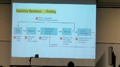
Fluid in body Extracellular > outside cells Itravascular fluid (blood plasma) Cerebrospinal fluid (brain jelly) Interstitial fluid (surrounds cells) Intracellular >in cytoplasm of cells. 2/3 of our liquid in cells
We lose waer thrue sweating respiration evaporation
2 types of thirst Osmometric thirst Intercellular -When solute (salt) concentration of interstitial fluid (liquid outside cell) increases Outside draws water thru osmosis (high to low concentartion of water) -Taking water out of cell Or of u lose water cuz of evaporation u again do osmosis
Cells shrink triggering osmoreceptors (found in OVLT (outside blod brain barrier))
Volumetric thirst Intravascular thirst (blood plasma) Loss of blood volume =hypovolemia)
Caused by evaporation, bleeding, vomitting, diarrhea
Detected 2 wayas Kidney Cells detect low blood volume Release angiotensin stimultinh drinkunh n sat aperite Angiosen dtected by subfornical organ (sfo) Heart When blood volume decrases the stretch sensitive baroreceptors in atria tell brain (68)
Both sfo and ovlt go to median preoptic nucleus Takes info n tells brain to drink |
|
|
Eating homeostasis aka hunger |
Fuel for body
Metabolism
2 fuel resoviour Short Sustain fuel for few hours Liver Glucose fuel Glycogen stored fuel >When ur full ur tummy makes glucose o glycogen Using inusilin (from pancreas) extra stored in liver >When glucose fall (empty stomach) we need more glucose so pancrease make glucagon (makes glycogen to glucose)
Long Made of adipose tissue (fat) Made on triglyceride (glycerol n 3 fatty acids) For prolonged fasting (sleep?) Fat cells make tryglceride to fuel that cells use Activated when hugry goes fasting when full makes absorbitive
2 phases of metabolism Fasting phase When stomach empty Glucose lvl low More glucacon Low insulin Liver makes glycogen to glucose Can go into the long term resevoir
Absorbitive phase Have food in tummy More glucose Insuilin secreted Excess glucose turns into glycogen n stored or in adipose tissue both long n short
|
|
|
Hormones in hunger n regions |
Starts eating Ghrelin Peptide hormone released by gastrointestinal system (stomache ) Goes thru blood to to receptor in hypothalmus (makes us huungry) Ghrelin released in fasting phase
Stopp eatingPyy (peptide yy3-36) Small intestine after meal, proportinal to calories Introducing pyy makes u eat less
Insulin Increase in absorbtive phase Put insulin in brain makes u eat less
Leptin Peptide hormone made by adipose Ob mouse Low metabolism Overeats Incrd weight Make no leptin >When u inject leptin They eat less increase metaboiam
Regions
Lateral hypothamus Regulate hunger in genera Neurons make Mch (melanin concentrating hormone) and orexin Lateral hypothalmus activated by arcuate nucleus Release neuropeptide y (npy) And agouti related peotien (agrp) Accurate nucleus activated by ghrelin
Venromedia hypothalmus Satiety Atop eating Leptin inhibits accurate nucleus
|