Reading...
![]()
Play button
![]()
Play button
![]()
Use LEFT and RIGHT arrow keys to navigate between flashcards;
Use UP and DOWN arrow keys to flip the card;
H to show hint;
A reads text to speech;
116 Cards in this Set
- Front
- Back
|
Dualistic Approach
|
Mind and Body sepration
|
|
|
Psychological Approach
|
The mind is exlusively responsible
|
|
|
Biological Monolism
|
Biology is exclusively responsible
|
|
|
What are the three parts of the Brain.
|
Forebrain, Midbrain and Hindbrain
|
|
|
Neuropsychology
|
The branch of psychology that deals with the relationship between the nervous system, especially the brain, and cerebral or mental functions such as language, memory, and perception.
|
|
|
Glial Cells
|
Continue to divide and multiply. They carry out many important functions for normal brain function, including insulating nerves cells with myelin.
|
|
|
Ectoderm
|
The embryonic tissue that is used to develop the nervous system.
|
|
|
How big is the brain? How much does the brain weigh?
|
The adult human brain weighs between 1300 g and 1400 g (approximately 3 lbs). A newborn human brain weighs between 350 and 400 g.
|
|
|
Central Nervous System
|
The central nervous system is divided into two parts: the brain and the spinal cord.
|
|
|
Cerebral Cortex
|
Functions:
Thought Voluntary movement Language Reasoning Perception |
|
|
"cerebellum"
|
Functions:
Movement Balance Posture |
|
|
Brain stem
|
Functions:
Breathing Heart Rate Blood Pressure |
|
|
Hypothalamus
|
Functions:
Body Temperature Emotions Hunger Thirst Circadian Rhythms |
|
|
Thalamus
|
Functions:
Sensory processing Movement |
|
|
Limbic System
|
Functions:
Emotions |
|
|
Hippocampus
|
Functions:
Learning Memory |
|
|
Basal Ganglia
|
Functions:
Movement |
|
|
Midbrain
|
Functions:
Vision Audition Eye Movement Body Movement |
|
|
One way to divide the brain
|
Telencephalon
Diencephalon Mesencephalon Metencephalon |
|
|
FRONTAL LOBE
|
Located in front of the central sulcus.
Concerned with reasoning, planning, parts of speech and movement (motor cortex), emotions, and problem-solving. |
|
|
PARIETAL LOBE
|
Located behind the central sulcus.
Concerned with perception of stimuli related to touch, pressure, temperature and pain |
|
|
TEMPORAL LOBE
|
Located below the lateral fissure.
Concerned with perception and recognition of auditory stimuli (hearing) and memory (hippocampus). |
|
|
OCCIPITAL LOBE
|
Located at the back of the brain, behind the parietal lobe and temporal lobe.
Concerned with many aspects of vision. |
|
|
Prefrontal Cortex
|
Problem Solving, Emotion, Complex Thought
|
|
|
Motor Association Cortex
|
Coordination of complex movement
|
|
|
Primary Motor Cortex
|
Initiation of voluntary movement
|
|
|
Primary Somatosensory Cortex
|
Receives tactile information from the body
|
|
|
Sensory Association Area
|
Processing of multisensory information
|
|
|
Visual Association Area
|
Complex processing of visual information
|
|
|
Visual Cortex
|
Detection of simple visual stimuli
|
|
|
Wernicke's Area
|
Language comprehension
|
|
|
Auditory Association Area
|
Complex processing of auditory information
|
|
|
Auditory Cortex
|
Detection of sound quality (loudness, tone)
|
|
|
Speech Center
(Broca's Area) |
Speech production and articulation
|
|
|
Dorsal view
|
Means on top. Looking from above as well as behind
|
|
|
To separate the brain into right and left hemispheres, you need to cut the brain in the
|
"midsagittal plane".
|
|
|
Ventral view
|
Consider below or in front therefore you can say that x structure is ventral to y structure
|
|
|
Lateral view
|
To look at the brain on the side therefore you can see Fontral lobe, Central lobe, and Partial lobe.
|
|
|
Coronal section
|
take a cut right smack between the two ears
|
|
|
The CSF has several functions including:
|
Protection: the CSF protects the brain from damage by "buffering" the brain. In other words, the CSF acts to cushion a blow to the head and lessen the impact.
Buoyancy: because the brain is immersed in fluid, the net weight of the brain is reduced from about 1,400 gm to about 50 gm. Therefore, pressure at the base of the brain is reduced. Excretion of waste products: the one-way flow from the CSF to the blood takes potentially harmful metabolites, drugs and other substances away from the brain. Endocrine medium for the brain: the CSF serves to transport hormones to other areas of the brain. Hormones released into the CSF can be carried to remote sites of the brain where they may act. |
|
|
Cerebrospinal fluid (CSF)
|
The CSF is contained within a system of fluid-filled cavities called ventricles.
|
|
|
Ventricles
|
Irrigation system of the brain are filled with CSF.
|
|
|
Hydrocephalus
|
Under some pathological conditions, CSF builds up within the ventricles. This condition is called hydrocephalus. Hydrocephalus may result from:
Overproduction of CSF An obstruction at some point within the ventricular system Problems with CSF absorption |
|
|
The names of the cranial nerves
|
On Old Olympus Towering Top A Famous Vocal German Viewed Some Hops.olfactory, optic, oculomotor, trochlear, trigeminal, abducens, facial, vestibulocochlear, glossopharyngeal, vagus, spinal accessory, hypoglossal
|
|
|
The Blood Supply of the Brain
|
Although the brain is only about 2% of the total body weight in humans, it receives 15-20% of the body's blood supply. Because brain cells will die if the supply of blood which carries oxygen is stopped, the brain has top priority for the blood. Even if other organs need blood, the body attempts to supply the brain with a constant flow of blood.
|
|
|
neuron
|
a cell body called the soma which is like a central processing facility, a long thin projection called an axon which transmits signals and a branching collection of short finger-like projections called dendrites which receive the signals.
|
|
|
Inside the neurons
|
the signals move as electrical impulses
|
|
|
Synapse between two neurons
|
the signals that are transfered are through chemical reactions
|
|
|
how fast does a message from a neuron travel
|
Messages travel at different speeds depending on the type of neuron
|
|
|
synaptic transmission
|
requiring about half a millisecond each
|
|
|
Synapse
|
seperates dendrites and axons
|
|
|
Cell
|
Golgi body, microfilaments, lysosomes, nucleus, endoplasmic reticular, mitochondrion, microtubules
|
|
|
Intra & Extra cellular fluid
|
is composed of water molecules that are polar - one negatively charged and the other is positively charged.
|
|
|
Which molecules can pass through cell membrane?
|
Oxygen
|
|
|
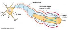
Myelin
|
Myelin coats and insulates the axon (except for periodic breaks called nodes of Ranvier), increasing transmission speed along the axon.
|
|
|
cell body (soma)
|
contains the neuron's nucleus (with DNA and typical nuclear organelles).
|
|
|
Glial cells
|
make up 90 percent of the brain's cells. Glial cells are nerve cells that don't carry nerve impulses. The various glial (meaning "glue") cells perform many important functions, including: digestion of parts of dead neurons, manufacturing myelin for neurons, providing physical and nutritional support for neurons, and more. Types of glial cells include Schwann's Cells, Satellite Cells, Microglia, Oligodendroglia, and Astroglia.
|
|
|
Into Brain
|
Oxygen
Carbohydrates Amino Acids Fats Hormones Vitamins |
|
|
Out of Brain
|
Carbon Dioxide
Ammonia Lactate Hormones |
|
|
Layers of the Brain
|
Skull
Dura Mater Arachnoid Layer Pia mater Subarachnoid space filled with CFS |
|
|
Neuron
|
Golgi body: packages protein molecules for transport
Microfilaments cell's skeleton Lyosomes enzymes that break down wastes Nuclues: executive containing chromosomes and genes Endoplasmic reticulum: folded layers of membrane where proteins are assembled Michondrion: gathers, stores and releases energy Microtubules transport molecules and help give the cell its shape |
|
|
intra and extracellular fluids
|
composed mainly of water in which salt and many other chemicals are dissolved
|
|
|
What contains the blue print for making proteins
|
Nucleus
|
|
|
Blue print
|
genes
|
|
|
Chromosones
|
giant molecular complexes which contain genes
|
|
|
DNA
|
deoxyribonucleic acid consisitng of four nucleotide bases
|
|
|
Four nucleotide bases
|
1. Adenine (A)
and 2. Thymine (T) = bind 3. Guanine (G) and 4. Cytosine (C) = Bind |
|
|
Gene
|
Segment of DNA that encodes the synthesis of particular type of protein molecule
|
|
|
Code
|
contains the sequence of nucleotides bases that direct the order in which amino acids (building blocks of protein) should be assembled to construct a particular type of protein.
|
|
|
Transcription
|
the transfer of genetic information from DNA into RNA. In case of protein-encoding DNA, transcription is the beginning of the process that ultimately leads to the translation of the genetic code into a funtional peptide or protein.
|
|
|
Translation
|
formation of amino-acid chains which occurs in the endoplasmic
DNA---RNA----Protein |
|
|
What mechanism enable substance to move in and out of the cells?
|
Protein act as:
1. Channels (e.g. K+) 2. Gates (NA+) 3. Transportation system |
|
|
restomg membrane potential
|
-70mV
|
|
|
Hyperpolarization
|
efflux of K+ ---- extracellular side of the membrane is more positive
|
|
|
Depolarization
|
influx of Na+ channels
|
|
|
Action potential
|
brief but extremely large change in polarity of an axon's membrane lasting about 1ms
|
|
|
threshold potential
|
electrical stimulation causes graded potential that causes the membrane potential to drop to -50mV
|
|
|
Absolute Refractory period
|
stimulation during depolarization or repolarization phases will not cause a new action potential
|
|
|
Saltatory conductionspeeds up conduction velocity
|
Saltatory conduction
|
|
|
neurotransmitter release from the pre-synaptic gap depends on two factors
|
1. Amount of Ca+ the enters the axon terminal in response to the action potential.
2. the number of vesicles that are docked at the membrane waitng to be released. |
|
|
Deactivation of the neurotransmitters
|
1. Diffusion from the synapse.
2. Enzymes in the synaptic gap de-activate the neurotransmitter. 3. Enzyme facilitate the re-absorption of the neuro transmitter into the axon for further use. 4. Neighboring glial cells absorb some of the neurotransmitters. |
|
|
CHOLINERGIC-SYSTEM
|
The system of nerve cells that uses acetylcholine as its neurotransmitter, nerve cells in the cholinergic system are damaged in the brains of Alzheimer patients.
|
|
|
adrenergic system
|
That system of organs and nerves in which catecholamines are the neurotransmitters.
All the nerve cells for which epinephrine and norepinephrine (and more broadly, other monoamines, dopamine, and serotonin) are the transmitter substances, as opposed to the cholinergic system, which consists of the nerve cells activated by acetylcholine. |
|
|
Dopaminergic pathways
|
1. The neurons of the dopaminergic pathways have axons which run the entire length of the pathway. The neuron's soma produces the dopamine, which is then transmitted via the projecting axons to their synaptic destinations.
2.are neural pathways in the brain which transmit the neurotransmitter dopamine from one region of the brain to another. |
|
|
serotonergic system
|
The system of nerve cells that uses serotonin as their neurotransmitter.
|
|
|
The three part division of the brain
|
Forbrain
brainstem spinal cord |
|
|
monists
|
that the mind and the body are simply two words for the same thing and the both are either material or non material
|
|
|
pyramidal cortex (corticospinal pathway)
|
leads from the cortex to the spinal cord, suggesting that the cortex sends intructions to the spinal cord to command movement of the muscle
|
|
|
anterior
|
Objects near the front are
|
|
|
posterior
|
those near the rear are
|
|
|
Ipsilateral
|
means on the same side
|
|
|
contralateral
|
means on the other side
|
|
|
sagittal
|
plane divides the body into left and right portions
|
|
|
pyramidal cell
|
is a type of a neuron. Pyramidal cells constititute the majority of the cells in the cerebral cortex. They are called pyramidal because the shape of their cell body (soma) is pyramid-like.
Pyramidal cells have large dendrite trees, with typically a few thousand synapses. They also have relatively long axons, with similar number of synapses. Thus they constitute the main processing power of the cortex. |
|
|
corpus callosum
|
The corpus callosum connects the left and right cerebral hemispheres
|
|
|
localization of function
|
Gall dev a general theory of how the brain might produce differences in individual abilities inot a theory of brain function
|
|
|
phrenology
|
Spurzheim called the study of the relation between the skull's surface features and a person's faculties
|
|
|
phernological map
|
the relation bet brain function and the skull surface.
|
|
|
Pierre Flourens (1794-1867)
|
to eat and drink difference areas of cortex had specialized function.
Found cerebellum coordinated locomotion |
|
|
Broca (1824-1880)
|
located speech in third convolution (gyrus) of the frontal lobe on the left side of the brain
1.) language was localized to a side of the brain-lateralization 2.) language was localized 3.) left hemiphere-dominant hemisphere- special role in laguage. |
|
|
Aphasia
|
is a loss or impairment of the ability to produce and/or comprehend language, due to brain damage.
|
|
|
Apraxia
|
is a neurological disorder characterized by loss of the ability to execute or carry out learned (familiar) movements, despite having the desire and the physical ability to perform the movements.
|
|
|
Wernicke
|
finding that the temporal lobe also was implicated in language disproved the strict localization view that language was localized to a part of the frontal lobe.
|
|
|
Alexia
|
or word blindness, is an acquired type of sensory aphasia where damage to the brain causes a patient to lose the ability to read. It is also called text blindness, or visual aphasia.
|
|
|
praxis
|
inability to make sequences of movements
|
|
|
Hughling-Jackson (1835-1911)
|
Hierarchical organization
Forebrain Brainstem Spinal cord |
|
|
Neuron hypothesis
|
nervous system is composed of discrete, autonomous units or
|
|
|
study of non human species
|
* studies directed toward understand the basic mechanism of the brain
*studies designed to produce models of human neurological disorders *studies designed to produce models of human neurological disorders *studies that aim to describe evolutionary dev of the brain |
|
|
gray matter
|
can be understood as the *parts of the brain responsible for information processing
*It forms the superficial parts of the brain and the deep parts of the spinal cord |
|
|
White matter
|
*can be understood as the parts of the brain and spinal cord responsible for information transmission
*White matter forms the bulk of the deep parts of the brain |
|
|
nucleus
|
a well-defined group of cell bodies
|
|
|
tract/fiber pathway
|
a large collection of axons projecting to or away from a nucleus or layer
|
|
|
nerves
|
the fiber and fiber pathway that enter and leave the central nervous system
|
|
|
superio/dorsal
|
above
frontal lobe central sulcus parietal lobe longitudinel fissure occipital lobe |
|
|
Medial (midsagittal section)
|
to the inside
frontal lobe central sulcus parietal lobe occipital lobe temperoal lobe brainstem cerrebellum |
|
|
Occipatal Lobe
|
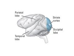
because of distinct stripes the visual cortex is sometimes called the striate cortex
|