Reading...
![]()
Play button
![]()
Play button
![]()
Use LEFT and RIGHT arrow keys to navigate between flashcards;
Use UP and DOWN arrow keys to flip the card;
H to show hint;
A reads text to speech;
24 Cards in this Set
- Front
- Back
|
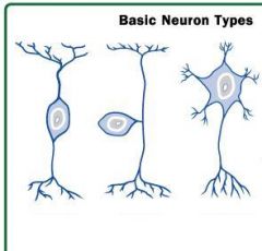
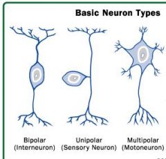
What characteristics distinguish nerve cells?
|
1. "owl's eye" nucleus - ie nucleolus is visible
2. Nissl bodies (RER's) make the cytoplasm look patchy 3. Angular border - like fblasts so look for the above 2 xtics |
|
|
|
|
|
How can you tell which part of the neuron is the axon?
|
There's no nissl bodies in the axon, so look for the light pink process.
|
|
|
How is a post-synaptic cell inhibited?
|
The NT causes the influx of Cl which will hyperpolarize the cell and make it less excitable.
|
|
|
What 4 things are in grey matter? What 3 things are in white matter?
|
Grey: soma, fibers, vessels, glial cells
White: fibers, vessels and glial cells |
|
|
What are 3 types of glial cells?
|
1. Oligodendroglia
2. Astrocytes 3. Ependyma |
|
|
What does an oligodendroglia cell do?
|
- secretes myelin for up to 50 neurons at a time. Myelin is multiple layers of cell membrane. It increases conduction speed
- covers multiple neurons but for only short distances, thereby creating gaps in the myelin known as Nodes of Ranvier |
|
|
What's the difference btwn white & grey matter?
|
No cell bodies in white matter!
|
|
|
What does an astrocyte do? How can it maintain its shape?
|
It sends out "foot processes" onto neuron, microvessels and the pia. This helps isolate the brain
Will also phagocytize stray NTs, and ions to help maintain homeostasis. Can phagocytose old cells but this can make glial scars which interrupt conduction. Has a well-developed cytoskeleton rich in intermediate fibers. |
|
|
What are ependyma cells?
Where are they found? What do they do (2)? |
Simple cuboidal epithelium, linked by tight junctions.
Line the ventricles, canals, and choroid plexuses. Play a role in CSF production & circulation. Also help make blood-brain barrier |
|
|
What is the choroid plexus?
|
A tuft of ependyma-covered blood vessels that project into the ventricles of the brain. The sites of CSF production.
|
|
|
What types of cells are found in sensory ganglia?
|
Pseudounipolar neurons - round profiles with central nucleus
Satellite cells - form a complete envelop around the neuron |
|
|
Which type of ganglia has NO synapses?
|
Sensory
|
|
|
What types of cells are in an autonomic ganlia? What do they look like?
|
multipolar neurons - angular profiles, eccentric nuclei
Only a few satellite cells - no complete envelop formed. |
|
|
Why do nerves follow a sinuous path?
|
So they have slack and can allow for movement
|
|
|
In what 2 ways are the cells of a nerve insulated?
|
Unmyelinated fibers can be supported in suface grooves of a schwann cell. Others are myelinated by a single schwann cell.
|
|
|
Define:
Endoneurium Perineurium Epineurium |
Endo- loose ordinary connective tissue that surrounds each fiber and its schwann cell
Peri - dense connective tissue that surrounds the fasicles (groups of fibers) Epi - loose ordinary connective tissue, surrounds a nerve (has more than one fasicle) |
|
|
How are the fasciles kept in isolation?
|
The perineum contains concentric layers of epitheliod cells and these have tight junctions - thus each fascile has its own isolated microenvironment.
|
|

In this x-section of the spinal cord - identify the layers of the meninges and the grey and white matter.
|

|
|

This is dura mater, what type of tissue is it? What covers the CNS?
|
Dense connective tissue - the dura, arachnoid and pia cover the CNS
|
|
|
What type of tissue is the arachnoid mater?
|
Loose connective - it is usually in contact with the dura mater (held there by the pressure of CSF)
|
|

|

|
|

Identify the nerve fiber, myelin sheath and neurolimma (what is that?) What else (1 thing) can you see?
|

Neurolimma - as the membrane of the oligodendrocyte winds around the nerve fiber, the cytoplasm is sequestered to the periphery, staining as a thin line.
|
|

|

These are satellite cells form almost a complete envelope around the cell bodies of pseudounipolar neurons in sensory ganglia
|