Reading...
![]()
Play button
![]()
Play button
![]()
Use LEFT and RIGHT arrow keys to navigate between flashcards;
Use UP and DOWN arrow keys to flip the card;
H to show hint;
A reads text to speech;
43 Cards in this Set
- Front
- Back
|
What stops happening at the blastula stage? What starts happening?
|
At the blastula stage, the embryo stops dividing symmetrically. Instead, it starts infolding and forming compartments.
|
|
|
What is the consequence of infolding? What is true about the resulting cells? What is another consequence of infolding?
|
As a consequence of infolding, you get endoderm, mseoderm, and ectoderm. At this point, these cells are FATED to become a certain cell type. Before this they were PLURIPOTENT.
Another consequence of infolding is the formation of the primitive groove, which runs along the anteriocaudal to the organism. |
|
|
What are the terms used to describe the coronal and saggital slices of an embryo?
|
Coronal slices are called LATERAL slices.
Saggital slices are called CRANIAL-CAUDAL slices. |
|
|
What is the notochord?
|
The notochord is a cord of mesodermal tissue that runs parallel to the neural groove/spinal cord.
|
|
|
What happened when the dorsal lip of the blastula was transplanted onto the bottom of another embryo? What did this show?
|
The dorsal lip grew into a new organism attached to the original embryonic organism (two tadpoles fuse together. This showed that the dorsal lip contained cells whose instructions were "make a body".
|
|
|
What is the cervical flexure?
|
The cervical flexure makes the brain and spinal cord perpendicular to each other.
|
|
|
What are the 3 original vesicles in the embryo, and what do they become?
|
From rostral to caudal, the 3 are the
1. proencephalon-->forebrain; telencephalon (cerebral cortex)+diencephalon (thalamus + striatum) 2. Mesencephalon-->midbrain; pons, cerebellum and midbrain. 3. Rhombencephalon--> hindbrain; medulla |
|
|
What kind of germ layer is CNS tissue made of? How does the CNS form from the primitive streak?
|
CNS tissue is endoderm. Primitive streak becomes neural groove, which becomes neural tube.
|
|
|
What are radial glia? What is the pial axis?
|
Radial glia are cells that extend from the surface of the ventricles to the basal surface of the developing brain. The radial glia are oriented along the pial axis.
|
|
|
What are the two kinds of cleavage? What do they result in?
|
Vertical cleavage occurs when the cells divide perpendicular to the pial axis. Then dividing, they look like an infinity sign. These cells reenter the cell cycle.
Horizontal cleavage is when the cells divide parallel to the pial axis, and look like the number 8. The bottom cell remains apical and reenters the cell cycle, but the top cell migrates away to form the cortex. |
|
|
What is the chemical basis of horizontal and vertical cleavage?
|
All apical cells have Notch and the north pole and Numb at the south pole. Numb is necessary to anchor the cell in the ventricular zone. Vertical cleavage along the longitudinal axis results in two cells with Numb, so they stay apical. Horizontal cleavage along the equator results in the top cell without Numb, so it migrates away.
|
|
|
How do radial glia interact with the migrating cells? What are the other terms for these migrating cells? Can these migrating cells continue to divide?
|
The migrating (i.e. Notch/neuroblast) cells associate with the radial glia, who guide it toward neocortex. No, these migrating cells cannot divide again once they leave the ventricular zone; cell division is arrested forever.
|
|
|
What are Notch cells called as they leave the radial zone? When do they begin to differentiate?
|
Notch cells are called neuroblasts. As they migrate, they are undifferentiated. Once they reach their destination, however, do they begin to differentiate.
|
|
|
What is true about dendritic growth?
|
The cell bodies stay the same size as the dendrites grow. The growth of dendrites is not mediated by radial glia, but is an intrinsic property of the neurons themselves.
|
|
|
How does the cortex grow during development?
|
The corrtex grows from the inside out. The deepest cell layers (e.g. Layer 6) form first, and then superficial layers form last.
|
|
|
What are HOX genes? What prior evidence do they explain.
|
Hox genes are a set of genes that are expressed differentially along the anteriocaudal axis during development. The differential Hox gene expression leads to differential gene expression, which results in the formation of different body parts. In short, the Hox genes contain information on how to build a body. Hox genes explain the dorsal lip transplant studies.
|
|
|
What is Wnt1? What happens in Wnt1 KO mutants?
|
Wnt1 is an organizer gene (same as Hox) that is expressed in a particular "organizer region" of the developing brain. The Wnt1 KO mutants do not develop a mes-/metencephalon, but retain everything else.
|
|
|
What is BMP? Describe its involvement in the differentiation of the developing ectoderm.
|
The developing ectoderm is naturally filled with BMP (bone-derived morphogenic protein). If left alone, the ectoderm will form epidermis by default. However, when BMP is blocked by BMP inhibitors, the ectoderm becomes neural tissue. With the further addition of retinoic acid, you get posterior neural plate. If not, you get anterior neural plate.
|
|
|
What are the 3 BMP inhibitors?
|
Chordin, noggin, and follistatin,
|
|
|
What is NeuroD?
|
NeuroD turns epidermal tissue into nervous tissue.
|
|
|
What protein interacts with BMP in the development of the neural tube? Why is this significant?
|
SHH (sonic hedgehog) interacts with BMP to help guide axons into their appropriate positions. SHH is on ventral half; BMP is on dorsal half. In fact, these molecules DETERMINE which half becomes dorsal and which becomes ventral.
|
|
|
What are the 6 mechanisms of axonal pathfinding?
|
1. Extracellular matrix adhesion
2. Cell Surface adhesion 3. Fasciculation 4. Chemoattraction 5. Contact inhibition 6. Chemorepulsion |
|
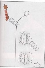
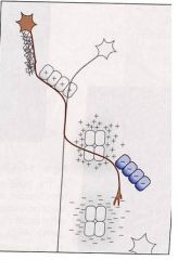
What mechanism is shown? Explain.
|
Extracellular matrix adhesion: axon grows along extracellular matrix components.
|
|
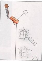
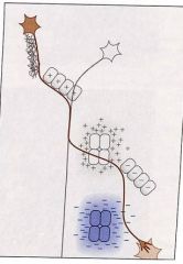
What mechanism is shown? Explain.
|
Cell Surface Adhesion: axon grows along the proteins on a cell surface (not the extracellular matrix)
|
|
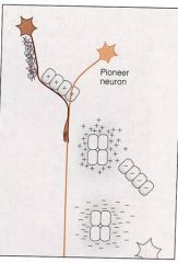
What mechanism is shown? Explain.
|
Fasciculation: pathfinder axon grows along pioneer axon.
|
|
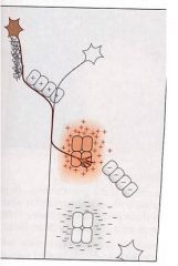
What mechanism is shown? Explain.
|
Chemoattraction: axon grows toward chemical.
|
|
What mechanism is shown? Explain.
|
Contact inhibition: (opposite of cell-surface adhesion) cell grows away from surface proteins on another cell.
|
|
What mechanism is shown? Explain.
|
Chemorepulsion: (opposite of chemoattraction) growth cone grows away from particular chemical.
|
|
|
In axonal pathfinding, which mechanisms are long-range and which short-range cues
|
The ones that involve contact with other cells or extracellular elements are short-range. The ones that involve chemicals are long-range.
|
|
|
What is the growth cone?
|
The growth cone is an amorphous structure at the end of a pathfinding axon. It detects chemicals, surface proteins, extracellular elements, etc., and changes its shape (via actin filaments) to change direction.
|
|
|
What is NGF?
|
NGF (neural growth factor) is a chemoattractant that is necessary for the proper development and organization of the nervous system.
|
|
|
How does differential receptor expression on the growth cone affect axonal pathfinding? Give an example.
|
If the growth cone expresses receptors that recognize chemoreceptive molecules, but then steps expressing these and instead expresses a receptor for a chemorepulsive molecule in the same region, you'll get a specific path. In developing spinal cord, netrinR causes chemoattaraction, then RoboR chemorepulses to Slit molecules, successfully guiding axon.
|
|
|
What is neural pruning? What determines which neurons get pruned/culled?
|
Neuronal pruning is the process by which some neurons die. The neurons that die are those that get the most trophic factors.
|
|
|
What do the studies in which a segment of the developing spinal cord is inverted show? What term is associated with this effect?
|
When the spinal cord segment is inverted, the motor neurons still find their appropriate targets. This is because all extending neurites/growth cones go through "sorting areas" that shuttle them to their appropriate locations.
|
|
|
What are the 4 steps by which the growth cones forms a synapse with a muscle fiber?
|
1. growth cone produces agrin when near muscle.
2. growth cone secretes agrin. 3. muscle detects agrin. 4. neuromuscular synapse forms. |
|
|
What is the end result of neuronal pruning at the neuron-muscle level? Why is this significant?
|
There is a 1:1 correspondence of neurons to muscle fibers. This allows for most precise control of muscle activity.
|
|
|
What is the role of agrin in formation of neuromuscular connections? What is the role of acetylcholine receptors? Summarize this information.
|
1. Agrin is necessary to FORM the connections.
2. Acetylcholine receptors are necessary to MAINTAIN the connections. (AChR blockade of formed synapses causes those synapses to disappear.) |
|
|
What is the role of sensory experience in the development of neuronal connections?
|
Sensory experience is necesary to progress from an immature state of no functional segregation to a mature state where functions are segregated into different regions. If you block sensory activty (i.e. deprivation), you never progress past immature state.
|
|
|
What is meant by a critical period?
|
A critical period is a window of time in which sensory input must be provided if the nerual connections are to form properly.
|
|
|
With the experiments involving visual sensory deprivation and the percentage of corresponding neurons in Layer III of visual cortex show? What happens after the sensory stimulation is restored to deprived side?
|
These experiments show that unilateral sensory deprivation during the critical period results in the failure of the corresponding cells to establish themselves in the cortex.
Even if sensory stimulation is restored to deprived side, there will be columns for each eye individually, but no columns that are shared by both eyes, which means that there is no integration of information from both eyes. |
|
|
What is meant by a "gradient" of gene expression in the developing brain?
|
In the cells of a certain region (the "epicenter), the concentration of a particular protein/molecule is very high. As you radiate outward, the concentration of that molecule in those cells decreases (this is the "gradient")
|
|
|
How do axons bundle together?
|
Individual axons bind to components in the extracellular matrix, and the axons bind to each other by chain-like connector proteins.
|
|
|
What 2 things are required for the switch in ocular dominance following unilateral sensory deprivation to occur?
|
You need projections from (1) the basal forebrain complex (acetycholine), and (2) the locus coeruleus (norepinephrine) to observe the shift in ocular dominance follwoing unilateral sensory deprivation.
|