Reading...
![]()
Play button
![]()
Play button
![]()
Use LEFT and RIGHT arrow keys to navigate between flashcards;
Use UP and DOWN arrow keys to flip the card;
H to show hint;
A reads text to speech;
37 Cards in this Set
- Front
- Back
|
Describe the metabolic demands of the human brain with respect to the rest of the body. How does the brain ensure it receives sufficient blood flow?
|
The human brain receives a disproportionate amount of metabolic attention despite its small size in relation to the rest of the body.
Cushings Reflex (i.e. fainting), etc. exist to ensure that blood always reaches the brain. |
|
|
How are the axes of the CNS different in rodents and primates? How is this reflected in the nomenclature?
|
In rodents, the brain and spinal cord are along the same axis. In primates, the brain is perpendicular to the spinal cord.
The nomenclature reflects this by replacing the rodent words rostral/caudal with primates words anterior/posterior. |
|

What view is this? Identify the central sulcus, primary motor cortex, and primary somatosensory cortex.
|

Dorsal view.
|
|
|
What is a gyrus? What is a sulcus? How are they related?
|
A gyrus is a raised area on the cortex. A sulcus is the depression formed by two adjacent gyri.
|
|
|
What is the principle reason why the brain has convolutions?
|
It is the best way to maximize content within a fixed space (the skull).
|
|

What view is this? What 2 fissures are present in this perspective? Identify them.
|

Lateral view. The central sulcus and the lateral/Sylvian fissure.
|
|

What view is this? Identify the corpus callosum and the pons. What are the functions of these two structures?
|

Medial/mid-saggital view.The corpus callosum connects the cerebral hemispheres. The pons connects the cerebellar hemispheres.
|
|
|
What hemispheres does the central sulcus separate? What hemispheres does the lateral/Sylvian fissure separate?
|
The central sulcus separates the frontal and parietal lobes.
The lateral/Sylvian fissure separates the parietal and temporal lobes. |
|

What view is this? Identify the olfactory nerves, optic chiasm, and medulla. What function does the medulla perform?
|
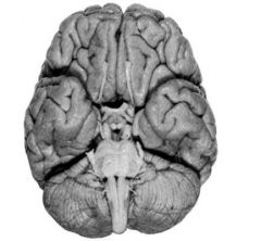
Ventral view. The medulla controls homeostatic functions like circulation and respiration.
|
|

How can you tell what dorsal/ventral is in a coronal spinal cord slice?
|

When oriented to look like an upside-down heart, dorsal is on top. The dorsal side also has gray matter touching the edges, but the ventral side doesn't.
|
|
|
How are dorsal/ventral referred to differently in the brain and the spinal cord?
|

BISSAP. (Brain- inferior/superior; spinal cord- anterior/posterior)
|
|
|
What is gray matter? What is white matter?
|
Gray matter is cell bodies. White matter is axon tracts.
|
|

Identify the 3 main structural features of the cerebellum. What is the function of the cerebellum
|

The left and right cerebellar hemispheres, and the vermis. The cerebellum smooths movements and controls balance.
|
|

What structure is this? What view is this? Identify the pointed structures.
|

Top to bottom: superior colliculi, inferior colliculi, cerebellar peduncles, floor of the fourth ventricle (normally covered by cerebellum).
|
|
|
What kind of impairment does Broca's aphasia cause? Why does this make sense, given the affected brain area. Why was the discovery of this area significant?
|
Broca's aphasia is characterized by an ability to understand language but an inability to speak it. This makes sense given that it results from a lesion to the MOTOR cortex. This case demonstrated that there is localization of function within the brain.
|
|
|
How were 18th and 19th century wars conducive the neuropsychology research?
|
Bullet wounds in the brain allowed for precise localization of brain functions. Also, syphilis caused analogously circumscribed brain damage.
|
|
|
What was phrenology?
|
Phrenology was an early, failed attempt to attribute function to particular brain areas. It relied on the false assumption that bumps on the skull corresponded to brain morphology.
|
|
|
What did the case of Phineas Gage demonstrate?
|
The case of Phineas Gage, who suffered circumscribed lesions in his frontal lobes, demonstrated that the executive role of the frontal lobes. This was evidence that the different lobes had different functions.
|
|
|
What is the fifth lobe of the brain? Where is it located?
|
The fifth is the insula, which control limbic/gustatory functions.
|
|
|
Damage to what region of what lobe will result in hemispatial neglect. Hemispatial neglect indicates a failure of what? What conclusion does this warrant?
|
Damage to the posterior parietal lobe will result in hemispatial neglect. Hemispatial neglect is a disorder of the "WHERE" pathway.
Therefore, the posterior parietal lobe is part of the "WHERE" pathway. |
|
|
Damage to what area of what lobe will result in object perception agnosia (e.g. Dr. P, the man who mistook his wife for a hat). This kind of object agnosia is a failure of what? What conclusion does this warrant?
|
Damage to the inferior temporal lobe results in object agnosia. Object agnosia is a disorder of the "WHAT" pathway.
Therefore, the inferior temporal lobe is involved in the "WHAT" pathway. |
|
|
In what region were the single-cell recordings of monkeys viewing faces done? Why is this relevant?
|
These highly specific "face cells" were found in the inferior temporal cortex. It supports the theory that the inferior temporal cortex is involved in object recognition/the "WHAT" pathway if cells like this are found here.
|
|
|
What are the four layers of the meninges? What is their spatial relationship to one another?
|

Dura mater --> Arachnoid membrane --> Sub-arachnoid space --> Pia mater.
|
|
|
Where in the meninges do you find blood vessels? Where does the cerebrospinal fluid flow?
|
The blood vessels are found in the arachnoid membrane.
The cerebrospinal fluid is found in the sub-arachnoid space. |
|
|
What is the purpose of cerebrospinal fluid (CSF)? Where is the source of CSF?
|
Cerebrospinal fluid buffers the brain and spinal cord from trauma. It also provides nutrients and immune defense.
All CSF is produced by epithelial cells in the choroid plexus, which is in the lateral ventricles and third ventricle. |
|
|
What are the arachnoid granulations? Where are they found?
|
The arachnoid granulations are structures on the top of the skull that absorb CSF back into general circulation.
|
|
|
What is the lumbar puncture? Why is it useful?
|
The lumbar puncture involves withdrawing a sample of cerebrospinal fluid from the lumbar region of one's spine. This test is a simple, non-invasive way to diagnose neurological diseases that cause changes in CSF content.
|
|
|
What are dermatomes?
|
Dermatomes are the areas of skin that are served by the dorsal root of one spinal segment. A retrograde virus like chickenpox that infects a particular dorsal root ganglion will affect only that root's dermatome.
|
|
|
Where does the spinal cord actually end? What is below it?
|
The spinal cord actually ends in the lumbar region, but axon tracts still extend down into the sacral region.
|
|
|
Where are the two bulges in the spinal cord located? What do they correspond to?
|
There is a cervical enlargement and a lumbar enlargement. These correspond to extra motor and sensory projections coming to and from the arms and legs, respectively.
|
|
|
Describe the functional localization of the spinal cord?
|
The dorsal half receives sensory projections. The ventral half emits motor projections.
Remember it with the mnemonic, "DS", for dorsal=sensory. |
|

Identify the middle cerebral artery. Why is it important? Identify the circle of Willis.
|

The middle cerebral artery is important because it supplies blood to 2/3 of the cortex. It is upstream of the carotid.
|
|

Identify the pointed structure.
|
Middle cerebral artery.
|
|

Name the other two cerebral arteries besides the middle cerebral artery. Identify them on this diagram.
|

|
|
|
What is the difference between a thrombosis and an aneurysm? How are they similar?
|
Both thrombosis and aneurysm cause strokes.A thrombosis is a clot in an artery that prevents blood flow to the areas upstream. An aneurysm is a swelling of a blood vessel; when it bursts, the upstream areas are deprived of blood. Even if it doesn't bursts, its expansion can impinge on other areas.
|
|
|
How can you identify a stroke on an MRI scan?
|
Since white areas correspond to fluid, a large white region indicates a place where blood is pooling.
On some scans, however, the blood accumulation is represented by inappropriately DARK areas. |