![]()
![]()
![]()
Use LEFT and RIGHT arrow keys to navigate between flashcards;
Use UP and DOWN arrow keys to flip the card;
H to show hint;
A reads text to speech;
42 Cards in this Set
- Front
- Back
|
Sagittal Plane |

Name the plane that divides CNS left and right |
|
|
Transverse, horizontal, or axial plane |
Name the plane that separates superior and inferior |
|
|
Coronal Plane |

Name the plane that divides brain or spinal cord into anterior and posterior parts |
|
|
Dura Mater ( Outer Meninges) |
Top |
|
|
Falx Cerebral (Part of Dura Mater that covers medial Surface of Cerebral hemisphere) |
A |
|
|
Arachnoid Mater. Middle Meninges. |
Top |
|
|
Pia Mater. Innermost Meninges. |

Bottom. |
|
|
Longitudinal (Sagittal) Fissure |

Separates right an left cerebral hemisphere |
|
|
Lateral (Sylvian) Sulcus/Fissure |
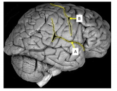
Divides Temporal lobe inferiorly from parietal and frontal lobe. A. |
|
|
Central Sulcus |

Vertical and Perpendicular to lateral Sulci. B |
|
|
Parietooccipital Sulcus |
Separates parietal and occipital lobes |
|
|
Superior Frontal Gyrus |

1 |
|
|
Middle Frontal Gyrus |

2 |
|
|
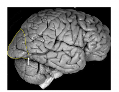
Inferior Frontal Gyrus |

3. Dominates in language (left) and contains Broca's motor speech area |
|
|
Clinical Correlate: What action would a Broca's aphasia patient have difficulty performing? |
Speaking Fluently |
|
|
Precentral Gyrus |

Contains primary motor cortex. 1 |
|
|
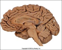
Orbital Gyri. Collection of gyri on inferior surface of frontal lobe. |

2 |
|
|
Gyrus Rectus. Medial and Ventral elevation of frontal lobe. |

1 |
|
|
Olfactory Bulb. Contains cell bodies of 2nd order cell bodies and receives info from olfactory bulb. |
6 |
|
|
Olfactory Tract. Fiber tract leaving bulb posteriorly projecting to primary olfactory region of temporal lobe. |

7 |
|
|
Postcentral Gyrus. Poserior to central sulcus and contains primary somatosensory cortex. |

2 |
|
|
Supramarginal Gyrus. Surrounds posterior parts of lateral fissure. |

3 |
|
|
Angular Gyrus. Posterior to Supramarginal Gyrus. |

Written 1 |
|
|
Superior Temporal Gyrus. Inferior to lateral fissure. |

Written 1 |
|
|
Middle Temporal Gyrus. Middle Temporal Gyrus. |
Written 2. |
|
|
Inferior Temporal Gyrus. Inferior Temporal Gyrus. |
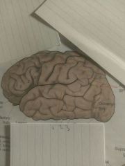
Written 3 |
|
|
Transverse Temporal Gyri. Extending medially into lateral fissure. Location of primary auditory center. Wernicke's area. |

1 |
|
|
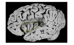
Parahippcampal Gyrus. Inferior surface, lateral diencephalon. |

4 |
|
|
Uncus |

Hook-Shaped. Rostral end of para |
|
|
Clinical Significance: Uncal Herniation |
Damage can cause olfactory hallucinations |
|
|
Cuneus. Superior to calcarine fissure. |

3 |
|
|
Calcarine Sulcus/Fissure. Extending to posterior lobe. |

2 |
|
|
Lingual Gyrus. Inferior to calcarine. |

1 |
|
|
Cingulate Gyrus. Medial area of temporal lobe encircling corpus callosum |
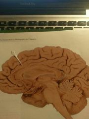
1 |
|
|
Insular Gyri |
Interpretation of tasting and visual sensations |
|
|
Cuneus Gyrus |
B |
|
|
Superior temporal gyrus |

|
|
|
Name structure |
|
|
|
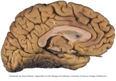
Cingulate sulcus |
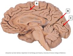
A |
|
|
Parietooccipital sulcus |

B |
|
|
Calcarine sulcus (seperates lingual and cuneus gyrus |

C |
|
|
Inferior frontal gyrus |
Name structure |