Reading...
![]()
Play button
![]()
Play button
![]()
Use LEFT and RIGHT arrow keys to navigate between flashcards;
Use UP and DOWN arrow keys to flip the card;
H to show hint;
A reads text to speech;
99 Cards in this Set
- Front
- Back
|
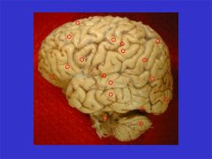
The orbital frontal cortex is part of the granular prefrontal cortex involved in regulation of emotions, control of emotions and motivation. Damage will alter personality.
[Orbital frontal cortex] |

Identify this region, what functions does it control?
|
|
|
The opercular part of the inferior frontal gyrus in the dominant (usually left) hemisphere (along with the triangular portion of the inferior frontal gyrus) is the motor area of language generation (Broca's area).
[Opercular part, inferior frontal gyrus] |

Name this area, what functions take place here?
|
|
|
The triangular part of the inferior frontal gyrus in the dominant (usually left) hemisphere (along with the opercular portion of the inferior frontal gyrus) is the motor area of language generation (Broca's area).
[Triangular part, inferior frontal gyrus] |

Name area and function?
|
|
|
The middle frontal gyrus is part of the lateral frontal lobe. The posterior part includes premotor areas (planning of movement) while the frontal eye fields and dorsolateral prefrontal cortex are progressively more anterior. The latter is involved in strategic planning of higher motor and cognitive tasks. Lesions effect the ability to perform delayed spatial tasks.
[Middle frontal gyrus] |
Identify area and associated function(s)
|
|
|
The Sylvian (lateral) fissure is a deep fissure that separates the opercular portions of the frontal and parietal lobes from the opercular portion of the temporal lobe. The insular cortex lies at the depth of the lateral fissure.
[Sylvian (lateral) fissure] |

Identify fissure, what does it separate? What lies at the depth of this fissure?
|
|
|
The central sulcus defines the border of the precentral (primary motor) and postcentral (primary sensory) gyri. It usually doesn't quite reach the lateral fissure inferiorly, while it usually extends over the dorsal aspect of the hemisphere onto its medial side.
[Central sulcus] |

What is the name of this structure? What border does it define?
|
|
|
The preoccipital notch is the indentation on the inferior surface of the brain that is at the location of the petrous ridge of the temporal bone. It defines the location where the temporal lobe meets the occipital lobe.
[Preoccipital notch] |

What is the name of this indentation? With what osteologic structure does it correspond? It defines the meeting of what two lobes?
|
|
|
The postcentral gyrus is the primary sensory cortex with strong input from somatic sensory areas of the thalamus. There is a topography (somatotopy) with the face represented ventrally and the lower limb dorsally. The foot is represented on the medial aspect of the cortex.
[Postcentral gyrus] |

What is this structure? What is its function? From were does it receive input? What is the somatotopy of this region?
|
|
|
The precentral gyrus is the primary motor cortex with strong axonal projections from pyramidal neurons to motor areas of the brain stem and spinal cord. There is a topography (somatotopy) with the face represented ventrally and the lower limb dorsally.
[Precentral gyrus] |

Identify the structure? functions?
|
|
|
spinal cord enlargements are at the level of:
|
the cervical and lumbar vertebrae
|
|
|
spinal cord ends at what level?
|
L1/L2 intervertebral disc
|
|
|
where is the lumbar spinal cord found?
|
at the level of the lowest thoracic vertebra
|
|
|
conus medullaris
|
lowest tapering portion of the spinal cord
|
|
|
filum terminale interna
|
continuation of the pia mater from the end of the conus medularis
|
|
|
cauda equina consists of what?
|
lumbar and sacral nerve roots arrising from the conus medularis
|
|
|
structures marking points of exit of the nerve roots from the spinal cord
|
DORSOLATERAL
and VENTROLATERAL SULCI (small grooves on dorsolateral and ventrolateral portions of the spinal cord |
|
|
where is the anterior median sulcus? what important structure is associated with it?
|
groove on anterior surface of spinal cord
in which the ANTERIOR SPINAL ARTERY runs |
|
|
dorsal median sulcus
|
very small
|
|
|
where is the dorsal intermediate sulcus?
|
ONLY PRESENT IN UPPER PART OF SPINAL CORD
located between the: -DORSAL MEDIAN SULCUS and -DORSOLATERAL SULCUS |
|
|
What spinal cord division is between the median and posterolateral sulci?
|
DORSAL FUNICULUS
|
|
|
The lateral funiculus is between what structures?
|
DORSOLATERAL SULCUS
and VENTROLATERAL SULCUS |
|
|
What structure is found between the VENTROLATERAL SULCUS and the ANTERIOR MEDIAN SULCUS?
|
VENTRAL FUNICULUS
|
|
|
What is found in the dorsal, lateral and ventral funiculi?
|
WHITE MATTER (in which the majority of nerve tracts that ascend and descend the spinal cord run)
|
|
|
the dorsal funiculus contains almost exclusively what?
|
the dorsal columns
|
|
|
the DORSAL COLUMN
-where is it located? -what is its function? |
located in the dorsal funiculus of the spinal cord
an ASCENDING TRACT, carries very specific sensations from the body up toward the brain (some division between the sensory fibers coming from the legs and those from the arms) |
|
|
The posterior intermediate sulcus separates what?
|
the (medial) fibers from the legs from the (lateral) fibers of the arms
(fibers are located within the DORSAL COLUMN (tract) of the DORSAL FUNICULUS of the SPINAL CORD) |
|
|
damage to the posterior part of the spinal cord can affect what functions?
|
vibratory sense, well-localized touch sensation and joint position sense
in the LIMBS! |
|
|
Basic internal structure of the spinal cord
|
gray matter, districuted in an H-shape, with a central canal appearing in the x-piece
|
|
|
central canal of spinal cord is a remnant of what embryologic structure?
|
the lumen of the NEURAL TUBE
|
|
|
ventral horn
|
part of the H (gray matter) in spinal cord that protrudes forward
contains: large ALPHA MOTOR NEURONS that give rise to ALPHA MOTOR FIBERS innervating muscle |
|
|
dorsal horn
|
part of the H (gray matter) in spinal cord that protrudes back
|
|
|
most posterior portion of the dorsal horn is called what? why?
|
SUBSTANTIA GELATINOSA
bc of its appearance using stains for fibers |
|
|
small folds making up the majority of the cerebellum
|
FOLIA
|
|
|
a deep division that separates the anterior lobe of the cerebellum from the posterior lobe
|
PRIMARY FISSURE
|
|
|
midline division of the cerebellum
|
VERMIS
|
|
|
lateral portion of the cerebellum
|
hemisphere(s)
|
|
|
the FLOCCULUS is part of what?
where is it located? |
two small lobes of the cerebellum
siutated posteriorly and inferiorally to the VERMIS and HEMISPHERES |
|
|
what connects the cerebellum to the brainstem?
|
the 3 cerebellar peduncles (consist of large white matter connections, provide input and output from the cerebellum)
|
|
|
Inferior Peduncle (aka Restiform Body) is located where?
|
appears at the dorsolateral side of the brainstem (appears as an upward continuation from the spinal cord)
|
|
|
MIDDLE CEREBELLAR PEDUNCLE (brachium pontis) connects what structures?
where is it seen? |
connects PONS out to the CEREBELLUM
obvious from ventral or lateral surface of brainstem |
|
|
SUPERIOR CEREBELLAR PEDUNCLE (brachium conjunctivum)
connects what structures? where is it seen? |
connects the cerebellum down to the upper portion of the brainstem
difficult to see without removing the cerebellum (largely surrounded on the lateral side by the middle cerebellar peduncle) |
|
|
middle cerebellar peduncle is connected to what large protuberance on the ventral side of the brain
|
BASIS PONTIS or BASAL PONS
|
|
|
what structures divide the brainstem into 3 portions?
|
the MIDDLE CEREBELLAR PEDUNCLE
and the BASAL PONS |
|
|
what are the 3 portions of the brainstem?
|
MEDULLA (spinal cord to the...)
PONS MIDBRAIN (extends rostrally from the pons) |
|
|
External features of the MEDULLA
|
INFERIOR CEREBELLAR PEDUNCLE
GRACILE and CUNEATE TUBERCLES TUBER CINEREUM INFERIOR OLIVE PRE-OLIVARY SULCUS (CNXII) POST-OLIVARY SULCUS (CNIX & CNX) PYRAMIDS (w/ DECUSSATION) |
|
|
TUBER CINERIUM
(where is it found?) |
a small prominence running along the sides of the medulla (just lateral to cuneate tubercle)
situated over a band of fibers and nucleus associated with FACIAL SENSATIONS) |
|
|
forms large protruberance on lateral (/ventro-lateral) aspect of the rostral medulla
|
INFERIOR OLIVE
|
|
|
what emanates from the post-olivary sulcus?
|
CN IX and CNX
GLOSSOPHARYNGEAL and VAGUS Nerves |
|
|
what emanates from the preolivary sulcus?
|
CNXII
the HYPOGLOSSAL NERVE |
|
|
most ventral structure running the length of the brainstem
|
PYRAMIDS (to either side of midline)
|
|
|
Medullary Pyramids contain
|
important bundles of fibers that arise from the CEREBRAL CORTEX and extend down to the SPINAL CORD
*critical for FINE/SKILLED MVMTS |
|
|
Pyramidal Decussation
|
area of medulla at which the two pyramids seem to be joined (sight of crossing of fibers)
|
|
|
what is located on the rostral side of the dorsal brainstem?
|
the FOURTH VENTRICLE
|
|
|
OBEX
|
caudal-most end of 4th Ventricle
|
|
|
What is visible in the floor of the ventricle?
|
a number of ridges (mostly over various cranial nerve nuclei)
|
|
|
What nerves exit at Medullary-Pontine Junction?
|
ABDUCENS NERVE (CNVI)
FACIAL NERVE (VII) VESTIBULOCOCHLEAR (CNVIII), portions wrap around inferior cerebellar peduncle |
|
|
what structures form the ACOUSTIC TUBERCLE
|
portion of VESTIBULOCOCHLEAR NERVE (CNVIII) wrapping around the INFERIOR CEREBELLAR PEDUNCLE (around the medullary-pontine junction)
|
|
|
the ventral pons is dominated by what?
|
the BASAL PONS
|
|
|
Where does the trigeminal nerve exit?
|
the lateral side of the MIDDLE CEREBELLAR PEDUNCLE
|
|
|
FACIAL COLLICULUS
|
"hill" prominance on floor of 4th centricle
|
|
|
distinctive bands of fibers on either side of the midline of the midbrain
|
CEREBRAL PEDUNCLES (or CRUS CEREBRI)
contain most of the nerve fibers leaving the cerebral cortex heading for the brainstem or spinal cord (IMPORTANT!) |
|
|
interpeduncular fossa is located where?
what emanates from here? |
between the CEREBRAL PEDUNCLES
from which the OCULOMOTOR NERVE (CNIII) emanates |
|
|
CORPORAL QUADRIGEMINA
|
four bumps on dorsal surface of the midbrain:
SUPERIOR COLLICULI (visual and other reflexes) and INFERIOR COLLICULI (hearing) |
|
|
Brachium of the inferior and superior colliculi
|
laterally running ridges from the inferior and superior colliculi
|
|
|
Visible portions of the hypothalamus
|
MAMILLARY BODIES (prominances on posterior aspect)
INFUNDIBULUM (midline, extending to pituitary) LAMINA TERMINALIS (on the rostral side of the infundibulum [most rostral portion of developing nervous system from embryology]) |
|
|
OPTIC CHIASM is just rostral to what diencephalic structure?
|
the INFUNDIBULUM
site of crossing of many nerve fibers from the eye |
|
|
EPITHALAMUS
located where? consists of? |
dorsal side of diencephalon
consists of: PINEAL GLAND (midline) and HABENULAR NUCLEI (swellings where attachments from pineal gland to back of brain are made) |
|
|
the diencephalon comprises the walls of what structure?
|
the THIRD VENTRICLE
(most of diencephalon is located on either side of the 3rd ventricle) |
|
|
HYPOTHALAMIC SULCUS
|
small groove that runs from anterior to posterior along the wall of the third ventricle
|
|
|
major functions of the hypothalamus
|
endocrine regulation
regulation of feeding autonomic regulation regulation of sexual behaviors |
|
|
massa intermedia
|
or thalmic adhesion
small connection between the dorsal thalamus and hypothalamus (NO FUNCTIONAL SIGNIFICANCE) |
|
|
stria medullaris thalami
|
a ridge toward the dorsal side of the thalami (running for anterior to posterior to meet the HABENULA)
|
|
|
the THALAMUS is what type of mass?
|
a very large NUCLEAR MASS
the origin of many of the afferent fibers projecting to the cerebral cortex most information (sensory & even motor feedback information) required by the cerebral cortex passes through the THALAMUS |
|
|
MEDIAL AND LATERAL GENICULATE BODIES
|
two prominances located just rostral to the colliculi of the brainstem
chief sites of relay of auditory (medial) and visual (lateral) information to the cerebral cortex respectively |
|
|
Sylvian fissure
|
aka LATERAL SULCUS divides the FRONTAL and PARIETAL LOBES above from the TEMPORAL LOBE below (longer in left hemisphere of brain) (transverse plane)
|
|
|
Central Sulcus
|
separates PARIETAL LOBE from FRONTAL LOBE
(coronal plane) |
|
|
Parieto-occipital sulcus
|
?? separates parietal and occipital lobes of brain?
|
|
|
post posterior lobe of brain
|
occipital lobe
|
|
|
lobe anterior to central sulcus
|
frontal lob
|
|
|
lobe ventral to the lateral fissure
|
temporal lobe
|
|
|
parietal lobe is between
|
the frontal and occipital lobes
|
|
|
precentral gyrus is located where?
contains many what? stimulation will result in... |
just anterior to the central sulcus
contains many neurons whose axons wil run down into the spinal cord, controlling movement electrical movements of this ?gyrus? will result in MOVEMENTS OF THE FACE AND EXTREMITIES |
|
|
postcentral gyrus
-location? -what happens in this area? -stimulation of this area will result in... |
located just behind the central sulcus
area of termination of many of the somatic sensory fibers coming from the thalamus stimulation of this area will result in sensations projected onto certain areas of the body |
|
|
Transverse gyri (of Heschl)
and ISLE OF REIL |
small sulci that run into the depths of the lateral fissure
deeper in the lateral fissure is the INSULA or "isle of Reil" another group of gyri |
|
|
CALCARINE SULCUS
where is it located? |
in occipital lobe (medial side) runs from neer posterior tip of occipital lobe anteriorward toward the parieto-occipital sulcus
gyri on either side (portions of cortex) intially receive visual information |
|
|
what is the name of the large connection between the two sides of the cerebral hemispheres?
|
CORPUS CALLOSUM
|
|
|
parts of the CORPUS COLLOSUM
|
rostral-most ROSTRUM
GENU BODY large posterior SPLENIUM |
|
|
what follows the corpus collosum around its dorsal surface
|
CINGULATE GYRUS
|
|
|
SUBCALLOSAL REGION
|
portion of ??CINGULATE GYRUS or CORTEX??? directly ventral to ROSTRUM of CORPUS COLLOSUM
|
|
|
What separates the two lateral venticles?
|
SEPTUM PELLUCIDUM (thin membrane)
|
|
|
thickening at inferiormost portion of the septum pellucidum
|
FORNIX
|
|
|
FORNIX represents what type of pathway? where does it go?
|
WHITE MATTER PATHWAY
follows the ventricle posteriorly and laterally toward the temporal lobe at rostral portion of septum pellucidum, fornix swings ventrally toward hypothalamus |
|
|
ANTERIOR COMMISSURE
|
tract that connects the two sides of the brain
|
|
|
how does CSF pass from the lateral ventricle to the third ventricle?
|
must pass ventral to the fornix, via the FORAMEN OF MONROE
|
|
|
Choroid plexus is located where? What does it produce?
|
rough and irrecular tissue, extending through the foramen of MONROE into the LATERAL VENTRICLE
responsible for production of CSF |
|
|
floor of the lateral ventricle is composed primarily of what?
|
dorsal surface of the thalamus
|
|
|
STRIA TERMINALIS
|
groove at the lateral edge of the thalamus
|
|
|
Caudate nucleus
Head of the Caudate Body of the Caudate |
???
|
|
|
which lobes does the lateral ventricle invade?
|
temporal and occipital lobes
|