Reading...
![]()
Play button
![]()
Play button
![]()
Use LEFT and RIGHT arrow keys to navigate between flashcards;
Use UP and DOWN arrow keys to flip the card;
H to show hint;
A reads text to speech;
1022 Cards in this Set
- Front
- Back
- 3rd side (hint)
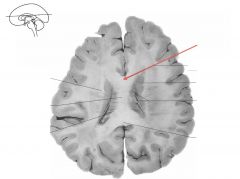
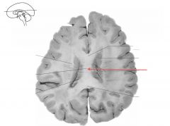
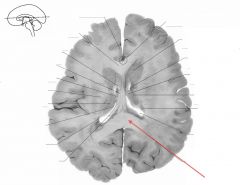
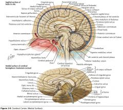
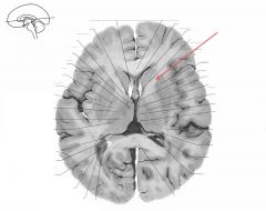
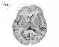
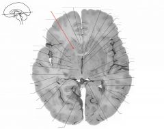
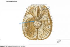
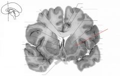
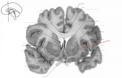
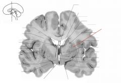
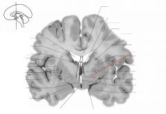
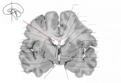
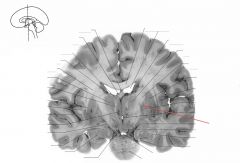
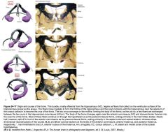
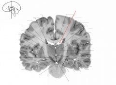
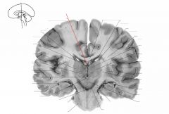
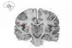
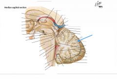
Red arrow.
|

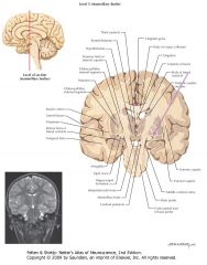
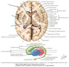
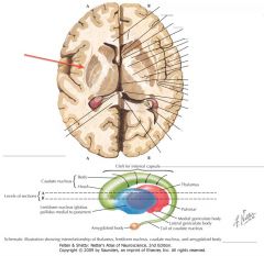
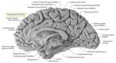
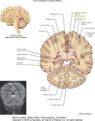
Body of the Corpus Callosum (toward the Genu)
|
|
|

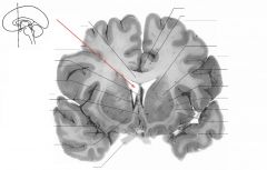
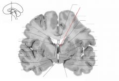
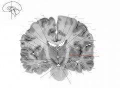
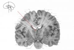
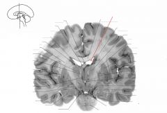
What sulcus is indicated by the white arrow?
|

The Cingulate Sulcus.
|
|
|

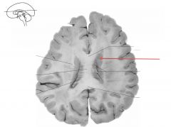
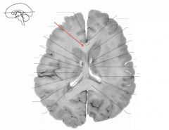
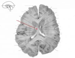
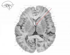
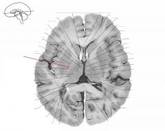
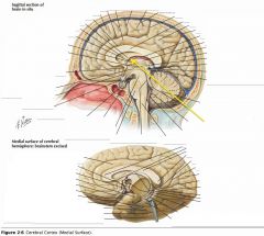
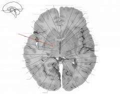
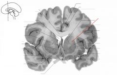
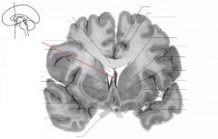
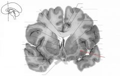
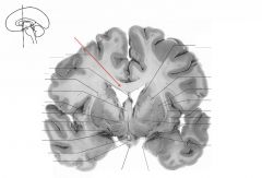
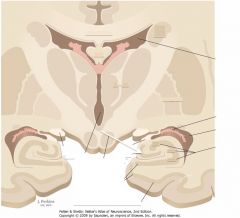
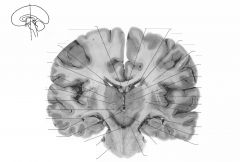
Red arrow.
|

Body of the Corpus Collosum
(Towards the Splenium) |
|
|

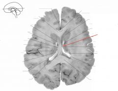
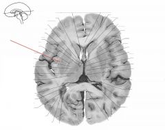
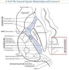
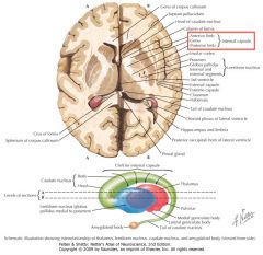
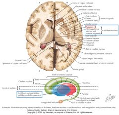
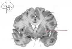
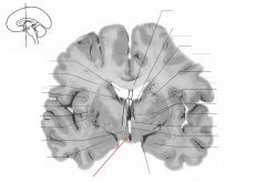
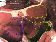
What portion of the Internal Capsule is indicated by the purple arrow?
|

The Anterior Limb
|
|
|
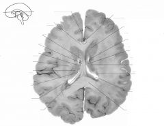
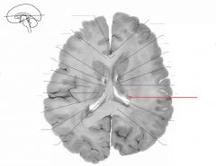
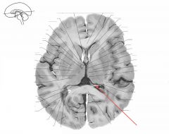
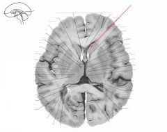
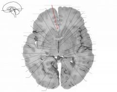
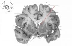
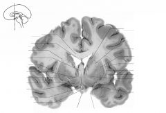
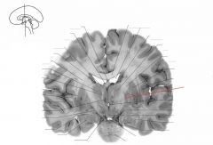
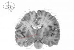
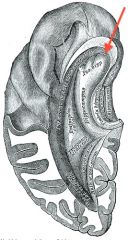
Red arrow.
|
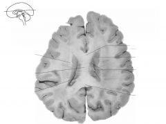
Caudate Nucleus
|
|
|
|
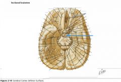
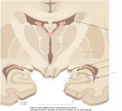
How many cell layers are in the Cerebral Neocortex?
|
Six:
1. Molecular, 2. External Granular, 3. External Pyramidal, 4. Internal Granular, 5. Internal Pyramidal, and 6. Multiform layers. |
|
|
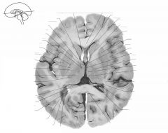
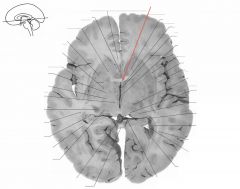
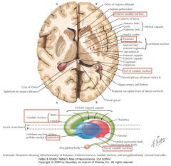
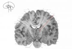
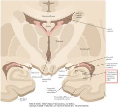
Red arrow.
|

Corpus Callosum.
|
|
|

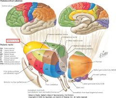
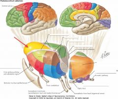
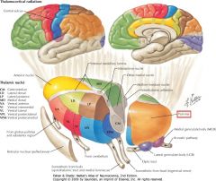
What are the divisions of the Diencephalon?
|
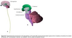
1. Thalamus
2. Subthalamus, 3. Hypothalamus, and 4. Epithalamus. |
|
|
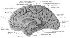
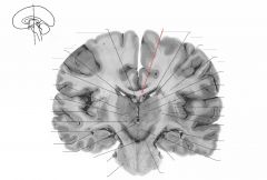
Red arrow.
|
Genu of the Corpus Callosum.
|
|
|

What sulcus is indicated by the white arrow?
|

The Sulcus of the Corpus Callosum (Callosal Sulcus)
|
|
|
Red arrow.
|

Splenium of the Corpus Callosum.
|
|
|

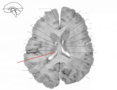
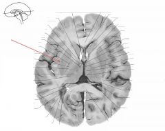
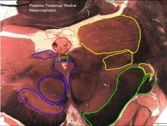
What portion of the Internal Capsule is indicated by the purple arrow?
|
The Genu.
|
|
|
Red arrow.
|

Body of Fornix
|
|
|

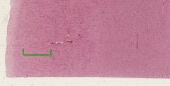
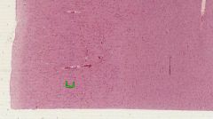
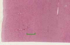
Which Neocortex layer is indicated by the green bracket?
What cells are found there? |

Layer I, the Molecular layer.
It contains relatively few neurons (those being interneurons), mostly consists of interconnecting axons and dendrites. |
|
|

Red arrow.
|

Crus of Fornix.
|
|
|

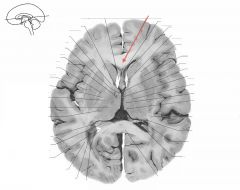
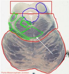
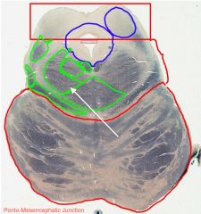
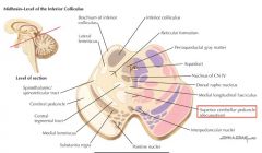
White arrow.
|

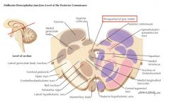
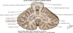
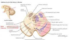
Superior Colliculus.
|
|
|

Red arrow.
|

Head of Caudate Nucleus.
|
|
|

What gyrus is indicated by the white arrow?
|

The Cingulate Gyrus.
|
|
|
Red arrow.
|

Tail of Caudate Nucleus.
|
|
|

What portion of the Internal Capsule is indicated by the purple arrow?
|

The Posterior Limb,
between the Globus Pallidus and the Thalamus. |
|
|
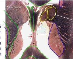
Red arrow.
|

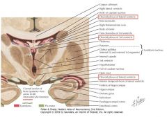
Anterior Nucleus of Thalamus.
|
|
|
Which Neocortex layer is indicated by the green bracket?
What cells are found there? |
Layer II, the External Granular Layer.
It contains many small neurons, with both pyramidal and granular cells. |
|
|

Red arrow.
|

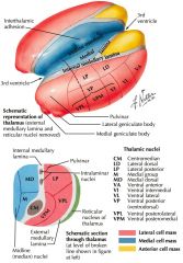
Lateral Thalamic Nuclei.
|
|
|

What collection of internal cell fibres of the Thalamus is indicated by the white arrow?
|

The Internal Medullary Lamina.
|
|
|
Red arrow.
|

Dorsomedial Nucleus of Thalamus.
|
|
|

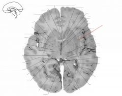
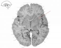
What structure is indicated by the white arrow?
|

The Uncus
|
|
|

Red arrow.
|

Lateral Ventricle.
|
|
|

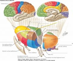
What portion of the Internal Capsule is indicated by the purple arrow?
What types of fibres pass through it? |
The Retrolenticular Limb.
The Optic Radiations from the Lateral Geniculate Bodies pass through it on their way to the Occipital Lobe. |
|
|

Red arrow (space)
|

Anterior Horn of Lateral Ventricle.
|
|
|
Which Neocortex layer is indicated by the green bracket?
What cells are found there? |

Layer III, the External Pyramidal Layer.
It consists mainly of small to medium-sized pyramidal cells. |
|
|
Red arrow.
|

Genu of the Corpus Callosum.
|
|
|
What is the Spinal Lemniscus?
Where does it terminate? |

It is a continuation of the Spinothalamic tract, which could not be visualized at medullary and pontine levels.
It terminates (mainly) in the Ventral Posterolateral Thalamic Nucleus. |
|
|

Red arrow.
|

Splenium of the Corpus Callosum.
|
|
|

White arrow.
|

Ventral Anterior Thalamic Nucleus (of the Ventral Tier of the Lateral Thalamic Nuclear Group).
|
|
|
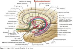
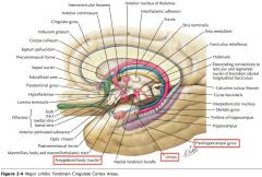
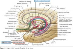
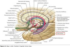
What are the Cingulate Gyrus, the Parahippocampal Gyrus and the Uncus collectively called?
|

The "Limbic Lobe." (along with some associated structures)
|
|
|
Red arrow.
|

Column of the Fornix.
|
|
|

What fibres pass through the Anterior Limb of the Internal Capsule?
|

It includes:
1) Frontopontine Fibres, 2) Anterior Thalamic Radiations (Medial and anterior thalamic projections to the frontal and cingulate cortex) and, 3) Descending fibres from the Frontal Eye Fields. |
|
|
Red arrow.
|
Crus of the Fornix.
|
|
|
Which Neocortex layer is indicated by the green bracket?
What cells are found there? |
Layer IV, the Internal Granular Layer.
It consists of a large number of Stellate ("star-shaped") neurons, also called Granule Cells. |
|
|

Red arrow.
|

Genu of the Internal Capsule.
|
|
|
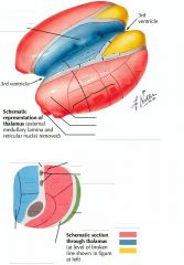
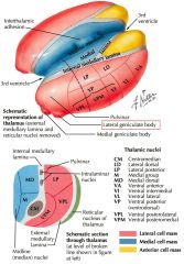
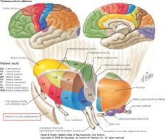
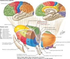
How many nuclear groups does the Thalamus divide into?
What are they? |
Three:
1. Anterior, 2. Medial, and 3. Lateral Nuclear Groups. |
|
|
Red arrow.
|

Anterior Limb of the Internal Capsule.
|
|
|

What does the Limbic System do?
|

It is involved with emotion and certain kinds of memory.
|
|
|
Red arrow.
|

Posterior Limb of the Internal Capsule.
|
|
|

What fibres pass through the Genu of the Internal Capsule?
|
Corticonuclear Fibres (projection fibres), projecting to the motor nuclei of the cranial nerves.
|
|
|
Red arrow.
|

Head of Caudate Nucleus.
|
|
|
Which Neocortex layer is indicated by the green bracket?
What cells are found there? |

Layer V, the Internal Pyramidal Layer.
It consists of medium to large-sized Pyramidal Cells. |
|
|
Red arrow.
|

Tail of Caudate Nucleus.
|
|
|

White arrow.
|

Cerebral Aqueduct.
|
|
|

Red arrow.
|

The Putamen
|
|
|

What information does the Uncus receive?
|

It receives olfactory input
|
|
|
Red arrow.
|

Globus Pallidus
|
|
|
What fibres pass through the Posterior Limb of the Internal Capsule?
|
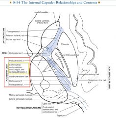
It contains:
1) The Superior Thalamic Radiations (VA, VL, and VP projections to motor and sensory cortices) 2) Corticospinal projections, 3) Others. (Corticotegmental [yellow box], Parietopontine, and Pallidothalamic* fibres) |
*Connecting the Globus Pallidus Medius to the Thalamus.
|
|
Red arrow.
|

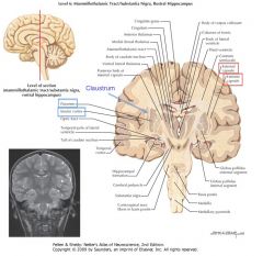
Claustrum.
|
|
|
Which Neocortex layer is indicated by the green bracket?
What cells are found there? |

Layer VI, the Multiform Layer.
It contains neurons with a variety of shapes, including Fusiform and Pyramidal Cells. |
|
|
Red arrow. (white matter)
|

External Capsule
|
|
|
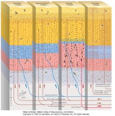
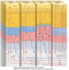
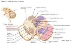
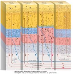
What is a "specific" thalamic nucleus?
|

It is a nucleus receiving input from defined sensory or motor pathway that projects to restricted, clearly delineated cortical regions.
|
|
|

Red arrow.
|

Anterior Nucleus of Thalamus.
|
|
|

What fibres pass through the Posterior Limb of the Internal Capsule?
|

It contains:
1) The Superior Thalamic Radiations (VA, VL, and VP projections to motor and sensory cortices) 2) Corticospinal projections, 3) Others. (Corticotegmental [yellow box], Parietopontine, and Pallidothalamic* fibres) |
*Connecting the Globus Pallidus Medius to the Thalamus.
|
|
Red arrow.
|

Dorsomedial Nucleus of Thalamus.
|
|
|

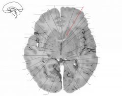
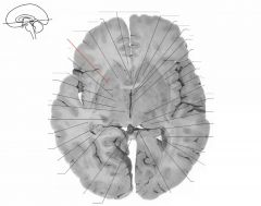
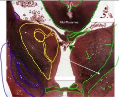
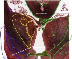
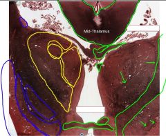
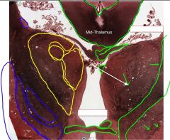
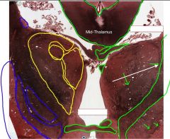
What region of the Corpus Callosum is indicated by the yellow arrow?
|

The Genu
|
|
|
Red arrow.
|

Lateral Nuclear Group of Thalamus.
|
|
|

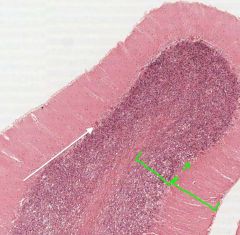
What layer of the Cerebellar Cortex is indicated by the white arrow?
|

The Molecular Layer.
|
|
|
Red arrow. (white matter)
|

Internal Medullary Lamina of the Thalamus.
|
|
|

What fibres pass through the Posterior Limb of the Internal Capsule?
|
It contains:
1) The Superior Thalamic Radiations (VA, VL, and VP projections to motor and sensory cortices) 2) Corticospinal projections, 3) Others. (Corticotegmental [yellow box], Parietopontine, and Pallidothalamic* fibres) |
*Connecting the Globus Pallidus Medius to the Thalamus.
|
|
Red arrow.
|

Anterior Horn of the Lateral Ventricle.
|
|
|
Is this (golgi-stained) Cortex Homotypical or Heterotypical?
If Heterotypical, is it Granular or Agranular? |
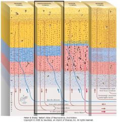
It is Homotypical. You can make out the six different layers
|
(although I have a hard time figuring out which is which...)
|
|
Red arrow. (space)
|

Posterior Horn of the Lateral Ventricle.
|
|
|
What collection of fibres is indicated by the yellow arrow?
|

The Fornix.
|
|
|
Red arrow.
|

Genu of the Corpus Callosum.
|
|
|

Where are all of the Specific Thalamic Nuclei located?
|

They are all in the Lateral Nuclear Group.
|
|
|
Red arrow.
|

Anterior Commissure.
|
|
|
What portion of the Internal Capsule is indicated by the purple arrow?
What fibres does it carry? |

The Sublenticular Limb. It carries:
1) Auditory Radiations (from the MGN to the Temporal Lobe) and, 2) Temporopontine Radiations. |
|
|
Red arrow. (white matter)
|

Fimbria of the Fornix (or, alternatively, of the Hippocampus)
|
|
|
|
What is the distinguishing feature of a Heterotypical Agranular Cortex?
|
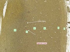
Agranular Cortices are typically Primary Motor Cortex. They can be distinguished by the Giant Pyramidal Cells of Betz, in Layer V, which can be up to 100 microns across, and are much bigger than all other neurons.
|
|
|
Red arrow. (look at the symmetrical structure)
|

Column of the Fornix.
|
|
|
What portion of the Fornix is indicated by the white arrow?
|

The Body.
|
|
|
Red arrow.
|

Anterior Limb of the Internal Capsule.
|
|
|

White arrow.
|

Periaqueductal Gray Matter.
|
|
|
Red arrow. (white matter)
|

Posterior Limb of the Internal Capsule.
|
|
|
What types of fibres are carried by the Internal Capsule?
|
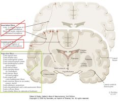
Projection Fibres.
(as opposed to Commissural Fibres, and Association Fibres) |
|
|
Red arrow.
|

Head of the Caudate Nucleus.
|
|
|
|
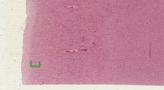
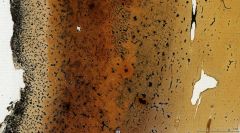
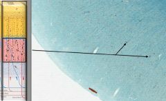
How can you identify Heterotypical Granular Cortex from the Primary Visual Cortex?
|
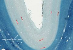
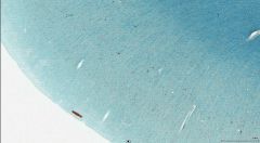
The Visual Cortex is the only cortex with the Stria Gennari (shown in iron hematoxylin, which stains axons blue).
The Stria arrises from the Optic Radiations off of the Lateral Geniculate Body of the Thalamus. |
The Stria is named for Francesco Gennari, an Italian medical student and keener.
|
|

Red arrow.
|

Tail of the Caudate Nucleus.
|
|
|

What portion of the Fornix is indicated by the white arrow?
|

One of the Columns.
|
|
|
Red arrow.
|

Putamen
|
|
|

What is the name of the white matter surrounding the Thalamus?
|
The External Medullary Lamina.
|
|
|

Red arrow.
|

Globus Pallidus, External Segment
|
|
|
|
How can you distinguish the External and Extreme Capsules?
What fibres do they carry? |
The External Capsule is located between the Putamen and the Claustrum, and the Extreme Capsule is between the Claustrum and the Insular Cortex.
Not sure: Some sources describe both as having Association Fibres (between two areas of the cortex), and others suggesting that the External Capsule includes Projection Fibres to the Putamen. |
|
|
Red arrow.
|

Globus Pallidus, Internal Segment.
|
|
|
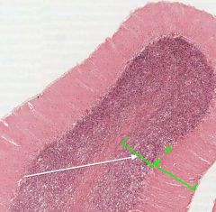
Is this Cortex Homotypical or Heterotypical?
If Heterotypical, is it Granular or Agranular? |
This is an Agranular Heterotypical Cortex. You can tell it's a primary motor cortex by the presence of Giant Pyramidal Cells of Betz, indicated.
|
|
|
Red arrow.
|

Claustrum
|
|
|

What septum is indicated by the white arrow?
|

The Septum Pellucidum.
|
|
|

Red arrow.
|

Claustrum
|
|
|

White arrow.
|
Medial Lemniscus
|
|
|
Red arrow. (white matter)
|

External Capsule
|
|
|
|
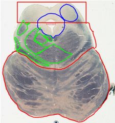
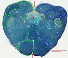
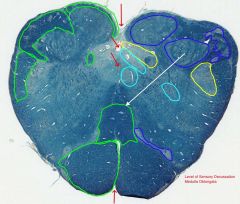
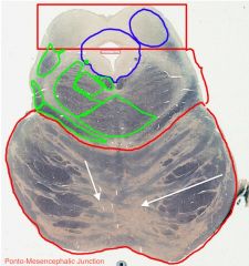
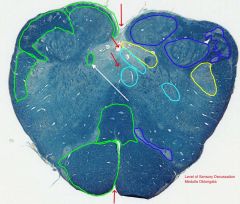
Anatomically, what's the difference between Ganglia, Nuclei, and Cortex?
|
Ganglia: Neuronal cell bodies and dendrites (grey matter) in the PNS.
Nuclei: Grey matter in the CNS that is surrounded by white matter. (blue arrows) Cortex: Grey matter in the outer layer of the CNS. (red arrow) |
|
|
Red arrow.
|
Extreme Capsule
|
|
|

Is this Cortex Homotypical or Heterotypical?
If Heterotypical, is it Granular or Agranular? |
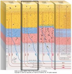
It is Homotypical, i.e. all 6 layers are ("more or less") readily distinguishable.
Starting with the Molecular Layer most superficially, try to count all six layers by changes in the density of the cell bodies. Layers 2 and 4, the Granular layers, have more densely packed cell bodies. Go back to the question slide and examine the layers within the red bracket until you're comfortable identifying all six layers. |
|
|

Red arrow.
|

Posterior Horn of Lateral Ventricle.
|
|
|

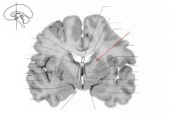
What gyrus is indicated by the blue arrow?
|

The Parahippocampal Gyrus.
|
|
|

Red arrow.
|
Head of Caudate Nucleus.
|
|
|
What types of fibres are found in the External Medullary Lamina?
|

It contains:
1. Thalamocortical, and 2. Corticothalamic Fibres. So, fibres to and from the cortex and thalamus. |
|
|

Red arrow.
|

Tail of Caudate Nucleus.
|
|
|
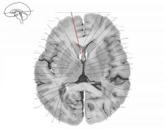
What structure is indicated by the purple arrow?
What is its overall shape? |
The Head and Tail of the Caudate Nucleus.
It is a C-shaped structure that wraps around the Lentiform Nucleus and tapers to its tail towards the Amygdala. |
|
|

Red arrow.
|

Body of Corpus Callosum.
|
|
|
What structure is indicated by the blue arrow?
|

The Uncus
(a prominent bump on the Parahyppocampal Gyrus) |
|
|
Red arrow.
|

Anterior Limb of the Internal Capsule
|
|
|

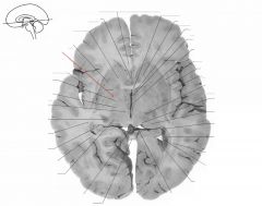
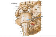
White arrow.
|

Red Nucleus
|
|
|
Red arrow.
|

Head of the Caudate Nucleus
|
|
|
What do the Caudate and the Putamen form together?
|

The "Neostriatum," (Striatum)
as opposed to the "Paleostriatum" (Pallidum), which refers to the Globus Pallidus. |
|
|
Red arrow.
|

Putamen
|
|
|
What structure is indicated by the blue arrow?
|

A Mamillary Body
|
|
|
Red arrow.
|

Septum Pellucidum
|
|
|
What system is the Anterior Nuclear Group considered a part of?
|
It's considered part of the Limbic System.
|
|
|
Red arrow.
|

Anterior Horn of the Lateral Ventricle
|
|
|
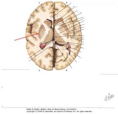
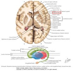
What structure is indicated by the red arrow?
What does it form with the Globus Pallidus? With the Claustrum? With the Caudate Nucleus? |
The Putamen.
With the Globus, it forms the Lentiform Nucleus. With the Caudate, it forms the Neostriatum. All three form the Corpus Striatum. The Claustrum is on its own. It has no special friends. |
|
|
Red arrow.
|

External Capsule
|
|
|

What are the Mamillary Bodies?
|

Two hypothalamic nuclei that receive input from the limbic lobe via the fornix.
|
|
|

Red arrow.
|
Claustrum
|
|
|

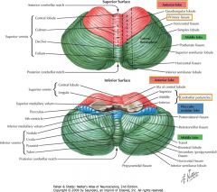
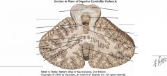
What cerebellar feature is indicated by the white arrow?
|
The Vermis.
|
|
|
Red arrow.
|

Extreme Capsule
|
|
|
What grey matter is indicated by the red arrow?
What is it bordered by? |

The Claustrum.
Bordered medially by the External Capsule, and laterally by the Extreme Capsule. We don't know its function, but it is extensively connected to the cerebral cortex. |
|
|
Red arrow.
|

Body of Corpus Callosum.
|
|
|

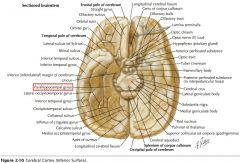
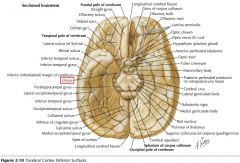
What is the region of cortex immediately in front of and below the Rostrum of the Corpus Callosum?
|
The Septal Area, including the "Parolfactory Gyrus" and the Septal Nuclei.
|
The Septal Area projects to the Hypothalamus and Brainstem.
|
|

Red arrows. (white matter)
|

Anterior Commissure.
|
|
|
What group of nuclei is indicated by the red arrow?
Where does it receive afferents from? Where does it project to? |
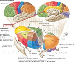
The Anterior Nuclear Group.
It receives afferents from the Mammillary Bodies. Its axons project to the Cingulate Gyrus. |
|
|
Red arrow.
|

Column of Fornix.
|
|
|

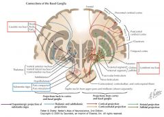
What are the components of the (modern) Basal Ganglia?
They've all got bits showing in this image. |
1. The Caudate Nucleus,
2. Putamen, 3. Globus Pallidus, 4. Subthalamic Nucleus, and 5. Substantia Nigra. Note: does not include the Claustrum. It has no friends. |
|
|
Red arrow.
|

Anterior Limb of Internal Capsule.
|
|
|
What two sulci bound the Cingulate Gyrus?
|
The Cingulate Sulcus, and
The Callosal Sulcus. |
|
|
Red arrow.
|

Head of Caudate Nucleus.
|
|
|

White arrow.
|

Substantia Nigra
|
|
|

Which portion of the Lateral Ventricle is indicated by the white arrow?
|

The Anterior Horn,
which extends into the Frontal Lobe. |
|
|
Red arrow.
|

Globus Pallidus (undifferentiated)
|
|
|
What input does the cortex of the Cingulate Gyrus receive?
|

It receives input fro the Anterior Nucleus of the Thalamus.
|
|
|
Red arrow.
|
Claustrum
|
|
|
Which Thalamic Nucleus is indicated by the yellow arrow?
|

The Medial Dorsal (or Dorsomedial) Nucleus.
|
|
|

Red arrow.
|

External Capsule.
|
|
|

Which portion of the Lateral Ventricle is indicated by the white arrow?
|
The Body.
|
|
|
Red arrow.
|

Extreme Capsule.
|
|
|
What is the anterior portion of the Parahippocampal Gyrus called?
|

It is the Entorhinal Area.
|
|
|
Red arrow.
|

Body of Corpus Callosum.
|
|
|

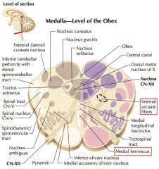
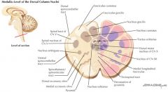
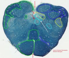
What is the source of the Internal Arcuate Fibers?
|
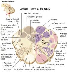
The Internal Arcuate Fibres arise from the Nuclei Cuneatus and Gracilis.
|
|
|

Red arrow.
|

Column of Fornix.
|
|
|
Which portion of the Lateral Ventricle is indicated by the white arrow?
|

The Inferior Horn,
which extends into the Temporal Lobe. |
|
|
Red arrow.
|

(Genu of) Internal Capsule
|
|
|
What is located immediately beneath the Uncus?
|
The Amygdaloid Body, or simply the Amygdala.
|
|
|
Red arrow.
|
Head of Caudate Nucleus.
|
|
|
What is the largest nucleus in the Medial Cell Mass of the Thalamus?
|

The Medial Dorsal (or Dorsomedial) Nucleus
|
|
|
Red arrow.
|

Putamen
|
|
|

Which portion of the Lateral Ventricle is indicated by the white arrow?
|

The Posterior Horn,
which extends into the Occipital Lobe. |
|
|

Red arrow.
|

Globus Pallidus, External Segment
|
|
|
What are the Entorhinal Area, the Uncus, and the Amygdala involved in?
|
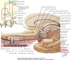
Both olfactory and limbic functions. (Olfactory shown)
|
|
|
Red arrow.
|

Internal Segment of the Globus Pallidus
(or Medial Globus Pallidus) |
|
|

White arrow.
|

Basis Pedunculi (or Crus Cerebri).
|
|
|
Red arrow.
|

Anterior Nucleus of the Thalamus.
|
|
|

Which portion of the Lateral Ventricle is indicated by the white arrow?
|

The Trigone, where the Body, Inferior Horn, and Posterior Horn meet, and there's a little triangle.
|
|
|
Red arrow.
|

Hypothalamus
|
|
|

Where do the Mammillary Nuclei receive inputs from?
Where do they project to? |
They receive inputs from the Hippocampal Formation through the Fornix.
They project to the Anterior Nucleus of the Thalamus (along the Mammillothalamic Tract) (They also project to the Brainstem along the Mammillotegmental Tract) |
|
|
Red arrow.
|

Body of Lateral Ventricle.
|
|
|

Where does the Dorsomedial (or Medial Dorsal) Thalamic Nucleus receive its afferents from?
|
The Amygdala and the Corpus Striatum (among others).
(Amygdala efferent pathways shown) |
|
|
Red arrow.
|

Body of the Corpus Callosum.
|
|
|

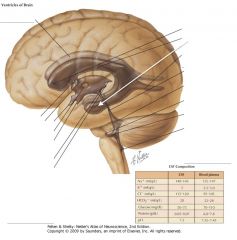
What ventricle is indicated by the white arrow?
|

The Third Ventricle.
|
|
|

Red arrow.
|
Body of the Fornix
|
|
|

What is the pathway connecting the Mammillary Bodies and the Anterior Thalamic Nuclei?
|

The Mammillothalamic Tract.
|
|
|
Red arrow.
|

Posterior Limb of the Internal Capsule
|
|
|
White arrow.
|

Fasciculus Gracilis
|
|
|

Red arrow.
|

Posterior Limb of the Internal Capsule
|
|
|

What space is indicated by the white arrow?
What ventricles does it connect? |

The Interventricular Foramen (of Munro).
It connects a Lateral Ventricle to the Third Ventricle, and there's one on each side. |
|
|

Red arrow.
|

Posterior Limb of the Internal Capsule
|
|
|
What is the source of fibres for the Fornix?
|
It consists of fibres arising from the Hippocampal Formation.
|
|
|

Red arrow.
|

Posterior Limb of the Internal Capsule
|
|
|

Where does the Dorsomedial Nucleus project its axons too?
What is it thought to be involved with? |

It is connected (reciprocally) to the Prefrontal Cortex.
It is thought to be involved in affective states, judgement, and some aspects of memory. |
|
|

Red arrow.
|

Posterior Limb of Internal Capsule
|
|
|

What space is indicated by the white arrow?
What ventricles does it connect? |

The Cerebral Aqueduct (of Sylvius)
It connects the Third and Fourth Ventricles. |
|
|
Red arrow.
|

Body of Caudate Nucleus.
|
|
|
Does the Septum Pellucidum contain any neurons?
|
Yes, it contains both grey and white matter, including some neurons of the septal nuclei.
|
|
|
Red arrow.
|

Putamen
|
|
|
White arrow.
|

Basis Pedunculi (or Crus Cerebri).
|
|
|
Red arrow.
|

Lateral Globus Pallidus
(External Segment of Globus Pallidus) |
|
|

Which ventricle is indicated by the white arrow?
|

The Fourth Ventricle.
|
|
|
Red arrow.
|

Globus Pallidus.
Likely External Segment. |
|
|
|
What is the name of the thin membrane stretching between the Fornix and the Corpus Callosum?
|

The Septum Pellucidum.
|
|
|
Red arrow.
|

Medial Globus Pallidus
(Internal Segment of Globus Pallidus) |
|
|

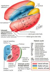
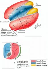
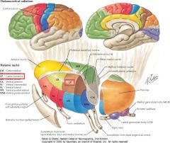
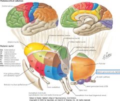
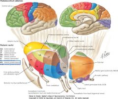
What are the two tiers of the Thalamic Lateral Nuclear Group?
Which tier contains Specific Nuclei? |

The two tiers are the:
1) Dorsal (LD, LP, and Pulvinar), and 2) Ventral (VA, VL, VI, VL, VPM, VPL, MGB, LGB) tiers. The Ventral Tier consists of specific nuclei. |
|
|
Red arrow.
|

Lateral Nuclear Group of Thalamus.
(Ventral Anterior Nucleus) |
|
|

How is the Fourth Ventricle continuous with the Subarachnoid Space?
|
Through the two lateral Foramina of Luschka, and the medial Foramen of Magendi.
|
|
|
Red arrow.
|

Anterior Nucleus of Thalamus.
|
|
|

What is the posterior portion of the Fornix called?
|

The Crura (legs).
(s. Crus) |
|
|
Red arrow.
|

Dorsomedial Nucleus.
|
|
|

What sulcus is indicated by the blue arrow?
|

The Lateral Sulcus (or Sylvian Fissure)
|
|
|

Red arrow. (white matter)
|

Internal Medullary Lamina of the Thalamus.
(Mammillothalamic Tract) |
The Internal Medullary Lamina (IML) separates the Dorsomedial Nucleus from the Lateral Group. As the Mammillothalamic Tract passes through this portion of the Thalamus, it forms part of the IML.
|
|

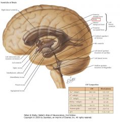
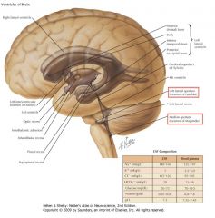
What fills the Ventricles of the Brain? Where is it produced?
|
Cerebrospinal Fluid (CSF).
It's produced in the Choroid Plexus, which can be found in each ventricle. |
|
|

Red arrow.
|

Mammillary Bodies of the Hypothalamus.
|
|
|

What is the middle section of the Fornix called?
|
The Body
|
|
|
Red arrow.
|

Septum Pellucidum.
|
|
|
Which Thalamic Nucleus is indicated by the white arrow?
|
The Lateral Dorsal Nucleus
|
|
|
Red arrow.
|

Body of Lateral Ventricle.
|
|
|

What is the divergent anterior portion of the Fornix?
|

The Columns.
|
|
|

Red arrow.
|

Body of Corpus Callosum.
|
|
|
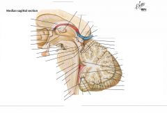
White arrow.
|
Pineal Body.
|
|
|

Red arrow.
|

Dorsomedial Nucleus.
|
|
|
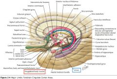
What bundle of axons is indicated by the white arrow?
What does it connect? |
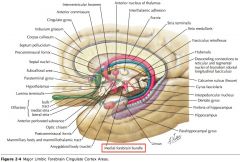
The Medial Forebrain Bundle.
It connects the Septal Area to the Hypothalamus and the Brainstem. |
|
|
Red arrow.
|

Body of the Fornix.
|
|
|

Which Thalamic Nucleus is indicated by the white arrow?
|
The Lateral Posterior Nucleus
|
|
|
Red arrow.
|

Posterior Limb of the Internal Capsule.
|
|
|
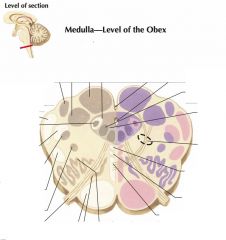
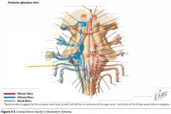
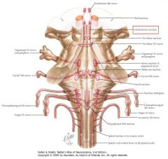
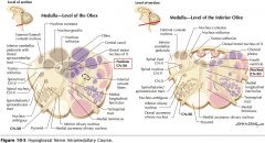
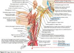
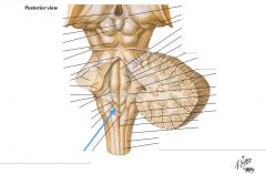
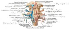
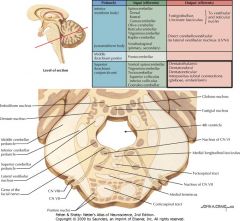
Which cranial nerves exit the Medulla Oblongata?
|
CN IX, X, XI, XII.
|
|
|

Red arrow.
|
Body of Caudate Nucleus.
|
|
|
Which Thalamic Nucleus is indicated by the white arrow?
|
The Pulvinar
(from latin for "cushion") |
|
|
Red arrow.
|

Putamen.
|
|
|

What bundle of axons is indicated by the white arrow?
What do they connect? |
The Mammillothalamic Tract.
It connects the Mammillary bodies and the Anterior Thalamic Nucleus. |
|
|
Red arrow.
|

Dorsomedial Nucleus of Thalamus
|
|
|

White arrow.
|

The Pulvinar Nuclei.
|
|
|

What (purple) structure is indicated by the white arrow?
|

The Hippocampus (Hippocampal Gyrus).
|
|
|

Red arrow.
|

Dorsomedial Nucleus.
|
|
|
Which Thalamic Nucleus is indicated by the white arrow?
|
The Ventral Anterior Nucleus.
|
|
|
Red arrow.
|

Lateral Nuclear Group of Thalamus.
(Ventral Lateral Nucleus) |
|
|
What (blue) structure is indicated by the white arrow?
|

The Dentate Gyrus.
|
|
|

Red arrow. (white matter)
|

Internal Medullary Lamina of Thalamus.
|
|
|

What are the superior and inferior extents of the spinal cord?
|

Superior: the Foramen Magnum.
Inferior: Interspace between L-1 and L-2 vertebrae. |
|
|
Red arrow.
|

Body of Lateral Ventricle.
|
|
|
What region is indicated by the white arrow?
|
The Hippocampus (Hippocampal Gyrus)
|
|
|
Red arrow.
|

Body of Corpus Callosum.
|
|
|

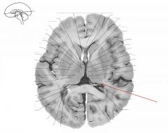
Which Thalamic Nucleus is indicated by the white arrow?
|

The Lateral subdivision of the Ventral Posterior Nucleus
(or Ventroposterolateral nucleus) |
|
|
Red arrow.
|

Body of Fornix.
|
|
|
What structure is indicated by the white arrow?
|

The Dentate Gyrus
|
|
|

Red arrow.
|

Posterior Limb of Internal Capsule.
|
|
|

White arrow.
|

Medial Geniculate Body (or Nucleus).
|
|
|
What is the most anterior portion of the Hippocampus called?
|
The Pes Hippocampi, because it looks slightly like a cat's paw.
|
|
|

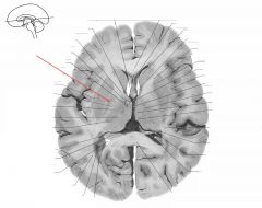
Which Thalamic Nucleus is indicated by the white arrow?
|

The Medial subdivision of the Ventral Posterior Nucleus.
(or, the Ventral Posteromedial) |
|
|

Red arrow.
|

Body of the Caudate Nucleus.
|
|
|

How is the cortex of the Hippocampus and the Dentate Gyrus different from the Neocortex?
|
They consist of only 3 cell layers, instead of the Neocortex's six.
|
|
|
Red arrow.
|

Body of the Caudate Nucleus.
|
|
|
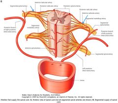
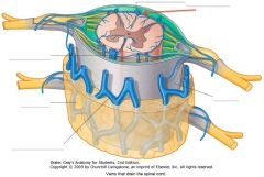
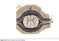
What artery is indicated by the blue arrow?
|

The Anterior Spinal Artery.
|
|
|
Red arrow.
|

Putamen.
|
|
|
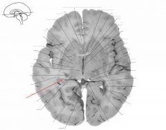
What collection of fibres is indicated by the white arrow?
|
The Fimbria of the Fornix (also of the Hippocampus).
|
|
|
Red arrow.
|

Dorsomedial Nucleus of Thalamus.
|
|
|

Which Thalamic Nucleus is indicated by the white arrow?
|
The Ventral Lateral Nucleus.
|
(The Ventral Intermedial is not distinguished in the lab manual)
|
|
Red arrow.
|
Lateral Nuclear Group
(Ventral Posterolateral Nucleus) |
|
|
White arrow.
|

Lateral Geniculate Body (or Nucleus).
|
|
|

Red arrow.
|

Internal Medullary Lamina.
|
|
|

Which Thalamic Nucleus is indicated by the blue arrow?
|

The Reticular Nucleus,
located between the external medullary lamina and the Internal Capsule. |
|
|

Red arrow.
|

Body of the Lateral Ventricle.
|
|
|
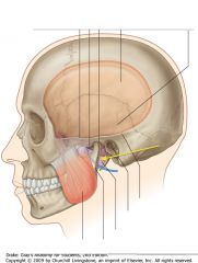
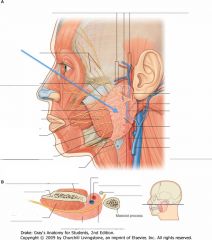
What is the pink region indicated by the yellow area?
|
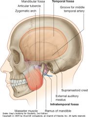
The Infratemporal Fossa
|
|
|

Which Thalamic Nucleus is indicated by the blue arrow?
|
Medial Geniculate Body
(From latin: "genu" knee) |
|
|

White arrow.
|

Corpus Callosum
|
|
|
What input does the Ventral Posterolateral Nucleus receive?
|
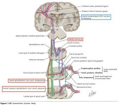
It receives somatosensory pathways from the Medial Lemniscal and Spinothalamic pathways.
|
|
|

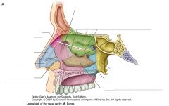
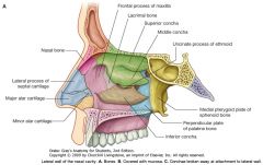
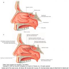
What is the skeleton of the nose composed of?
|

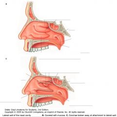
The skeleton of the external nose is largely cartilaginous, except for the Nasal Bones.
|
|
|

What input does the Medial division of the Ventral Posterior Thalamic Nucleus receive?
(The Ventral Posteromedial Nucleus) Where does it project to? |
Trigeminothalamic Pathways.
It projects to the Postcentral Gyrus. |
It also receives the taste pathways from the Rostral Nucleus Solitarus, and projects to the Sensory Cortex. (14.11, Netter Neuroanatomy)
|
|

White arrow.
|

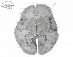
Crus of the Fornix.
|
|
|
Where does the Ventral Anterior Nucleus receive input from?
What region of the Neocortex is it connected to? |
The Globus Pallidus and the Substantia Nigra.
It is (reciprocally) connected to the Frontal Lobe, particularly the Motor Cortex. |
|
|
|
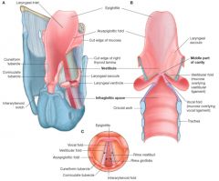
What is the space between the laryngeal inlet and the vestibular folds?
|

The Vestibule
|
|
|

Where does the Ventral Lateral Thalamic Nucleus receive its inputs from?
What regions of the Neocortex is it connected to? |

It receives input from the Globus Pallidus and the Cerebellum (Dentate Nucleus, not shown).
It is connected to motor and pre-motor regions of the Frontal Lobe. |
It also receives input from the Substantia Nigra.
|
|
White arrow.
|

Column of the Fornix.
|
|
|
Where does the Lateral Geniculate Body (LGB) receive input from?
What part of the Neocortex is it connected to? |
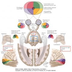
It receives visual input via the Optic Tract.
It projects to the Visual Cortex in the ipsilateral Occipital Lobe. |
|
|
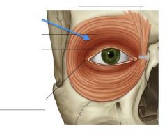
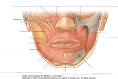
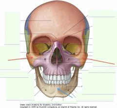
What muscle is indicated by the blue arrow?
|

Orbicularis Oculi
|
|
|

Where does the Medial Geniculate Body receive input from?
What area of the Neocortex does it connect with? |
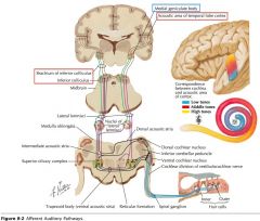
It receives auditory input via the Inferior Colliculus.
It's connected to the Auditory Cortex in the Temporal Lobe. |
|
|
White arrow.
|

Internal Capsule.
|
|
|

What inputs does the Reticular Nucleus receive?
Where does it project to? |
It receives collateral fibres of Thalamocortical and Corticothalamic fibres.
It only projects to other Thalamic Nuclei, regulating their functions. (it is the only thalamic nucleus to not project to the Cortex) |
|
|

What bones make up the Vault of the skull?
|

1. Frontal Bone
2. Parietal Bones (separated by Sagittal Suture). 3. Occipital Bone (singular) 4. Temporal Bones 5. Sphenoid Bone. |
|
|

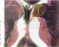
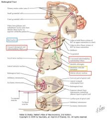
What Thalamic Nuclei are indicated by the white arrows?
Where do they receive inputs from? What cortices do they connect to? |
The Intralaminar Nuclei.
They receive input from a variety of sources, including the brainstem Reticular Formation, and are connected to the Frontal and Parietal Cortices. They are thought to influence alertness and levels of consciousness. |
|
|

White arrow.
|

Anterior Commissure.
|
|
|

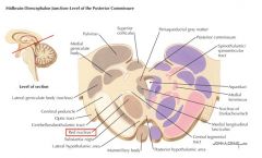
What structure is indicated by the green arrow?
What part of the Diencephalon is it a part of? |

The Pineal Body.
It's part of the Epithalamus. |
|
|
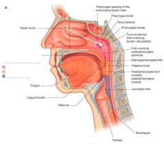
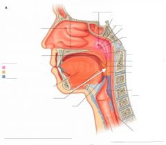
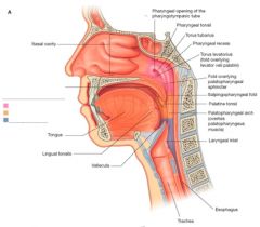
What subdivision of the Pharynx is indicated by the white arrow?
|

The Nasopharynx
|
|
|
What is the Pineal Body involved in?
|
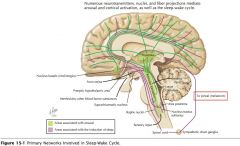
It secretes melatonin, involved in regulating sleep-wakefulness cycles, as well as gonadal maturation.
|
|
|

White arrow.
|

Third Ventricle.
|
|
|
Where do the Habenular Nuclei receive input from?
|
They receive input from the Septal Nuclei via the Stria Medullaris Thalami.
|
|
|

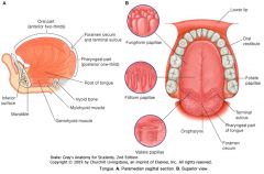
What feature of the tongue is indicated by the white arrow?
What boundary do they mark? |

Vallate Papillae
The 8-12 vallate papillae mark the juncture between the oral anterior 2/3 and the posterior pharyngeal 1/3. |
|
|
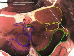
What nucleus is indicated by the white arrow? What section of the Diencephalon is it a part of?
What activity is it involved with? |
The Subthalamic Nucleus, of the Subthalamus.
It is involved in motor control. |
Lesions in the Subthalamic Nucleus can produce Hemiballismus.
|
|

White arrow.
|

Medial Segment of Globus Pallidus.
|
|
|
What section of the Diencephalon is indicated by the white arrow?
(all the coloured bits) Broadly, what is it involved in? |

The Hypothalamus.
It is involved with the control of visceral function and the maintenance of homeostasis, while being intimately associated with the Limbic System. |
|
|

White arrow.
|

Spinal Lemniscus.
|
|
|

White arrow.
|

Lateral Segment of Globus Pallidus.
|
|
|
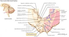
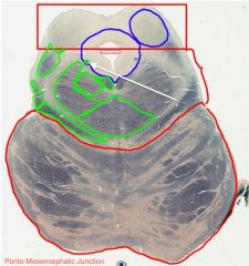
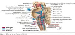
Where does the Trochlear Nucleus project to?
|

Motor neurons in the Trochlear Nucleus give rise to axons which arch dorsally and caudally, around the periaqueductal grey, then cross and exit caudal to the Inferior Colliculus, to go and innervate the Superior Oblique muscle of the contralateral eye.*
|
*Note, CN IV is the only CN which exits the Brainstem dorsally.
|
|
White arrow.
|

Putamen.
|
|
|
What layer of the Cerebellar Cortex is indicated by the white arrow?
What type of cells are found in this layer. |

The Granule Cell layer.
2 types: 1) Granule Cells, with axons going to the Molecular layer, and 2) The larger Golgi Cells, with dendrites coming from the Molecular Layer. (There is a much smaller number of Golgi Cells) |
|
|

White arrow.
|
Stria Medullaris Thalami.
|
|
|

What cerebellar fissure is indicated by the white arrow?
|

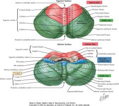
The Primary Fissure
|
|
|

White arrow.
|

Dorsomedial Thalamic Nucleus
|
|
|
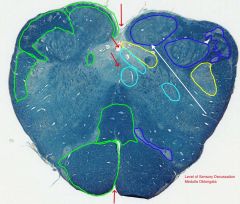
What do the Internal Arcuate Fibers form?
|
They cross at the Sensory Decussation and form the contralateral ascending Medial Lemniscus.
|
|
|
White arrow.
|

Internal Medullary Lamina.
|
|
|
White arrow.
|

Fasciculus Cuneatus
|
|
|

White arrow.
|

Ventral Lateral Thalamic Nucleus, (in the Lateral Thalamic Nuclear Group)
|
|
|

What feature is indicated by the white arrow?
|
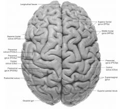
The Longitudinal Fissure.
|
|
|
White arrow.
|

External Medullary Lamina
|
|
|
What cranial nerve is indicated by the orange arrow?
|

CN IX, the Glossopharyngeal Nerve.
|
|
|

White arrow.
|

The Corpus Callosum (specifically the Rostrum).
|
|
|
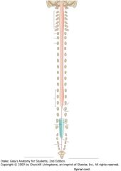
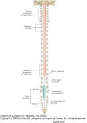
What are the two locations of the spinal cord where the diameter of the cord is increased?
|

The Cervical Enlargement, between C4 and T1, and
The Lumbo-Sacral Enlargement between the L3 and S3. |
|
|

White arrow.
|

The Fornix
|
|
|
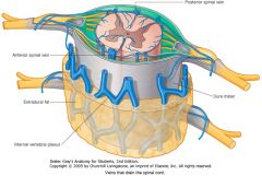
What artery is indicated by the blue arrow?
|

The Left Posterior Spinal Artery.
|
There are 2 posterior spinal arteries, but only one anterior.
|
|

White arrow.
|

Lateral Ventricle.
|
|
|

What nerve is indicated by the blue arrow?
|
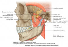
The Mandibular Nerve (V3 of CN V)
|
|
|

White arrow.
|

Choroid Plexus of the Lateral Ventricle
|
|
|
|
What forms the superior border of the nasal cavity?
|

The Anterior Cranial Fossa via the Cribriform Plate of the Ethmoid Bone.
|
|
|

White arrow.
|
Putamen
|
|
|
|
What are the superior and inferior boundaries of the middle part of the Larynx?
|
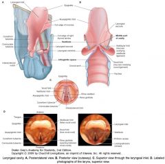
It is very thin:
between the Vestibular Folds (false vocal folds) superiorly, and the Vocal Folds inferiorly. |
|
|

White arrow.
|

Head of Caudate Nucleus.
|
|
|
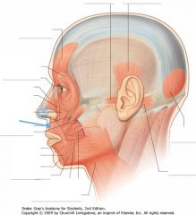
What muscle is indicated by the blue arrow?
|

The Orbicularis Oris
|
|
|

White arrow.
|

Anterior Thalamic Nucleus (of the Anterior Thalamic Nuclear Group).
|
|
|
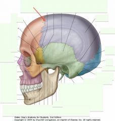
What structure (joint) is indicated by the red arrow?
|
The Coronal Suture
|
|
|
What subdivision of the Pharynx is indicated by the white arrow?
|

The Oropharynx
|
|
|

White arrow.
|

Septum Pellucidum
|
|
|

What feature of the tongue is indicated by the white arrow?
|

The Foramen Caecum, at the apex of the Terminal Sulcus.
|
|
|
White arrow.
|

Lateral Thalamic Nuclear Group
|
|
|

White Arrow.
|

Lateral Lemniscus.
|
|
|

White arrow.
|

Tail of the Caudate Nucleus.
|
|
|
What types of nerve fibres come off of the Oculomotor Nucleus?
|
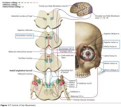
It provides Somatic motor innervation to the extrinsic muscles of the eye.
The parasympathetics derive from the Accessory Oculomotor Nucleus (of Edinger-Westphal). |
|
|

White arrow.
|

External Medullary Lamina.
|
|
|

What types of cells are found in the Molecular Layer?
|

2 types:
1) Stellate Cells: in the upper region of the layer. 2) Basket Cells: larger, and located adjacent to the Purkinje Cells. |
|
|
What cerebellar fissure is indicated by the white arrow?
|

The Horizontal Fissure.
|
|
|

What pathway is the Medial Lemniscus a part of?
|

It is part of the somatosensory pathway, which carries proprioceptive, fine touch and vibration sensation up the Dorsal Column (DC).
|
The DC consists of either the Fasciculus Cuneatus or Gracilis, depending on whether the upper or lower limbs are the source.
|
|

White arrow.
|

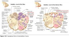
Nucleus Cuneatus (Cuneate Nucleus)
|
|
|
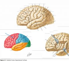
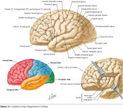
What lobe is indicated by the blue arrow?
|

The Frontal Lobe
|
|
|

What cranial nerve is indicated by the orange arrow?
|

CN X, the Vagus Nerve.
|
|
|

What is the reason for the cervical and lumbosacral enlargements?
|

The areas of enlargement correspond to the origins of the spinal nerves that innervate the upper and lower limbs, respectively.
|
|
|
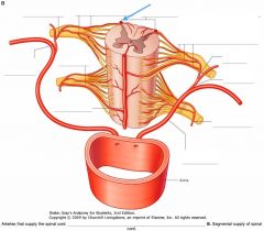
What artery is indicated by the blue arrow?
|
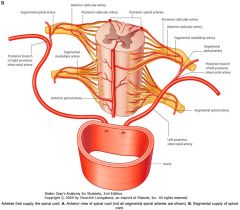
The Right Posterior Spinal Artery.
|
|
|
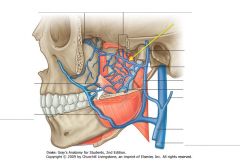
What veins are indicated by the yellow arrow?
|

The Pterygoid Plexus of veins.
|
|
|

What forms the lateral borders of the nasal cavity?
|

The Maxillary Sinuses and the Ethmoidal Sinuses (superior to the cross section shown)
|
|
|
|
What is the space inferior to the Vocal Folds called?
|

The Infraglottic Space
|
|
|
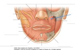
What muscle is indicated by the blue arrow?
What action does it perform? |

Levator Anguli Oris
Elevates the angle (corner) of mouth. |
|
|
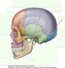
What structure (joint) is indicated by the red arrow?
|

The Lambdoid Suture
|
|
|

What subdivision of the Pharynx is indicated by the white arrow?
|
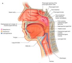
The Laryngopharynx
|
|
|

What is the embryologic origin of the Foramen Caecum?
|
It is a remnant of the embryological development of the thyroid gland.
It closes before birth but remains as a landmark. |
|
|

White Arrow.
|

Nucleus of the Inferior Colliculus.
|
|
|

What is the role of the Nucleus of Edinger-Westphal?
|
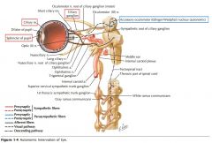
It provides the autonomic innervation of CN III, providing (presynaptic) parasympathetic innervation to the muscles of pupillary constriction.*
|
*(The constrictor pupillae and ciliary muscles.)
|
|

What cell layer is indicated by the white (and green) arrow?
|

The Purkinje Cell Layer.
|
|
|

What are the convolutions on the exterior surface of the cerebellum called?
|

Folia, (Folium, singular)
|
|
|
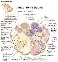
What is the function of the Lateral (Accessory) Cuneate Nucleus?
|
It is the source of the Cuneo-Cerebellar Fibers, which convey proprioceptive information to the Cerebellum from the arm.
It is analogous to the Nucleus Dorsalis (of the spinal cord) for the legs. |
|
|

White arrow.
|

Spinal tract of the Trigeminal Nerve.
|
|
|
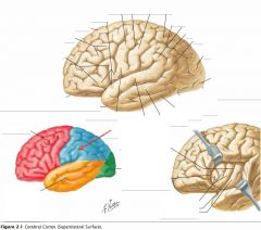
Which lobe is indicated by the red arrow?
|

The Parietal Lobe
|
|
|
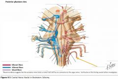
What cranial nerve is indicated by the orange arrow?
|

CN XI, the Spinal Accessory Nerve.
|
|
|
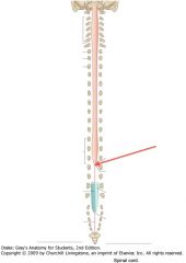
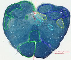
What is the point of the spinal cord indicated by the red arrow?
|

The Conus Medullaris, the point to which the spinal cord tapers.
|
|
|

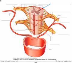
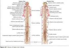
Where is the arterial blood flow from the Spinal Arteries derived from?
|
Most of the arterial blood flowing through the anterior and posterior apinal arteries is derived from the anterior and posterior Radicular Arteries.
|
|
|
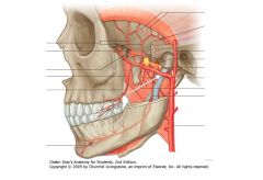
What is the artery indicated by the white arrow?
|

The Maxillary Artery
|
|
|
What forms the inferior border of the nasal cavity?
|
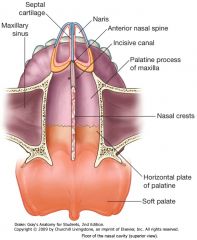
The Hard and Soft Palate
|
|
|

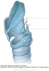
What is the name of the cartilage indicated by the white arrow?
|
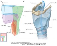
The Thyroid Cartilage.
|
|
|
What muscle is indicated by the blue arrow?
What action does it perform? |
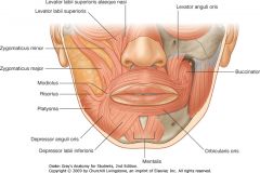
Levator Labii Superioris
Elevates the upper lip. |
|
|

What bone is indicated by the red arrow?
|

A Parietal Bone (paired, separated by the sagittal suture)
|
|
|
What separates the Pharynx from the vertebral column?
|

The pharynx is separated from the posteriorly positioned vertebral column by a thin retropharyngeal space containing loose connective tissue.
|
|
|

What is the name of V-shaped groove immediately posterior to the Vallate Papillae?
|

The Terminal Sulcus.
|
|
|

White Arrow.
|

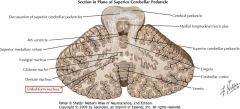
Medial Longitudinal Fasciculus.
|
|
|

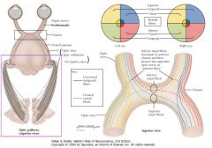
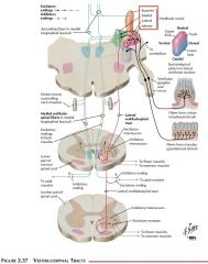
Where does the Nucleus of the Superior Colliculus receive inputs from?
|

A wide variety of sources, including the retina (shown), cerebral cortex and spinal cord.
|
|
|

How can you identify the Purkinje Cell Layer?
|

It is only one cell deep, but the cells are very large and it borders the Molecular and Granular Cell layers.
|
|
|

What is the relationship between the Horizontal Fissure and the Posterolateral Fissure?
|

They are continuous with each other.
The Horizontal Fissure is on the superior surface, and becomes the Posterolateral Fissure on the inferior surface. |
|
|
Where do the Corticospinal Tracts arise?
|

They originate from the Frontal and Parietal lobes.
|
|
|
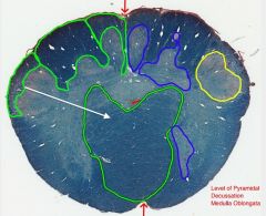
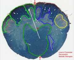
White arrow.
|
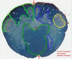
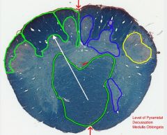
Decussation of the Pyramids.
|
|
|

Which lobe is indicated by the red arrow?
|

The Occipital Lobe
|
|
|

What cranial nerve is indicated by the orange arrow?
|

The Trigeminal Nerve, CN V
|
|
|
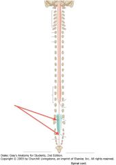
What is the structure indicated by the red arrows?
|
The Filum Terminale
|
|
|
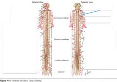
What artery is indicated by the blue arrow?
What vertebral areas does it supply? |
Right Vertebral Artery.
The vertebral arteries only supplies the upper cervical cord. |
|
|

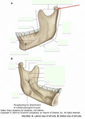
What part of the mandible is indicated by the red arrow?
What is indicated by the blue arrow? |

Red: Ramus
Blue: Body |
|
|
|
What defines the posterior border of the Nasal Cavity?
|

The Nasopharynx
|
|
|

What is the point on the Thyroid Cartilage indicated by the white arrow called?
|

The Laryngeal Prominence.
|
|
|

What muscle is indicated by the blue arrow?
What is it important? |

The Buccinator muscle.
It forms the muscular component of the cheek and is used when forcefully expelling air expanding the cheeks. |
|
|

What bone is indicated by the red arrow?
|

The Frontal Bone
|
|
|
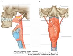
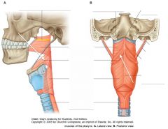
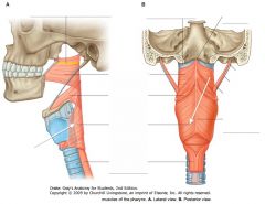
What muscle is indicated by the white arrow(s)?
|
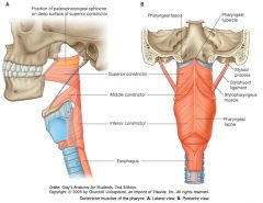
The Superior Constrictor
|
|
|

What is the name of the groove indicated by the white arrow?
|

The Median Groove
|
|
|

White Arrow.
|

Periaqueductal Gray Matter.
|
|
|
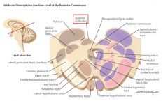
What projections arise in the Nucleus of the Superior Colliculus?
What role does the Superior Colliculus play? |
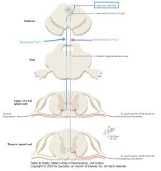
It gives rise to the Tectobulbar and Tectospinal (shown) Tracts.
It is involved in coordinating head and eye movements. |
|
|

What is the role of Purkinje Cells?
|

They provide the sole efferent path from the cerebellar cortex. Their axons project inhibitory signals onto the cells of the Cerebellar Nuclei.
|
|
|

What does the outer surface of the Cerebellum consist of?
|
The Cortex.
It is grey matter consisting of three cell layers. |
|
|
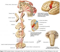
What do the Corticospinal Tracts form in the Medulla?
|
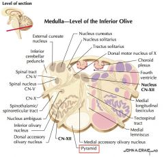
The Pyramids.
|
|
|

White arrow.
|

Fasciculus Gracilis.
|
|
|

Which lobe is indicated by the blue arrow?
|

The Temporal Lobe
|
|
|
What cranial nerve is indicated by the blue arrow?
|

CN VI, the Abducent Nerve
|
|
|

What is the Filum Terminale?
|

It is a connective tissue filament that extends from the tip of the Conus Medullaris to insert on the posterior surface of the coccyx.
|
|
|
What vein is indicated by the red arrow?
|
Posterior Spinal Vein.
|
|
|
What is the ridge indicated by the blue arrow?
|

The Symphysis Menti
|
|
|
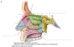
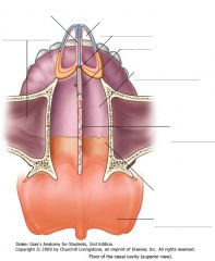
What structure is indicated by the white arrow?
|

The Superior Concha of the ethmoid bone.
|
|
|
What is the name of the cartilage indicated by the white arrow?
|
The Cricoid Cartilage
|
|
|
What gland is indicated by the blue arrow?
|

The Parotid Salivary Gland
|
|
|

What bone is indicated by the red arrow?
|

The Occipital Bone
|
|
|
What muscle is indicated by the white arrow(s)?
|

The Middle Constrictor Muscle.
|
|
|

What type of papillae are found at the location on the tongue indicated by the white arrow?
|

Foliate Papillae
|
|
|

White Arrow.
|

Cerebral Aqueduct
|
|
|
What is the extent of the Periaqueductal Grey matter?
What is it involved in? |

It extends the length of the Cerebral Aqueduct through the Mesencephalon.
It is involved in regulating pain transmission.* |
*Its exact function is not completely understood.
|
|

Where do the Purkinje Cells receive their input?
|

Their dendrites project into the Molecular Layer.
|
|
|

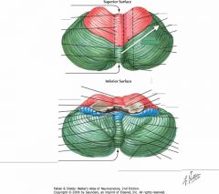
How many lobes are in the cerebellum?
What are they called? |
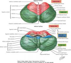
Three lobes:
1) Anterior Lobe 2) Posterior Lobe (or Middle Lobe) 3) Flocculo-Nodular Lobe. |
|
|
How many of the axons of the Corticospinal Tracts cross at the Decussation of the Pyramids?
|

Approximately 85% of the axons cross at the Decussation and descend the spinal cord through the Lateral Corticospinal Tracts.
|
The remainder descend ipsilaterally through the Anterior Corticospinal Tracts.
|
|
White arrow.
|
Fasciculus Cuneatus.
|
|
|

Which lobe is indicated by the blue arrow?
|

The Insula
|
|
|

What is the function of the Glossopharyngeal Nerve (CN IX)?
|
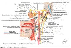
CN IX conveys general and taste sensation from the posterior 1/3 of the tongue, and innervates the pharyngeal muscles and the parotid gland.
|
|
|

What is the collection of nerve fibres indicated by the orange arrows?
|
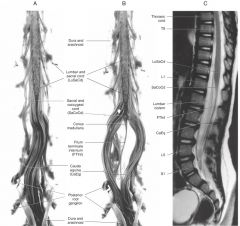
The Cauda Equina.
|
|
|
What vein is indicated by the red arrow?
|

Anterior Spinal Vein
|
|
|

What portion of the Mandible is spanned (roughly) by the red arrows?
|

The Body
|
|
|
What structure is indicated by the white arrow?
|

The Middle Concha of the Ethmoid Bone.
|
|
|
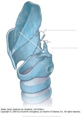
What is the name of the cartilage indicated by the white arrow?
|

The Arytenoid Cartilage
|
|
|

What muscle is indicated by the blue arrow?
|

The Masseter muscle
|
|
|

What bone is indicated by the red arrow?
|

The Sphenoid Bone (the greater wing of the bone is shown, more to see on interior of skull).
|
|
|
What muscle is indicated by the white arrow(s)?
|

The Inferior Constrictor Muscle.
|
|
|

What type of papillae are found at the location indicated by the white arrow?
|

Fungiform Papillae
|
|
|
White Arrow.
|

Fibers of the Trochlear Nerve.
|
|
|

What cells provide inputs to Purkinje Cells?
|

There are four inputs, 2 inhibitory and 2 excitatory.
1) Inhibitory (blue boxes): Basket and Stellate cells from the Molecular Layer. 2) Excitatory (red boxes): Granule cells from the Granule Cell Layer, and Climbing Fibers from the Inferior Olive of the Medulla. |
|
|

What lobe of the cerebellum is indicated by the orange arrows?
What are its boundaries? |
The Anterior Lobe.
It lies between the Primary Fissure and the Cerebellar Peduncles. |
|
|

What is the extent of the Hypoglossal Nerve Nucleus?
|

It extends the length of the Medulla.
|
|
|
White arrow.
|

Nucleus Gracilis
|
|
|

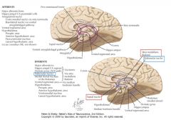
What is the functional role of the Frontal Lobe?
|

The cortex found in this region is associated with motor control, and, in the pre-frontal area, with judgment, the ability to formulate a plan of action in response to circumstances.
(Side 3) |
Also some aspects of memory and smaller functions.
|
|
|
Where does CN IX exit the brainstem?
|

The Glossopharyngeal Nerve exits the Medulla Oblongata just caudal to its junction with the Pons and dorsal to the Inferior Olive.
|
|
|

What is contained in the Cauda Equina?
|

The lumbar, sacral and coccygeal spinal nerves that continue down the vertebral canal, inferior to the Conus Medullaris.
|
|
|
Where do the anterior and posterior Spinal Veins drain?
|
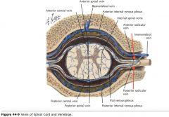
They drain into the anterior and posterior Radicular Veins at each spinal level.
|
|
|

Where does the Red Nucleus* receive input from?
|
It receives input from the cerebellum (shown) and cerebral cortex.
|
*So-named because of its pinkish appearance in a fresh brain.
|
|

What portion of the Ramus is indicated by the red arrow?
What is its function? |

The Coronoid Process.
It provides attachment for the Temporalis Muscle. |
|
|

What bone is indicated by the white arrow?
|
The Inferior Concha (a distinct bone, not part of the Ethmoid)
|
|
|
|
What is the space between the Vocal Folds called?
|

Rima Glottidis
|
|
|

What artery is indicated by the blue arrow?
|

The External Carotid artery.
|
|
|

What bone is indicated by the red arrow?
|

The Temporal Bone (Squamous part).
|
|
|

What nerves form the Pharyngeal Plexus?
|
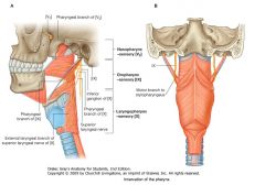
The pharyngeal branch of the Vagus (CN X), and
the pharyngeal branch of the Glossopharyngeal (CN IX). |
Also includes branches from the external laryngeal nerve and from the superior laryngeal branch of the vagus nerve [X].
|
|

What type of papillae are found at the location indicated by the white arrow?
|

Filiform Papillae.
|
|
|
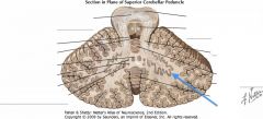
White Arrow.
|

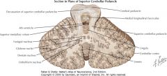
Decussation of the Superior Cerebellar Peduncle.
|
|
|
Which cells provide the strongest excitatory input to the Purkinje Cells?
|
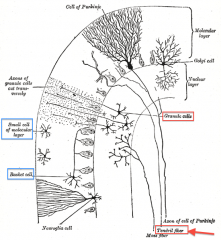
The Climbing Fibres from the Inferior Olive have the strongest excitatory input.
|
|
|
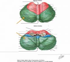
What lobe of the cerebellum is indicated by the orange arrows?
What are its boundaries? |

The Posterior (or Middle) Lobe.
It extends from the Primary Fissure posteriorly until the Posterolateral Fissure. |
|
|
Where does the Hypoglossal Nucleus appear in cross section?
|
It appears close to the midline, as is common for motor nuclei.
|
|
|
White arrow.
|

Sensory Decussation (or Decussation of the Medial Lemniscus)
|
|
|

What is the functional role of the Parietal Lobe?
|

The cortex in this region is associated with somatosensation, and with the integration of sensory information.
|
|
|

Which cranial nerves exit the Pons?
|

CNs V, VI, VII, VIII.
|
|
|
|
How many Spinal Nerves are there?
|
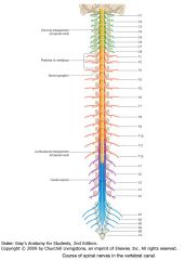
There are 31 pairs of spinal nerves:
C1-C8; T1-T12; L1-L5; S1-S5; Co |
|
|

What artery is indicated by the blue arrow?
|

The Internal Carotid.
|
|
|
What projections come off of the Red Nucleus?
|
It gives rise to (upper motor) projections to the Inferior Olive and the Cervical Spinal Cord.
|
|
|
What is the part of the Ramus indicated by the red arrow?
What are its two parts called? What does it articulate with? |
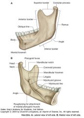
The Condylar Process.
It consists of a Head and Neck (seen in lower view). The Head articulates with the mandibular fossa of the temporal bone to form the temporomandibular joint. |
|
|

How do the Conchae control air flow in the nasal cavity?
|

They direct air flow to ensure contact with the largest surface of cilia and maintain temperature and humidity within the respiratory tract.
|
|
|

What is the structure indicated by the white arrows?
|
The Epiglottis
|
|
|

What is the foramen indicated by the red arrow?
What Cranial Nerve passes through it? |

The Stylomastoid Foramen
The Facial Nerve (CN VII). |
|
|
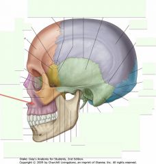
What is the bone indicated by the red arrow?
|

The Maxilla
|
|
|
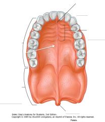
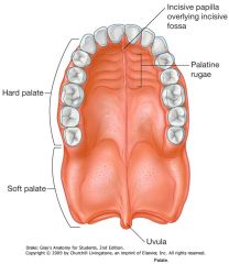
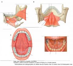
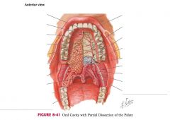
What portion of the palate is indicated by the white arrow?
|

The Hard Palate
|
|
|
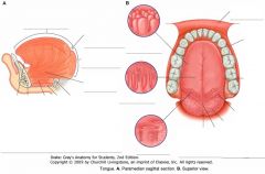
Which type of papillae contain taste receptors?
|

Foliate, Fungiform, and Vallate Papillae.
Filiform Papillae are mechanoreceptive only. |
|
|
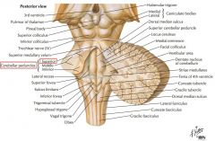
White Arrow.
|

Superior Cerebellar Peduncle.
|
|
|
How many Cerebellar Nuclei are there?
What are their names? |
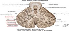
Four pairs. They are:
1) Fastigial, 2) Globose, 3) Emboliform, and 4) Dentate nuclei. |
|
|
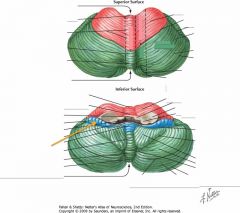
What lobe of the cerebellum is indicated by the orange arrow?
What are its boundaries? |

The Flocculo-Nodular Lobe
It is bordered posteriorly by the Posterolateral Fissure, and anteriorly by the Cerebellar Peduncles. |
|
|
|
How many Vestibular Nuclei are there?
What part of the brainstem are they in? |
Four: 1. Superior, 2. Inferior, 3. Lateral, and 4. Medial.
The Medial and Inferior nuclei are in the Rostral Medulla, and the Superior and Lateral nuclei are in the Caudal Pons. |
|
|

What is the functional role of the Occipital Lobe?
|

The cortex in this region is involved with vision and the processing of visual information.
|
|
|

Which cranial nerves exit the Mesencephalon?
|

CNs III, IV
|
|
|

What, generally, wraps around the spinal cord?
|
The three meningeal layers:
Pia Mater Arachnoid Outer Dura Mater |
|
|
What artery is indicated by the blue arrow?
|

A Vertebral Artery.
|
|
|

What type of nucleus is the Substantia Nigra?
What is it connected to? |
It is a motor nucleus.
It is reciprocally connected to the Corpus Striatum. |
|
|
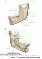
What is the feature indicated by the red arrow called?
|

The Mandibular Notch
|
|
|

What is the name of the space inferior to the Superior Concha?
|
The Superior Meatus
|
|
|

What is the space indicated by the white arrows called?
|

The Vestibule
|
|
|

What branch of the Facial Nerve is indicated by the blue arrow?
What muscles does it innervate? |

The Temporal branch
Frontalis and Orbicularis Oculi. |
|
|

What is the bone indicated by the red arrow?
|

The Zygomatic Bone
|
|
|

What portion of the palate is indicated by the white arrow?
|

The Soft Palate
|
|
|

What is the structure indicated by the white arrow?
|
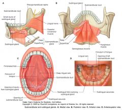
The Frenulum
|
|
|

Which cerebellar nucleus is indicated by the blue arrow?
|

The Fastigial Nucleus.
|
|
|

White Arrows.
|

Corticopontine and Corticospinal Fibers.
|
|
|

Which lobe of the Cerebellum is phylogenetically the oldest?
|

The Flocculo-Nodular Lobe.
|
|
|

What is the posterior surface feature marking the location of the Hypoglossal Nucleus?
|

In the Open Medulla, the Hypoglossal Trigone visibly shows the position.
In the Closed Medulla, the Gracile and Cuneate Nuclei obscure the position of the Hypoglossal Nucleus |
|
|
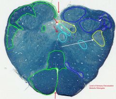
White arrow.
|

Spinal Nucleus of the Trigeminal Nerve
|
|
|

What is the functional role of the Temporal Lobe?
|

The cortex in this region is involved with receiving and processing auditory information, speech and olfaction. Also some involvement with the limbic system.
|
|
|
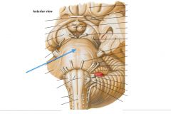
What structure is indicated by the blue arrow?
|

The Basilar Pons (or just pons)
|
|
|

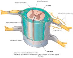
Which meningeal layer is indicated by the red arrow?
|

The inner Pia Mater, which closely invests the surface of the cord.
|
|
|

Where do the Vertebral Arteries enter the cranium?
|

Through the Foramen Magnum.
|
|
|

What happens to the Substantia Nigra during Parkinson's (grossly)?
|
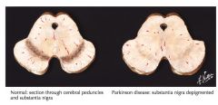
Neurons of the Substantia Nigra undergo degeneration in Parkinson's disease.
|
|
|

Where is the Mandibular Fossa of the temporal bone?
|

See the location of the red arrow.
|
|
|

What is the name of the space inferior to the Middle Concha?
What structures are found in it? |

The Middle Meatus.
The Bulla Ethmoidalis and the Hiatus Semilunaris |
|
|
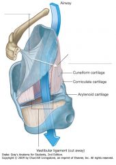
What structure is indicated by the white arrow?
|
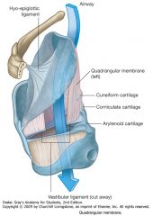
The Quadrangular Membrane
|
|
|

What branches of the Facial Nerve are indicated by the blue arrow?
What muscles do they innervate? |

The Zygomatic Branches.
Orbicularis Oculi |
|
|

What is the structure (i.e. part of a bone) indicated by the red arrow?
|

The Zygomatic Arch (or process) of the Temporal Bone
|
|
|
What bones make up the surface of the Hard Palate?
|

The palatine process of the Maxilla and the horizontal plate of the Palatine bone.
|
|
|
|
What structures lie lateral to the Frenulum, overlying the sublingual gland?
|

The Sublingual Folds
|
|
|

Which cerebellar nucleus is indicated by the blue arrow?
|
The Globose Nuclei
(Globose means, "Ball-shaped") |
|
|
White Arrows.
|

Pontine Nuclei.
|
|
|

What structures are indicated by the orange arrows?
|

They are the Flocculi (s. Flocculus) of the Flocculo-Nodular Lobe.
|
|
|

What two nuclei sit on either side to the Dorsal Vagal Nucleus?
|

Medially the Hypoglossal Nucleus.
Laterally the Solitary Nucleus (and tract). |
|
|

White arrow.
|

Hypoglossal Nucleus
|
|
|

What is the functional role of the Insula?
|

Possibly nociception and visceral function, but not certain.
|
|
|

What does the anterior surface of the Pons consist of?
|

Transversely arranged fibers which arise as axons of neurons in the pontine nuclei.
|
|
|
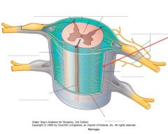
Which meningeal layer is indicated by the red arrow?
Where does it get its name? |

The Arachnoid Mater.
It is so named because of its delicate fibres that look like spider silk. |
|
|
Where do the Internal Carotid Arteries enter the cranium?
|

Through the Carotid Canal
|
|
|
What type of fibres pass through the middle region of the Basis Pedunculi (Crus Cerebri)*?
What fibres pass through the medial and lateral regions? |
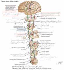
Corticospinal fibres pass through the middle region.
Corticopontine fibres pass through the outer two regions. |
*Don't call it the Cerebral Peduncle. Dr. Rutherford says the Cerebral Peduncle includes the Substantia Nigra and a couple other things, but Crus Cerebri is okay.
|
|

What feature of the Temporal Bone is indicated by the red arrow?
|

The Articular Eminence (or tubercle).
|
|
|
What is the space inferior to the Inferior Concha?
What duct opens into this space? |

The Inferior Meatus
The Nasolacrimal Duct opens into this meatus. |
|
|

What are the structures indicated by the white arrows?
|

The Vocal Folds.
|
|
|

What branches of the Facial Nerve are indicated by the blue arrow?
What muscles do they innervate? |

The Buccal Branches
The Buccinator and Levators Anguli Oris |
|
|

What is the bone indicated by the red arrow?
|

The Nasal Bone
|
|
|

What structure is indicated by the white arrow?
What muscles insert into it? |
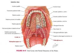
The Palatine Aponeurosis (Tendinous sheet) into which the Tensor and Levator Veli Palatini Muscles insert.
|
|
|
What gland is indicated by the white arrow?
|

The Sublingual Gland.
|
|
|

Which cerebellar nucleus is indicated by the blue arrow?
|
The Emboliform Nucleus.
|
|
|

What is the vernal portion of the Flocculo-Nodular Lobe called?
|
It is the Nodule.
|
|
|
What does the Dorsal Vagal Nucleus innervate?
|
It provides parasympathetic preganglionic fibers that innervate the viscera of the thorax and the abdomen.
(and apparently receives visceral afferents, but we don't go into that) |
|
|
White arrow.
|

Dorsal Motor Nucleus of the Vagus Nerve
|
|
|

What gyrus is indicated by the blue arrow?
|

The Precentral Gyrus.
|
|
|
What structure is indicated by the blue arrow?
|

The Vagal Trigone
|
|
|

Which meningeal layer is indicated by the red arrow?
|

The Dura Mater
|
|
|

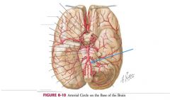
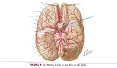
What is the structure represented by the dashed line called?
|

The Circle of Willis.
|
|
|

White Arrow.
|

Transverse Fibers of the Pons.
|
|
|
Where does the Superior Cerebellar Peduncle undergo its decussation?
|
At the level of the caudal midbrain.
|
|
|
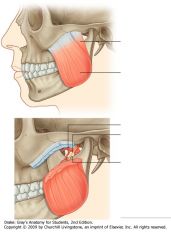
What muscle is indicated in this diagram?
What is it's primary function |
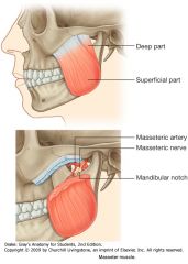
The Masseter Muscle
It elevates the Mandible. |
|
|

What structure on the Nasal Septum is indicated by the white arrow?
|
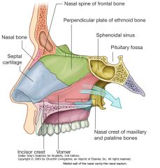
The Septal Cartilage
|
|
|

What structure is indicated by the white arrow?
|

The Vestibular Folds (or False Vocal Folds).
|
|
|

What branches of the Facial Nerve are indicated by the blue arrow?
What muscles do they innervate? |
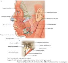
The Marginal Mandibular Branches
Depressors of lower lip |
|
|

What is the foramen indicated by the red arrow? What nerve does it carry?
|

The Mental Foramen.
It carries the V3 branch of Cranial Nerve V (the Mental Nerve). |
|
|

What structure is indicated by the white arrow?
|

The Pharyngotympanic (Auditory) Tube.
|
|
|

On which sublingual structures do the Submandibular Ducts appear?
|

The Sublingual Caruncles (or Papilla).
|
|
|
Which cerebellar nucleus is indicated by the blue arrow?
|
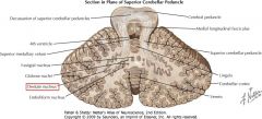
The Dentate Nucleus
|
|
|
How many regions does the Vermis divide into?
|

There are nine:
1) Lingula, 2) Centralis, 3) Culmen, 4) Declive, 5) Folium, 6) Tuber, 7) Pyramis, 8) Uvula, 9) Nodulus. (do not memorize) |
|
|

What is the vertical extent of the Vagal Dorsal Motor Nucleus?
|
The extent of the Medulla, similar to the Hypoglossal Nucleus.
|
|
|
White arrow.
|

Solitary Tract
|
|
|

What gyrus is indicated by the blue arrow?
|

The Superior Frontal Gyrus
|
|
|

What structure is indicated by the blue arrow?
|

The Hypoglossal Trigone
|
|
|

What is found in the space between the Arachnoid Mater and the Pia Mater.
|

That space, called the Subarachnoid Space, is filled with cerebrospinal fluid (CSF)
|
|
|

What artery is indicated by the blue arrow?
|

The Anterior Cerebral Artery.
|
|
|

What muscle is shown in this diagram?
Where does it form attachment? |
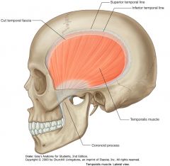
The Temporalis Muscle
Superiorly on the Temporal Fossa; inferiorly on the Coronoid Process of the Mandible. |
|
|

White Arrow.
|

Medial Lemniscus.
|
|
|
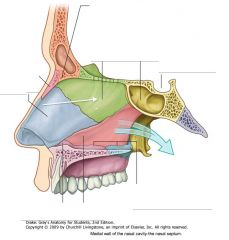
What structure on the Nasal Septum is indicated by the white arrow?
|

The Perpendicular Plate of the Ethmoid
|
|
|

What is the space between the Vocal Folds and the Vestibular Folds called?
|

The Ventricle of the Larynx.
|
|
|
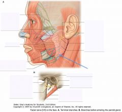
What branches of the Facial Nerve are indicated by the blue arrow?
What muscles do they innervate? |

The Cervical Branches
The Platysma (a muscle of facial expression that extends past the clavicle) |
|
|

What are the foramen indicated by the white arrows?
What do they carry? |

The Supraorbital Foramen, or notches.
Carry the V1 branch of CN V, the Supraorbital Nerve (and the associated vessels). |
|
|
What roles do the muscles of the soft palate perform?
|

Tensor: Stiffen the soft palate
Levator Veli Palatini: Elevate the soft palate to prevent the movement of food into the nasopharynx during swallowing. |
|
|
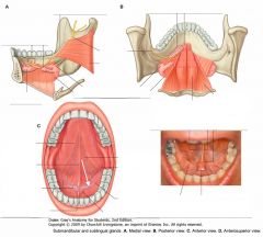
What duct is indicated by the white arrow?
Where does it open? |

The Submandibular Duct.
It opens on the Sublingual Papilla (Caruncle) |
|
|
What is the role of the cerebellar nuclei?
|
Almost all of the output from the cerebellum originates from cells in these nuclei.
|
|
|
What fissure is indicated by the blue arrow?
|
The Primary Fissure.
(Between the Culmen and the Declive) |
|
|

How can you locate the Nucleus Ambiguus in cross section?
|
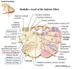
It's difficult to visualize, but two landmarks are that it's medial to the Spinal Tract of CN V, and dorsal to the Dorsal Accessory Olivary Nucleus, in the middle of the Medulla.
|
|
|

White arrow.
|

Medial Accessory Olivary Nucleus.
|
|
|

What gyrus is indicated by the blue arrow?
|
The Middle Frontal Gyrus
|
|
|

What structure is indicated by the blue arrow?
|

The Decussation of the Pyramids
|
|
|
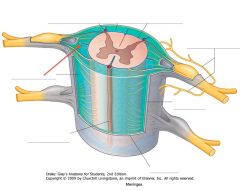
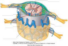
What structure is indicated by the red arrow (the darker blue bit)?
|

A Denticulate Ligament.
21 pairs of these ligaments provide attachment between the inner cord and the outer Dura Mater. |
They form roughly triangular projections of pia mater, with the base on the medial side, and the apex on the lateral side.
|
|

What artery is indicated by the blue arrow?
|

The (right) Middle Cerebral Artery.
|
|
|
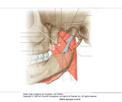
What muscle is indicated by the white arrow?
Where does it attach? |

The Medial Pterygoid Muscle
The deep head attaches to the medial surface of the lateral plate of the pterygoid process. The superficial head originates from the tuberosity of the maxilla. The two heads join to attach to the roughened medial surface of the ramus of the mandible (near the angle of the mandible). |
Both deep and superficial heads attach to the adjacent pyramidal process of the palatine bone.
|
|

White Arrow.
|

Spinal Lemniscus.
|
|
|

What structure on the Nasal Septum is indicated by the white arrow?
|

The Vomer
|
|
|
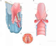
What is reflexively closed if a foreign object becomes trapped superior t the Vestibule of the Larynx?
|

The Rima Glottidis, the triangular opening between the two Vocal Folds.
|
|
|

What branche of the Facial Nerve is indicated by the blue arrows?
What muscles does it innervate? |

The Postererior Auricular nerve
The Occipitalis. |
|
|

What are the foramen indicated by the white arrows?
What do they carry? |
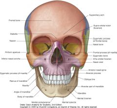
Infra-Orbital Foramen.
Carry the Infra-Orbital Nerve and vessels (V2 branch of CN V) |
|
|

What muscle is indicated by the white arrow?
|

The Tensor Veli Palatini, which tenses the soft palate.
|
|
|

What nerve is indicated by the white arrow?
|

The Lingual Nerve.
|
|
|

What are the inputs to the Cerebellar Cortex?
|
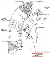
Only 2 kinds of fibres:
1) Climbing Fibres: from the Inferior Olive. 2) Mossy Fibres: from many places (cerebral cortex, vestibular nerve & nuclei, spinal cord, reticular formation, and cerebellar nuclei) |
|
|

White Arrow.
|

Trochlear Nucleus
|
|
|
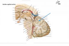
Which lobe is indicated by the blue arrow?
|

The Anterior Lobe, distinguished by its location anterior to the Primary Fissure.
|
|
|

What sort of neurons are found in the Nucleus Ambiguus?
|
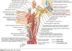
It is the source for lower motor neurons of CN IX, X, and XI.
It provides: CN IX: Somatic efferent to Stylopharyngeus muscle. CN X: Somatic efferent innervation to the muscles of the soft palate, the pharynx, the larynx and the upper third of the esophagus. It controls speaking and swallowing. CN XI: Fibers of CN XI from the Nucleus Ambiguus join X to innervate the larynx (often actually just considered part of X) |
It also provides parasympathetic preganglionic innervation to the heart.
|
|

White arrow.
|

Pyrimidal Tract.
|
|
|

What gyrus is indicated by the blue arrow?
|

The Inferior Frontal Gyrus
|
|
|
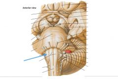
What structure is indicated by the blue arrow?
|

The Olive
|
|
|

What is contained in the "grey matter" of the spinal cord?
|
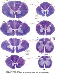
It contains collections of neurons and the synapses formed on them.
|
|
|

Where does the Middle Cerebral Artery supply?
|

It supplies the lateral surfaces of the frontal, parietal and upper portions of the temporal lobes, and the anterior region of the lateral surface of the occipital lobe.
|
|
|
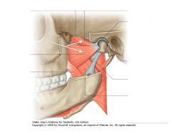
What muscle is indicated by the white arrows?
What action does it perform on the mandible? |

The Lateral Pterygoid Muscle.
The two heads act together to protrude the jaw. When the right and left act independently, they can produce a side to side movement of the jaw. |
|
|
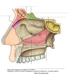
What structure is indicated by the white arrow?
|
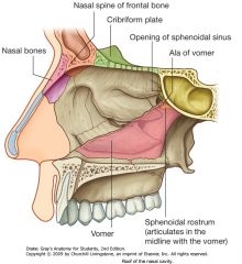
The Frontal Sinus
|
|
|
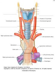
What cranial nerve supplies all of the efferent innervation of the intrinsic muscles of the Larynx?
|

The Vagus (CN X)
|
|
|
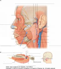
What structure is indicated by the blue arrow?
What muscle does it pierce? |
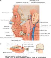
The Parotid Duct.
It crosses the Masseter muscle and pierces the Buccinator muscle. |
|
|

What are the foramen indicated by the red arrows?
What do they carry? |

The Mental Foramen.
Carry the Mental Nerve and vessels (V3 of CN V) |
|
|

What muscle is indicated by the white arrow?
|
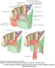
The Levator Veli Palatini, which elevates the soft palate.
|
|
|
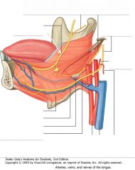
What nerve is indicated by the white arrow?
|

The Lingual Nerve
|
|
|
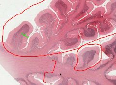
What cells do the Mossy Fibres synapse with?
|

The Granule Cells.
They form complex arrangements called "Rosettes." |
|
|
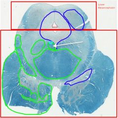
White Arrow.
|

Medial Longitudinal Fasciulus.
|
|
|
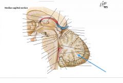
Which lobe is indicated by the blue arrow?
|

The Posterior (or Middle) Lobe, distinguished by its location dorsal to the Primary Fissure.
|
|
|
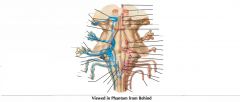
What is the vertical extent of the Nucleus Ambiguus?
|
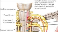
It extends rostrally to just below the Pontomedullary Junction, and caudally to the level of the Foramen Magnum.
|
|
|
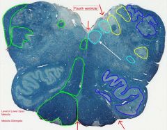
White arrow.
|

Nucleus of the Hypoglossal Nerve
|
|
|

What gyrus is indicated by the blue arrow?
|

The Postcentral Gyrus
|
|
|

What structure is indicated by the blue arrow?
|
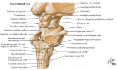
An Inferior Cerebellar Peduncle.
It's located adjacent to the fourth ventricle and projects into the cerebellum. |
|
|

What is the "white matter" of the spinal cord consist of?
How does it appear on slides? |

It consists of myelinated axons which ascend and descend the cord.
Because the myelin soaks up the stain, the white matter stains purple. |
|
|
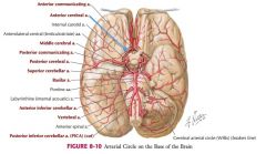
Where does the the Anterior Cerebral Artery supply?
|

It supplies the medial and superior surfaces of the frontal and parietal lobes.
|
|
|

What artery is indicated by the white arrow?
What is it a branch of? |

Inferior Alveolar artery.
A branch of the Maxillary Artery. |
|
|
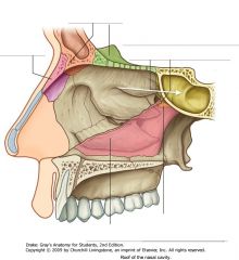
What structure is indicated by the white arrow?
|

The Sphenoidal Sinus
|
|
|
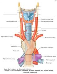
What nerves are indicated by the white arrows?
What intrinsic laryngeal muscle does it innervate? |
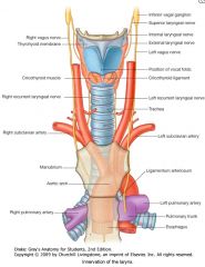
The External Laryngeal Nerve
The Cricothyroid Muscle |
|
|

What is the vessel indicated by the yellow arrow?
What does it branch from? |

The Facial Vein.
The Internal Jugular Vein. |
|
|

What bones are indicated by the red arrows?
|

Zygomatic Bones
|
|
|
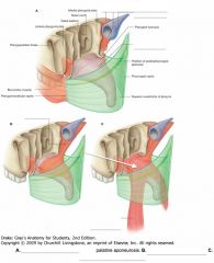
What muscle is indicated by the white arrow?
|

The Palatopharyngeus Muscle.
|
|
|

What nerve carries general sensory information from the anterior 2/3 of the tongue?
|

The Lingual Nerve (from V3)
|
|
|
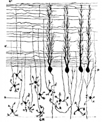
What cells do the Granule Cell axons synapse with?
|
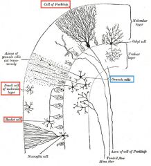
The Granule Cells form excitatory synapses with 3 cell types:
1) Purkinje Cells, 2) Stellate Cells, and 3) Basket Cells. |
|
|

White Arrow.
|

Periaqueductal Gray Matter.
|
|
|

What is the white matter seen on the medial surface of a hemisection called?
|

The Arbor Vitae (tree of life)
|
|
|
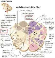
What does the Solitary Nucleus and Tract contain?
|
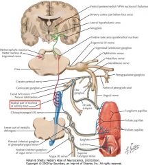
Sensory fibers derived from
CNs VII, IX, and X. Taste fibers synapse on the rostral portion of the solitary nucleus. General Visceral Afferent input terminate on the Caudal portion of the nucleus. |
|
|

White arrow.
|

Dorsal Motor Nucleus of the Vagus Nerve
|
|
|

What gyrus is indicated by the blue arrow?
|

The Supramarginal Gyrus
|
|
|
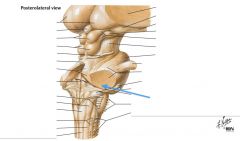
What are the Inferior Cerebellar Peduncles composed of?
|

They consist chiefly, but not exclusively, of pathways afferent to the cerebellum.
|
|
|
|
Which spinal levels have more white matter, the sacral or the thoracic?
|

Thoracic
White matter increases the more caudal on the spinal cord. |
|
|

What artery is indicated by the blue arrow?
|

The Posterior Cerebral Artery.
|
|
|

What artery is indicated by the white arrow?
What is it a branch of? |
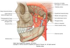
The Middle Meningeal Artery.
A branch of the Maxillary Artery. |
|
|
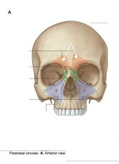
What sinuses are indicated by the white arrows?
|

The Frontal Sinuses.
|
|
|
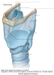
What connects the Thyroid cartilage to the Hyoid Bone?
|

The Thyrohyoid Membrane
|
|
|
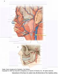
What is the vessel indicated by the yellow arrow?
What muscle on the neck can it be seen to pass over? |

The External Jugular Vein.
The Sternocleidomastoid muscle. |
|
|

What is the juncture (suture) indicated by the dashed circle (by the white arrow)?
What bones does it border? Why is it important clinically? |

The Pterion
The Frontal, Parietal, Temporal, and Sphenoid Bones. It is important because it is known as the weakest point of the skull |
|
|
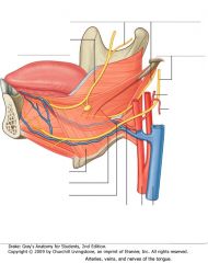
What nerve carries taste information from the anterior 2/3 of the tongue?
|

The Chorda Tympani (from CN VII) steals the fibres from the Lingual Nerve.
|
|
|

What is the source of the Parallel Fibres of the Molecular Layer?
|
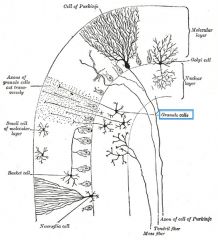
They are formed by divisions of the axons of the Granule Cells.
|
|
|

White Arrow.
|

Cerebral Aqueduct
|
|
|
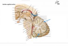
What is found at the terminal ends of the Arbor Vitae?
|

The Folia, which are covered in the gray matter of the cerebellar cortex.
|
|
|

What is the vertical extent of the Solitary Tract and Nucleus?
|
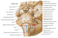
It extends rostrally to just above the level of CN X, and caudally to around the level of the Decussation of the Pyramids
|
|
|

White arrow.
|

Nucleus Cuneatus
|
|
|

What gyrus is indicated by the blue arrow?
|

The Angular Gyrus.
|
|
|

What structure is indicated by the blue arrow?
|

The Nucleus Gracilis (Gracile Tubercle)
|
|
|
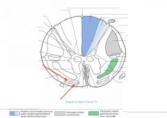
What funiculus are the tracts indicated by the red arrows part of?
|

They're part of the Anterior Funiculus.
|
|
|

What does the Posterior Cerebral Artery supply?
|

It supplies the medial, posterior, and portions of the lateral surfaces of the Occipital Lobe, and the inferior surfaces of the Temporal Love.
|
|
|
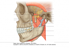
What nerve is indicated by the white arrow?
What is it a branch of? |

The Buccal Nerve
A branch of the Mandibular Nerve (V3) |
|
|

What sinuses are indicated by the white arrows?
|
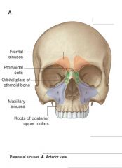
The Maxillary Sinuses
|
|
|

What muscle is indicated by the white arrows?
|

The Anterior belly of the Digastric Muscle
|
|
|

What vessel is indicated by the yellow arrows?
|

The Facial Artery.
|
|
|
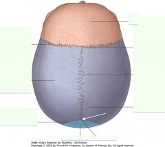
What is the juncture (suture) indicated by the white arrow?
|

The Lambda.
|
|
|

What nerve carries taste and general sensory innervation for the posterior 1/3 of the tongue?
|

The Glossopharyngeal Nerve (CN IX)
|
|
|
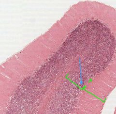
What is the role of the Golgi Cells in the Granule Cell Layer?
|
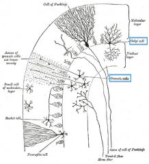
They act as inhibitory interneurons with the Granule Cells.
|
|
|

White Arrow.
|

Substantia Nigra
|
|
|
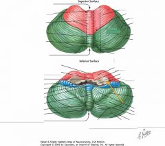
What structure is indicated by the orange arrow?
|
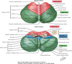
The Middle Cerebellar Peduncle.
|
|
|
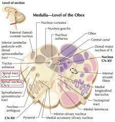
What type of input does the Spinal Trigeminal Tract (and Nucleus) receive?
|
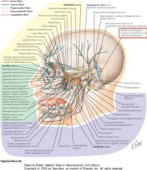
It receives General Afferent fibers from, primarily, CN V, as well as CN IX and X.
|
|
|

White arrow.
|

Lateral (or accessory) Cuneate Nucleus.
|
|
|

What gyrus is indicated by the blue arrow?
|

The Superior Temporal Gyrus.
|
|
|

What structure is indicated by the blue arrow?
|

The Fasciculus Gracilis
|
|
|

What is a Funiculus composed of?
(Three Funiculi are indicated) |

It consists of a group of tracts (bundles of axons), either ascending, descending, or both.
|
|
|

What artery is indicated by the blue arrow?
|

Superior Cerebellar Artery
|
|
|
|
What is the primary function of the Buccal Nerve?
|

It is predominantly a sensory division of V3, but may also carry motor innervation to the lateral pterygoid muscle.
|
|
|

What sinuses are indicated by the white arrows?
|

The Ethmoidal Sinuses (or cells)
|
|
|
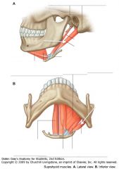
What muscle is indicated by the white arrows?
|

The Posterior Belly of the Digastric Muscle.
|
|
|

What artery is indicated by the yellow arrows?
What is it a terminal branch of? |

The Superficial Temporal Artery.
The External Carotid Artery. |
|
|

What is the juncture (suture) indicated by the white arrow?
|

The Bregma
|
|
|
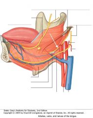
What nerve is indicated by the white arrow?
|

The Glossopharyngeal Nerve (CN IX)
|
|
|

White Arrow.
|

Decussation of the Superior Cerebellar Peduncle.
|
|
|
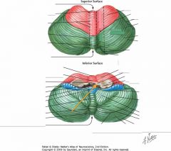
What structure is indicated by the orange arrow?
|

The Superior Cerebellar Peduncle.
|
|
|
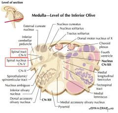
What is the vertical extent of the Spinal Trigeminal Nucleus and Tract?
|
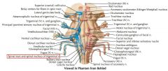
It extends rostrally to the mid-pons, and caudally down into the cervical spine.
|
|
|
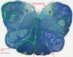
White arrow.
|
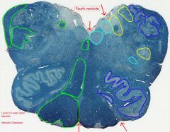
The Solitary Tract
|
|
|

What gyrus is indicated by the blue arrow?
|

The Middle Temporal Gyrus
|
|
|
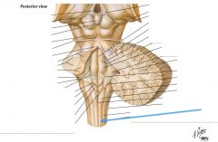
What information is carried by the fasciculi gracilis and cuneatus?
|

They transfer information concerning proprioception, fine touch and vibration sense.
|
|
|

What structure is indicated by the number 2 on the diagram?
|

The Posterior (Dorsal) Horn of the grey matter.
|
|
|
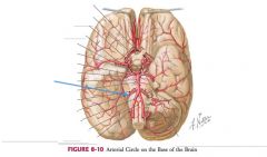
What artery is indicated by the blue arrow?
|

The Anterior Inferior Cerebellar Artery.
|
|
|

What nerve is indicated by the white arrow?
What does it innervate? |

The Lingual Nerve, a branch off of V3.
It carries general and special sensory fibres from the anterior 2/3 of the tongue. |
It carries the sensation to the Chorda tympani (From CN VII)
|
|
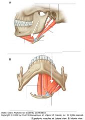
What muscle is indicated by the white arrows?
|

The Mylohyoid Muscle
|
|
|
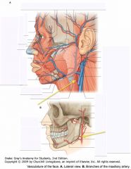
What artery is indicated by the yellow arrows?
|
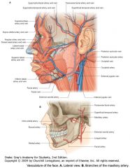
The External Carotid Artery.
|
|
|

What is the suture indicated by the white arrow?
|
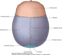
The Sagittal Suture
|
|
|

White Arrow.
|

Basis Pedunculi (or Crus Cerebri).
|
|
|

What structure is indicated by the red arrow?
|
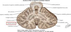
The Dentate Nucleus
|
|
|

What is the role of the Vestibular Nuclei?
|
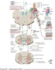
They mediate information about balance and equilibrium, and coordinates head and eye movement in part by projecting axons via the Medial Longitudinal Fasciculus (MLF).
|
|
|

White arrow.
|

The Solitary Nucleus
|
|
|

What gyrus is indicated by the blue arrow?
|

The Inferior Temporal Gyrus.
|
|
|
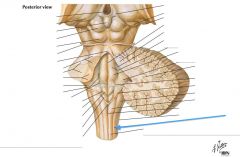
What structure is indicated by the blue arrow?
|

The Fasciculus Cuneatus
|
|
|
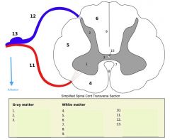
What structure is indicated by the number 1 on the diagram?
|
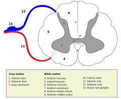
The Anterior (Ventral) Horn of the grey matter.
|
|
|
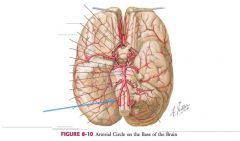
What artery is indicated by the blue arrow?
|
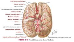
The Posterior Inferior Cerebellar Artery.
|
|
|
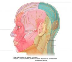
What nerve is indicated by the white arrow?
|
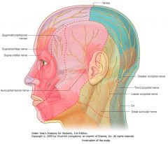
The Auriculotemporal Nerve, a branch off of the Mandibular Nerve (V3).
|
|
|

What bones make up the framework of each orbit?
|

There are seven:
1. Maxilla; 2. Zygomatic; 3. Frontal; 4. Lacrimal; 5. Sphenoid; 6. Ethmoid; 7. Palatine. |
|
|
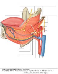
What nerve is indicated by the white arrow?
|

The Hypoglossal Nerve (CN XII)
|
|
|

White Arrows.
|

Location of the Corticopontine Fibers.
|
|
|

Where do the axons of the Dentate Nucleus go?
|

They exit the cerebellum through the Superior Peduncle.
From there they project to the thalamus. |
|
|
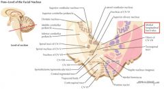
What is the source of axons for the Medial Longitudinal Fasciculous?
|

The Vestibular Nuclei.
|
|
|

White arrow.
|

Spinal Tract of the Trigeminal Nerve.
|
|
|

What gyri are indicated by the blue arrow?
|

The Short Gyri of the Insula.
|
|
|
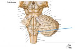
What structure is indicated by the blue arrow?
|

The Nucleus Cuneatus (Cuneate Tubercle)
|
|
|
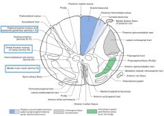
What is contained in the Nuclei (not in the cellular sense) of the grey matter? (e.g. indicated by blue boxes)
|

Nuclei consist of nerve cell bodies, the dendrites arising from the cell bodies and the axon terminals that synapse with them.
In 3D, the nuclei are actually columns of neurons that go up over several cord segments. |
|
|

Where does the brainstem derive its blood supply?
|
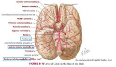
The brainstem is supplied by branches of the Inferior Cerebellar Arteries (Anterior and Posterior) as well as groups of the Central Arteries and the Pontine Arteries.
|
The Posteromedial and Posterolateral groups of the Central Arteries.
|
|

What nerve is indicated by the white arrow?
What foramen does it enter? |

The Inferior Alveolar Nerve, a branch off of the posterior Mandibular (V3).
It enters the Mandibular Foramen. |
|
|
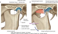
What is this joint called?
|

This is the Temporomandibular Joint.
A. Mouth closed B. Mouth open |
|
|

What artery is indicated by the white arrow?
|
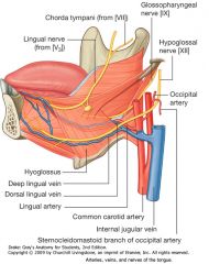
The Lingual Artery, off of the external carotid.
|
|
|
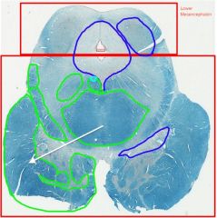
White Arrow.
|

Location of the Corticospinal Fibers.
|
|
|

What spinal tracts enter the cerebellum through the Superior Cerebellar Peduncle?
|
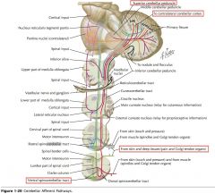
The Ventral Spinocerebellar Tracts.
(It also receives afferents from the mesencephalic nucleus of CN V, and the locus ceruleus) |
|
|

What type of signal is carried by the Medial Longitudinal Fasciculus?
|
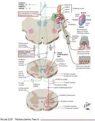
It carries mainly vestibular information that coordinates activities like the vestibulo-ocular reflex and gaze-holding.
|
|
|

White arrow.
|

Spinal Nucleus of the Trigeminal Nerve
|
|
|

What gyrus is indicated by the blue arrow?
|

The Long Gyrus of the Insula
|
|
|

What nerve is indicated by the blue arrow?
|

The Trigeminal Nerve (CN V)
|
|
|

What Nucleus of grey matter is indicated by the red arrow(s)?
What does it do? |

The Substantia Gelatinosa (Rolandi)
The neurons here process incoming pain sensations, but don't give axons conveying the information higher. |
|
|

Where does the Cerebellum derive its blood supply?
|
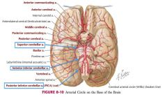
The Cerebellar Arteries.
Superior and Anterior Inferior off the Basilar. Posterior Inferior off of the Vertebral Arteries. |
|
|
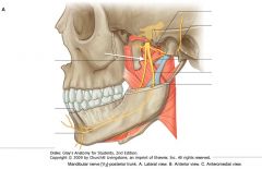
What nerve is indicated by the white arrow?
|
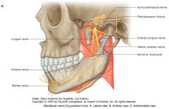
The Lingual Nerve, a sensory branch off of the posterior Mandibular Nerve (V3).
|
|
|
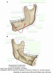
What portion of the Mandible is spanned (roughly) by the red arrows?
|

The Body
|
|
|

What vein does the blood from the tongue drain into?
|

The Internal Jugular Vein
(through the Dorsal and Deep Lingual Veins) |
|
|

White arrow.
|

Medial Lemniscus
|
|
|

What are the two components of the Inferior Cerebellar Peduncle?
|

The medial portion is the Justarestiform Body, and the Restiform Body forms the lateral portion. They cannot be distinguished grossly.
|
|
|

What is the function of the Tectospinal Tract?
|

It is involved in coordinating head and eye movements, similar to the MLF.
|
|
|
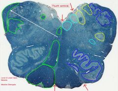
White arrow.
|

Inferior Cerebellar Peduncle.
|
|
|

What sulcus is indicated by the blue arrow?
|

The Precentral Sulcus.
|
|
|

Where does CN V arise?
|

From the region where the fibers of the Basilar Pons become continuous with the Middle Cerebellar Peduncle.
|
|
|
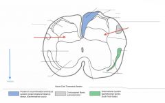
What Nucleus of grey matter is indicated by the red arrow(s)?
What happens here? |
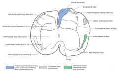
The Nucleus Proprius.
Neurons here give rise to axons which cross the cord in the Ventral White Commissure and ascend contralaterally in the spinothalamic tracts. |
|
|

How many groups of Central Arteries are there?
|
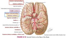
Four:
1. Anteromedial (off Anterior Communicating and Cerebral) 2. Anterolateral (off Middle Cerebral) 3. Posteromedial (off Posterior Communicating and Posterior Cerebral) 4. Posterolateral (Off the Posterior Cerebral Artery) |
|
|
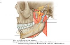
What nerve is indicated by the white arrow?
|

The Auriculotemporal Nerve, a sensory branch of the Mandibular Nerve (V3).
|
|
|
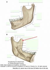
What portion of the Ramus is indicated by the red arrow?
What is its function? |

The Coronoid Process.
It provides attachment for the Temporalis Muscle. |
|
|

What nerve carries somatic motor innervation to the tongue?
|

The Hypoglossal Nerve (CN XII)
|
|
|

White arrow.
|

Spinal Lemniscus
|
|
|
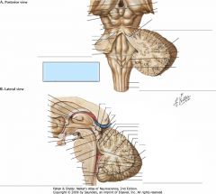
How do vestibulo-cerebellar fibers enter the cerebellum?
|

Through the Justarestiform Body of the Inferior Cerebellar Peduncle.
|
They project from the Vestibular Ganglia directly to the cerebellum as well as from the Vestibular Nuclei. As well, the Justarestiform Body carries efferent fibers from the cerebellum to the vestibular nuclei.
|
|
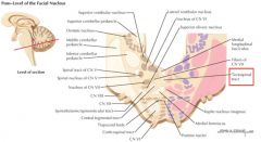
What is the source of axons for the Tectospinal Tract?
|
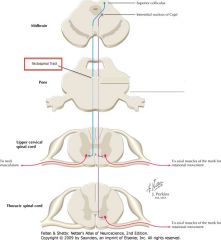
It arrises from the contralateral Superior Colliculus.
|
|
|

White arrow.
|

Principal Olivary Nucleus
|
|
|

What sulcus is indicated by the blue arrow?
|

The Postcentral Sulcus
|
|
|

What does CN V supply?
|
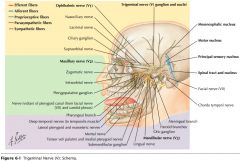
It supplies motor innervation to the muscles of mastication and sensory innervation to the structures of the face and mouth.
|
|
|
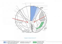
What do the neurons of the Nucleus Dorsalis do?
|
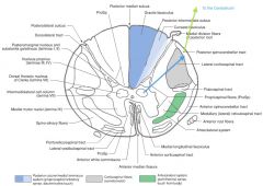
These neurons give off axons that ascend the cord in the Posterior Spinocerebellor Tract (see blue arrow), carrying proprioceptive info from the legs and trunk to the cerebellum (green arrow).
|
|
|
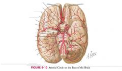
What are the Central Arteries?
|

Small arterial branches arising from the circle of Willis (or proximal branching arteries), that penetrate the base of the brain to supply deeply placed structures.
|
|
|

What is the part of the Ramus indicated by the red arrow?
What are its two parts called? What does it articulate with? |

The Condylar Process.
It consists of a Head and Neck (seen in lower view). The Head articulates with the mandibular fossa of the temporal bone to form the temporomandibular joint. |
|
|
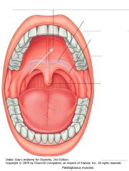
What section of the roof of the mouth is indicated by the white arrow?
|

The Hard Palate.
|
|
|

White arrow.
|

Oculomotor Nucleus.
|
|
|
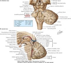
What is carried by the Restiform Body of the Inferior Cerebellar Peduncle?
|
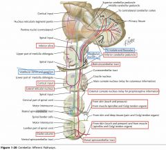
Fibers afferent to the cerebellum from (and through) the medulla oblongata (not including vestibular input).
|
|
|

What is the extent of the Tectospinal Tract?
|

It descends from the contralateral Superior Colliculus all the way through the brainstem and to the cervical spinal cord.
|
|
|

White arrow.
|

Medial Accessory Olivary Nucleus.
|
|
|

What sulcus is indicated by the blue arrow?
|

The Superior Temporal Sulcus
|
|
|

What nerve is indicated by the blue arrow?
|

The Abducens Nerve (CN VI)
|
|
|

What Nucleus of grey matter is indicated by the red arrow(s)?
What levels have this nucleus? |

Nucleus Dorsalis (Clarke's Column)
It is found throughout the thoracic and upper lumbar regions of the cord. |
|
|

Where does the Anteromedial Group of the central arteries arise?
Where does it supply? |
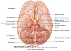
Chiefly from the Anterior Communicating and Anterior Cerebral Arteries.
Supply (portions of) the Hypothalamus. |
|
|

What is the feature indicated by the red arrow called?
|

The Mandibular Notch
|
|
|
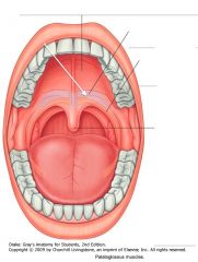
What section of the roof of the mouth is indicated by the white arrow?
|

The Soft Palate.
|
|
|

White arrow.
|

Autonomic Oculomotor Nucleus
|
|
|
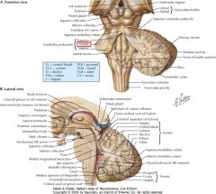
What is the source of fibers in the Middle Cerebral Peduncle?
|

It is exclusively axons derived from the Pontine Nuclei (afferent to the cerebellum), and a continuation of the Transverse Pontine fibers.
|
This pontocerebellar pathway is a continuation of the corticopontine fibres which carried information from the Primary Motor Cortex.
|
|
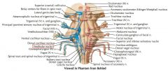
What signals do the Vestibular Nuclei receive?
|
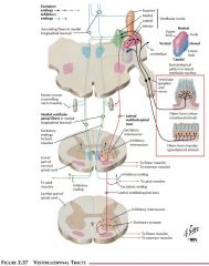
They receive input from the vestibular apparatus
(the semicircular canals and ampulllae, and the saccule and utricle). |
|
|

White arrow.
|

Medial Longitudinal Fasciculus.
|
|
|

What sulcus is indicated by the blue arrow?
|

The Inferior Temporal Sulcus
|
|
|

What nerve is indicated by the blue arrow?
|

The Facial Nerve (CN VII)
|
|
|
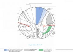
What Nucleus of grey matter is indicated by the red arrow(s)?
Where is it found? |

The Intermediolateral Cell Column (Lateral Horn).
It extends from the upper lumbar regions to the thoracic regions of the cord (similar to Nucleus Dorsalis). |
|
|
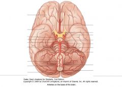
Where do the Anterolateral Group of the central arteries arise?
What do they supply? |
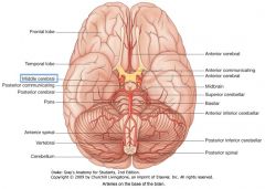
The Middle Cerebral Arteries.
Supply much of the "corpus striatum," and "the anterior limb, genu and much of the posterior limb of the internal capsule." |
|
|
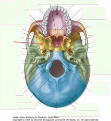
What is the bone indicated by the red arrow?
|

The Sphenoid Bone
|
|
|
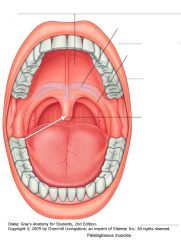
What structure of the mouth is indicated by the white arrow?
|

The Uvula
|
|
|

White arrow.
|

Nucleus of the Superior Colliculus.
|
|
|
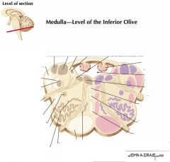
How many nuclei are included in each Inferior Olive?
|

Three:
1. Principal, 2. Dorsal Accessory, and 3. Medial Accessory Olivary Nuclei. |
|
|

White arrow.
|

Tectospinal Tract
|
|
|

What structure is indicated by the blue arrow?
|

The Medulla Oblongata
|
|
|
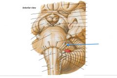
What nerve is indicated by the blue arrow?
|

The Vestibulo-cochlear Nerve (CN VIII) (also called the Auditory Nerve)
|
|
|

What is contained in the Intermediolateral Cell Column?
|

This Nucleus consists of sympathetic preganglionic motor neurons whose axons exit with the Anterior Root (blue arrow), then enter the sympathetic chain of ganglia (green arrow) via the White Ramus Cummunicantes (orange arrow).
|
|
|

Where do the Posteromedial group of the central arteries arise?
What do they supply? |
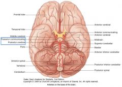
The Posterior Communicating Arteries and the Posterior Cerebral Arteries.
Supply much of the thalamus and hypothalamus, the subthalamus, and some medial portions of the midbrain. |
|
|
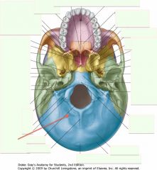
What is the bone indicated by the red arrow?
|

The Occipital Bone
|
|
|
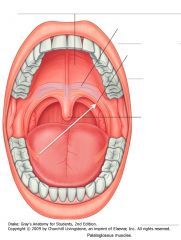
What muscle is indicated by the white arrow?
|

The Palatoglossus Muscle.
|
|
|
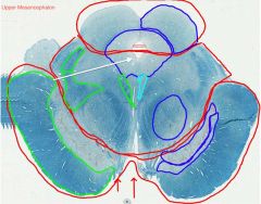
White arrow.
|

Periaqueductal Gray Matter.
|
|
|
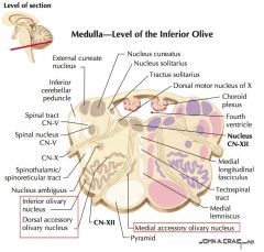
What sources give input to the Inferior Olives?
|

They receive inputs from:
1. Spinal, 2. Brainstem, 3. Cerebellar, and 4. Cerebral cortical sources. |
|
|

White arrow.
|

Pyramid
|
|
|
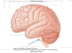
What structure is indicated by the blue arrow?
|
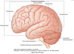
The Pons
|
|
|

What nerve is indicated by the blue arrow?
|

The Oculomotor Nerve (CN III)
|
|
|

What Nucleus of grey matter is indicated by the red arrow(s)?
|
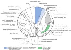
The Medial Anterior Horn group of motor neurons.
|
|
|

Where does the Posterolateral Group of central arteries arise from?
What do they supply? |

The Posterior Cerebral Artery.
Supply the posterior portion of the thalamus, as well as the tectum and some lateral portions of the midbrain. |
|
|
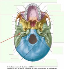
What is the bone indicated by the red arrow?
|

The Temporal Bone (one on each side)
|
|
|

What muscle is indicated by the white arrow?
|

The Palatopharyngeus Muscle, forming the structure of the palatopharyngeal arch.
|
|
|

White arrow.
|
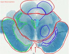
Cerebral Aqueduct.
|
|
|

What fibres do the Olivary Nuclei give off? Where do they go?
|
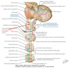
They give rise to Olivocerebellar Fibres, which cross the midline and enter the Cerebellum via the Inferior Cerebellar Peduncle.
|
|
|

White arrow.
|

Inferior Vestibular Nucleus.
|
|
|

What structure is indicated by the blue arrow?
|

The Cerebellum
|
|
|
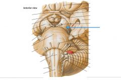
What nerve is indicated by the blue arrow?
|

The Trochlear Nerve (CN IV)
|
|
|
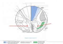
What is the function of the neurons in the Anterior Horn?
|

They're the nuclei of somatic motor neurons.
|
|
|

What veins are indicated by the red arrows?
|
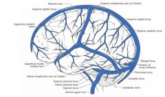
Superior Cerebral Veins
|
|
|

What is the protuberance (process) indicated by the red arrow?
|
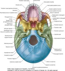
The Mastoid Process (of the Temporal Bone)
|
|
|

What is the protuberance (process) indicated by the red arrow?
|

The Mastoid Process (of the Temporal Bone)
|
|
|
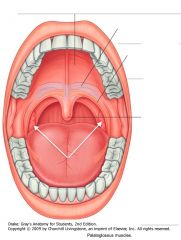
What structures are indicated by the white arrows?
|

The Palatine Tonsils
|
|
|

White arrow.
|

The Red Nucleus
|
|
|

What is the Reticular Formation?
|
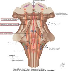
An area of intermingled grey and white matter occupying the central region of the brainstem throughout its extent.
It is actually groups of nuclei and their afferent and efferent connections. |
|
|
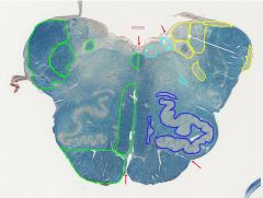
White arrow.
|

Medial Vestibular Nucleus
|
|
|

What sulcus is indicated by the white arrow?
|

The Cingulate Sulcus.
|
|
|
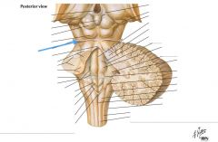
What nerve is indicated by the blue arrow?
|

The Trochlear Nerve (CN IV)
|
|
|

How far up the spinal cord do you find the Anterior Horn Motor Neurons Nucleus?
Where is it bigger? |

It's all up the spinal cord.
In the cervical swelling, there is both a Medial and Lateral Nucleus. It is also bigger in the area of the lumbo-sacral swelling. |
|
|

What vein is indicated by the red arrow?
|

The Superior Anastomotic Vein (of Trolard)
|
|
|

What is the canal indicated by the red arrow called?
|

The External Auditory Meatus (or canal)
|
|
|

What surface is indicated by the white arrow?
|
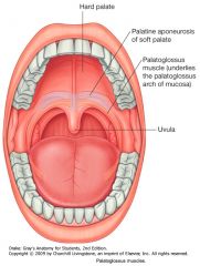
The Posterior Wall of the Oropharynx.
|
|
|
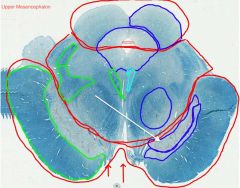
White arrow.
|

Compact Zone of the Substantia Nigra.
|
|
|
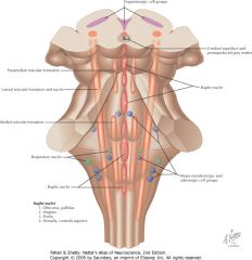
What does the Reticular Formation do?
|

It influences many things:
1) Level of consciousness (shown), 2) Coordination of somatic motor activity, 3) Control of respiration, 4) Modulation of pain transmission, and other functions. |
|
|

White arrow.
|

Dorsal Cochlear Nucleus.
|
|
|

What sulcus is indicated by the white arrow?
|

The Sulcus of the Corpus Callosum (Callosal Sulcus)
|
|
|
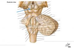
What structure is indicated by the blue arrow?
What is it involved with? |

One of the Superior Colliculi of the Corpora Quadrigemina.
It's involved with mediating visual reflexes. |
|
|
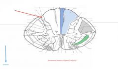
What tract of white matter is indicated by the red arrow?
|

The Dorsolateral Tract (Tract of Lissauer).
|
|
|

What vein is indicated by the red arrow?
|

The Inferior Anastomotic Vein (of Labbe)
|
|
|

What is the big hole indicated by the red arrow called?
What goes through that foramen? |

The Foramen Magnum
The spinal cord, vertebral arteries and CN XI |
CN XI is the Accessory Nerve.
|
|
|
Which extrinsic tongue muscle is innervated by the Vagus Nerve (CN X)?
|

The Palatoglossus
|
|
|

White arrow.
|

Reticular Zone of the Substantia Nigra.
|
|
|

Where does the Lateral Lemniscus derive its fibres?
|
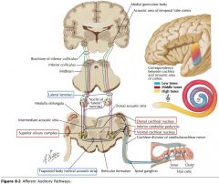
A variety of sources:
1) Dorsal and Ventral Cochlear Nuclei, 2) Nucleus of the Trapezoid Body, and 3) the Superior Olivary Nucleus. |
|
|

White arrow.
|

Ventral Cochlear Nucleus
|
|
|
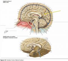
What sulcus is indicated by the yellow arrow?
|

The Parieto-occipital Sulcus
|
|
|

What structure is indicated by the blue arrow?
What is it involved in? |

One of the Inferior Colliculi.
It is involved with the auditory pathway. |
|
|

Where does the Dorsolateral Tract receive axons from?
Where do they go? |
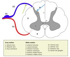
Axons of the lateral division of the dorsal root (green arrow) enter the spinal cord and travel up or down the Dorsolateral Tract before entering the grey matter and synapsing in the Dorsal Horn (blue arrow).
|
|
|
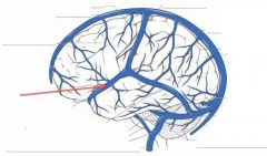
What vein is indicated by the red arrow?
|

The Superficial Middle Cerebral Vein
|
|
|

What is the hole indicated by the red arrow called?
What goes through it? |

The Jugular Foramen.
The Internal Jugular Vein and CN IX, X, and XI. |
|
|
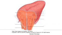
What muscle is indicated by the white arrow?
|

The Genioglossus
|
|
|

White arrow.
|

Basis Pedunculi (or Crus Cerebri)
|
|
|

What is the Lateral Lemniscus a portion of?
What information does it carry? |

It is a portion of the auditory pathway.
It conveys information related to hearing. |
|
|

White arrow.
|

Nucleus Ambiguus
|
|
|

What sulcus is indicated by the white arrow(s)?
|

The Calcarine Sulcus.
|
|
|

What is the function of the Abducens Nerve?
|
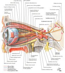
It provides motor innervation to the lateral rectus muscle of the eye.
|
|
|
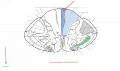
What Tract of white matter is indicated by the red arrow?
What signals does it carry? |

The Fasciculus Gracilis.
It carries sensation of fine touch, proprioception, and vibration from the lower limb. |
|
|
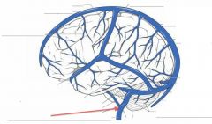
What vein is indicated by the red arrow?
|

The Internal Jugular Vein
|
|
|
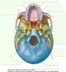
What is the hole indicated by the red arrow?
What does it carry? |

The Carotid Canal.
The Internal Carotid Artery and it's Sympathetics. |
|
|

What tongue muscle is indicated by the white arrow?
|
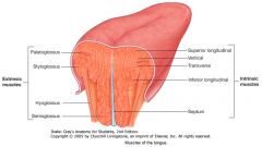
The Hyoglossus
|
|
|
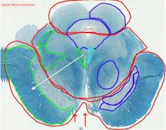
White arrow.
|

Location of Corticospinal Fibers.
|
|
|
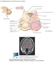
Where do the ascending fibres of the Lateral Lemniscus terminate?
|
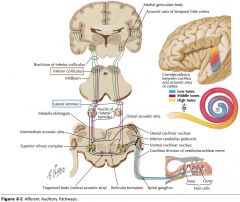
They terminate in the Inferior Colliculus.
|
|
|

White arrow.
|

Solitary Tract
|
|
|

What is the importance of the Calcarine Sulcus as a landmark?
|

The primary visual cortex is located on either side of this sulcus.
|
|
|

What structure is indicated by the blue arrow?
|
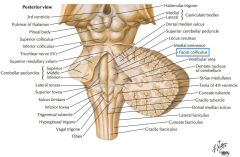
The Facial Colliculus.
|
|
|
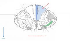
What Tract of white matter is indicated by the red arrow?
What signals does it carry? |

The Fasciculus Cuneatus.
Sensations of fine touch, proprioception, and vibration from upper limb. |
|
|

What is the hole indicated by the red arrow?
What travels through it? |

The Foramen Spinosum.
The Middle Meningeal Artery (a branch of the Maxillary Artery, itself off of the External Carotid). |
|
|

What tongue muscle is indicated by the white arrow?
|

The Styloglossus.
|
|
|
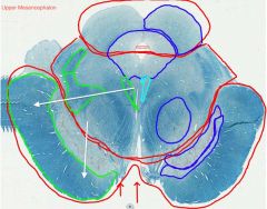
White arrows.
|

Location of Corticopontine Fibers.
|
|
|
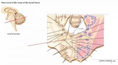
What nucleus is indicated by the red arrow?
What occurs at the blue arrow? |
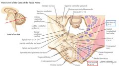
The Facial Motor Nucleus.
The Genu of the Facial Nerve, where the fibres of the Facial Nerve course around the Abducens Nucleus. |
|
|

White arrow.
|

Solitary Nucleus
|
|
|

What gyrus is indicated by the white arrow?
|

The Cingulate Gyrus.
|
|
|

What does the Facial Nerve innervate?
|
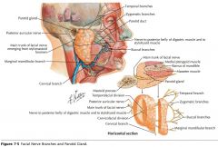
It provides taste to the anterior 2/3 of the tongue and motor innervation to the muscles of facial expression (among other things).
|
|
|
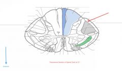
What Tract of white matter is indicated by the red arrow?
What signals does it carry? |
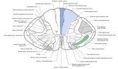
The Dorsal Spinocerebellar Tract.
It carries proprioceptive information from the lower limb to the cerebellum. |
|
|

What is the hole indicated by the red arrow?
What travels through it? |

The Foramen Ovale
Mandibular Nerve (CN V3) and lesser petrosal nerve of CN IX |
|
|
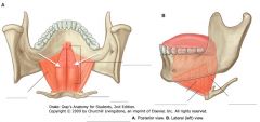
What muscles are indicated by the white arrows?
|

The Genioglossus Muscles.
|
|
|
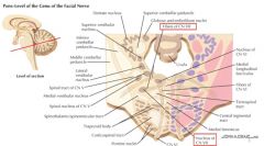
What does the Facial Nucleus innervate?
|
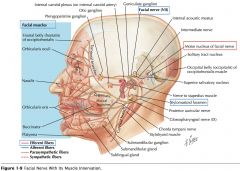
It provides motor innervation to the muscles of facial expression.
|
|
|

White arrow.
|

Spinal Tract of the Trigeminal Nerve.
|
|
|

What structure is indicated by the white arrow?
|

The Corpus Callosum
|
|
|

What is the function of the Vestibulocochlear Nerve?
|
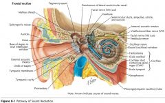
The Vestibular Root conveys information of balance and the position of the head.
The Cochlear Root mediates hearing. |
|
|

Where do the Dorsal Spinocerebellar Tracts' axons derive?
|
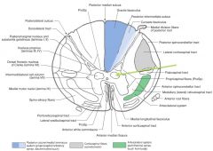
They come from the neuronal cell bodies in the Nucleus Dorsalis (Clarke's Column), on the same side of the spinal cord.
|
Clarke's Column is only located on the upper Lumbar through Thoracic spinal cord.
|
|
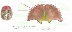
What is the name of the bone indicated by the red arrow? What is this "plate?"
|

This is the Orbital Plate (or part) of the Frontal Bone.
|
|
|

What movements do the Genioglossus Muscles control?
|
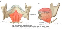
The Genioglossus Muscles depress and protrude the tongue.
|
|
|

What surface feature do the does the Genu of the Facial Nerve form?
|
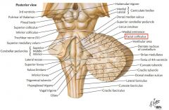
It forms the Facial Colliculus seen in the floor of the Fourth Ventricle.
|
|
|

White arrow.
|

Spinal Nucleus of the Trigeminal Nerve.
|
|
|
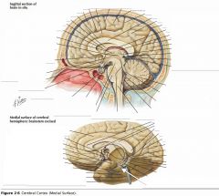
What gyrus is indicated by the white arrow?
|

The Parahippocampal Gyrus
|
|
|

What area is indicated by the blue arrow?
Why is it important? |
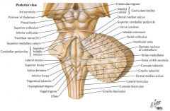
The Vestibular Area
It overlies the location of the vestibular nuclei. |
|
|
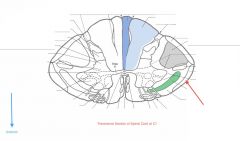
What Tract of white matter is indicated by the red arrow?
What signals does it carry? |

The Anterior Spinocerebellar Tract.
It carries proprioceptive information from the lower limb to the cerebellum. |
|
|

What is the name of the bone indicated by the red arrow? What is this "plate?"
|

The Cribriform Plate of the Ethmoid Bone.
|
|
|
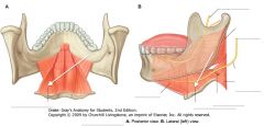
What tongue muscle is indicated by the white arrow?
|

The Hyoglossus
|
|
|

Where does the Facial Nerve exit the Brainstem?
|
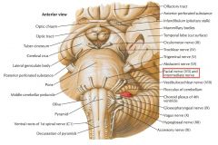
After encircling the Abducens Nucleus, they continue ventro-laterally toward their exit at the lateral margin of the ponto-medullary junction.
|
|
|

White arrow.
|

Medial Longitudinal Fasciculus (MLF)
|
|
|

What structure is indicated by the white arrow?
|

The Uncus
|
|
|
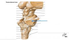
What structure is indicated by the blue arrow?
What does it do? |

The Middle Cerebellar Peduncle
It conveys information to the cerebellum derived from the cerebral cortex. |
|
|

Where does the Anterior Spinocerebellar Tract derive from?
|
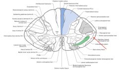
It derives its axons from the cell bodies on the other side of the spinal cord in the dorsal horn.
|
|
|

What are the little holes located on the plate indicated by the red arrow?
|

Those are the Olfactory Foramina in the Cribriform Plate, for Olfactory Nerves (CN I).
|
|
|

What action does the Hyoglossus perform?
|
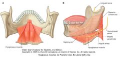
The Hyoglossus and the Styloglossus both retract the tongue.
The Hyoglossus also pulls down the sides of the tongue. |
|
|
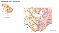
What nucleus is indicated by the red arrow?
|
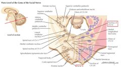
The Abducens Nucleus.
|
|
|

White arrow.
|

Tectospinal Tract
|
|
|

What are the Cingulate Gyrus, the Parahippocampal Gyrus and the Uncus collectively called?
|

The "Limbic Lobe." (along with some associated structures)
|
|
|

What structure is indicated by the blue arrow?
|

The Superior Cerebellar Peduncle
|
|
|
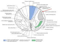
Where does the Posterior Spinocerebellar Tract (red arrow) enter the Cerebellum?
|

Through the Inferior Cerebellar Peduncle (see red line).
|
|
|

What is the bone indicated by the red arrow?
|

The (body of the) Sphenoid Bone
|
|
|
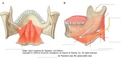
What muscle is indicated by the white arrows?
|

The Mylohyoid Muscle
|
|
|

What does the Abducens Nucleus innervate?
|

It provides motor innervation to the Lateral Rectus muscle of the eye.
|
|
|

White arrow.
|

Medial Lemniscus
|
|
|

What does the Limbic System do?
|

It is involved with emotion and certain kinds of memory.
|
|
|
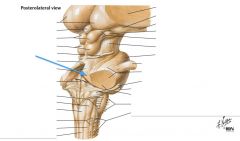
What type of information is carried on the Superior Cerebellar Peduncle?
|

It largely carries efferent fibers from the Cerebellum.
|
|
|

Where does the Anterior Spinocerebellar Tract (red arrow) enter the Cerebellum?
|
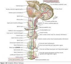
The Superior Cerebellar Peduncle (follow green line).
|
|
|

What is the feature of the Sphenoid indicated by the red arrow?
|

The Tuberculum Sellae of the Sella Turcica (Turkish Saddle).
|
|
|
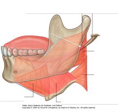
What tongue muscle is indicated by the white arrow?
|

The Styloglossus.
|
|
|

Where do the fibres of CN VI exit the Brainstem?
|
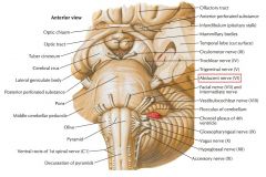
They exit medially on the ponto-medullary junction.
|
|
|

White arrow.
|

Dorsal Accessory Olivary Nucleus
|
|
|

What information does the Uncus receive?
|
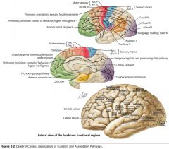
It receives olfactory input
|
|
|
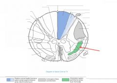
What Tract of white matter is indicated by the red arrow?
What are its divisions? |

The Anterolateral System
It's divided into the Anterior and Lateral Spinothalamic Tracts. |
|
|

What is the feature of the Sphenoid indicated by the red arrow?
|

The Hypophyseal Fossa, containing the pituitary gland (hypophysis).
|
|
|
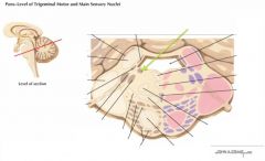
What nucleus is indicated by the green arrow?
|

The Locus Ceruleus.
|
|
|

White arrow.
|

Medial Accessory Olivary Nucleus.
|
|
|
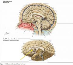
What region of the Corpus Callosum is indicated by the yellow arrow?
|

The Rostrum.
|
|
|
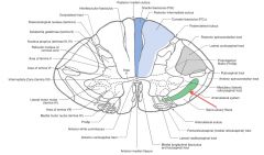
Where do the axons of the Anterolateral system (red arrow) derive from (both Lateral and Anterior Spinothalamic tracts)?
|

They both derive from the Nucleus Proprius on the opposing side (green arrow).
|
|
|

What is the feature of the Sphenoid indicated by the red arrow?
|

The Dorsum Sellae.
|
|
|

What nerve innervates the Styloglossus?
|
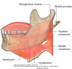
The Hypoglossal Nerve (CN XII)
|
|
|
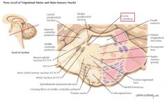
What system is the Locus Ceruleus a part of?
Where do its axons project? |
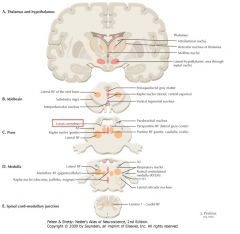
It's a component of the Reticular Formation.
It's neurons project to all levels of the nervous system with highly branched axons. |
|
|

White arrow.
|

The Principal Olivary Nucleus
|
|
|

What region of the Corpus Callosum is indicated by the yellow arrow?
|

The Genu
|
|
|

What sensations are carried by the Anterior Spinothalamic Tract (red arrow)?
|
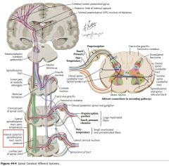
Light touch and pressure.
|
|
|

What part of the Temporal Bone is indicated by the red arrow?
|

The Petrous part of the Temporal Bone.
It is a pyramidal section of the bone wedged in between the sphenoid and occipital bones. |
|
|

What is the action of the Styloglossus?
|

Both the Styloglossus and the Hyoglossus retract the tongue.
|
|
|
|
How many nuclear groups are associated with the Trigeminal Nerve?
|
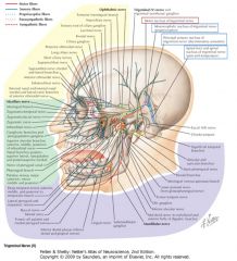
Four (3 sensory; 1 motor):
1) The Spinal Tract and Nucleus, 2) Principal Sensory Nucleus, 3) Mesencephalic Tract and Nucleus, and 4) the Trigeminal Motor Nucleus. |
|
|

White arrow.
|

Inferior Cerebellar Peduncle
|
|
|

What region of the Corpus Callosum is indicated by the yellow arrow?
|

The Body
|
|
|

Where do the neurons whose axons form the Lateral Spinothalamic Tract (red arrow) receive their input from?
How is this different from the Anterior Spinothalamic Tract? |
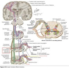
Afferent neurons from the dorsal root ganglion cells within a segment or two of the neurons in question (yellow arrows).
The anterior Spinothalamic Tract's input sensory cell bodies can be located several segments away. |
|
|
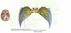
What part of the Temporal Bone is indicated by the red arrow?
|

The Squamous Part of the Temporal Bone.
|
|
|

What muscle is indicated by the white arrow?
|

The Mylohyoid Muscle
|
|
|
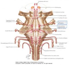
What does the Motor Nucleus of the Trigeminal Nerve innervate?
|
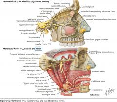
It provides innervation to the muscles of mastication, through the Mandibular division of the Trigeminal Nerve.
|
|
|
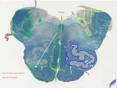
White arrow.
|

Pyramid
|
|
|
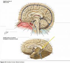
What region of the Corpus Callosum is indicated by the yellow arrow?
|

The Splenium
|
|
|
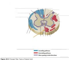
What tract of white matter is indicated by the orange arrow?
|
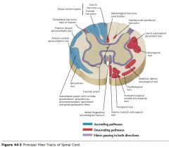
The Anterior Corticospinal Tract
|
|
|

What foramen is located at the point indicated by the red arrow?
What does it contain? |

The Optic Canal.
It contains the Optic Nerve (CN II) and the Ophthalmic Artery (a branch of the Internal Carotid that supplies blood to all the structures in the orbit). |
|
|
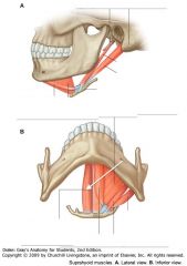
What muscle is indicated by the white arrow?
|

The Geniohyoid Muscle.
|
|
|
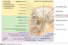
What is the role of the Principal Sensory Trigeminal Nucleus?
Where is it located? |
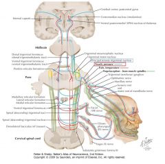
It mediates sensations of touch and pressure from various regions of the head.
It's located laterally at the mid-pontine level. |
|
|

White arrow.
|

Medial Lemniscus
|
|
|

What is the role of the Corpus Callosum?
|

It connects the right and left cerebral hemispheres, ensuring communication through commissural fibres.
|
|
|
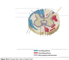
What tract of white matter is indicated by the orange arrow?
|

The Lateral Corticospinal Tract.
|
|
|

What structure is indicated by the red arrow?
What passes through it? |

The Superior Orbital Fissure.
CNs III, IV, VI and branches of the ophthalmic division (V1) of CN V. |
|
|
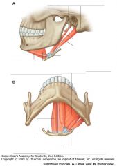
What muscles form the floor of the mouth?
|

The Mylohyoid and Geniohyoid muscles.
|
|
|
|
What does the Mesencephalic Tract of CN V contain?
|
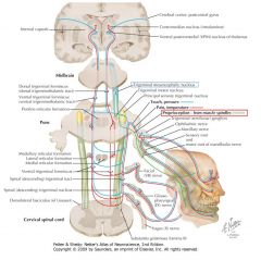
It consists of of axons of the pseudounipolar neurons which make up the Mesencephalic Nucleus.
|
|
|

White arrow.
|

Lateral Vestibular Nucleus
|
|
|

What collection of fibres is indicated by the yellow arrow?
|

The Anterior Commissure
|
|
|

What types of signals are carried by the Corticospinal Tracts (both Anterior and Lateral)?
|

Motor signals, they are often described as "Upper Motor Neurons."
|
|
|

What structure is indicated by the red arrow?
What passes through it? |

The Foramen Rotundum
The Maxillary Nerve (CN V2) |
|
|
|
What actions do the Mylohyoid and Geniohyoid muscles perform?
|

They contribute to elevating the hyoid bone (and therefore the larynx) during swallowing or depressing the Mandible, depending on the tone of the strap muscles.
|
|
|
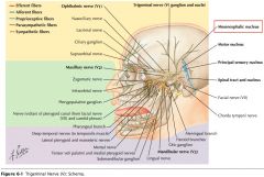
What type of sensation does the Mesencephalic Nucleus of CN V mediate?
|

It mediates Proprioceptive information, particularly from the Temporomandibular Joint and the teeth.
|
|
|

White arrow.
|

Superior Vestibular Nucleus
|
|
|
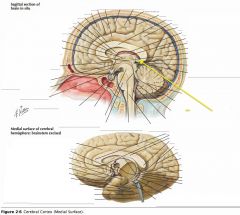
What collection of fibres is indicated by the yellow arrow?
|

The Posterior Commissure.
|
|
|

Where do the Lateral Corticospinal Tracts cross?
|

They cross at the Pyramidal Decussation in the Medulla.
|
|
|

What structure is indicated by the red arrow?
What passes through it? |

The Foramen Ovale
Mandibular Nerve (CN V3) and lesser petrosal nerve of CN IX. |
(The source of pre-ganglionic parasympathetics that synapse within the otic ganglion, post ganglionics are carried by the auriculotemporal nerve, part of CNV3, to the parotid gland) -TMI
|
|
|
Where do the Corticopontine Fibres originate?
|
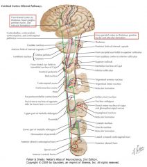
They originate from Frontal, Parietal, and to some extent Occipital regions of the Cerebral Cortex.
|
|
|

White arrow.
|

Medial Vestibular Nucleus
|
|
|

What collection of fibres is indicated by the yellow arrow?
|

The Fornix.
|
|
|
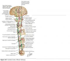
Which white matter tracts contains 75-90% of the pyramidal tract fibres that cross at the pyramidal decussation in the medulla?
|
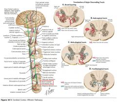
The Lateral Corticospinal Tracts.
|
|
|

What is the structure indicated by the red arrow?
What passes through it? |

The Foramen Spinosum.
The Middle Meningeal Artery. |
|
|

Where do the Corticopontine terminate?
Where does the pathway continue after that? |

They terminate by synapsing on the Pontine Nuclei.
The Pontine Nuclei project axons that cross the Pons as Transverse Fibres and enter the Cerebellum through the Middle Cerebellar Peduncle. |
|
|

White arrow.
|

Motor Nucleus of the Facial Nerve
|
|
|

What two lobes are connected by the Anterior Commissure?
|

The right and left Temporal Lobes.
|
|
|

Do the anterior Corticospinal Tracts cross before entering the spinal cord?
|
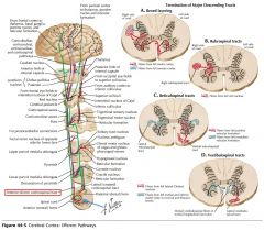
No, although they may (but not always) cross at the level they terminate, using the Ventral White Commissure (blue arrow).
|
|
|

What structure is indicated by the red arrow?
What passes through it? |

The opening of the Carotid Canal (or Groove)
The Internal Carotid Artery and sympathetics. |
|
|
|
Which of the Cerebellar Peduncles carries mostly efferent fibres from the Cerebellum?
|

The Superior Cerebellar Peduncle (it has very few afferent).
The Inferior also has some too, but not as much. |
|
|
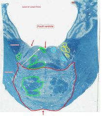
White arrow.
|

Genu (and fibers) of the Facial Nerve
|
|
|

What foramen is indicated by the yellow arrow?
|
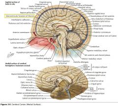
The Interventricular Foramen.
|
|
|

How low down the spinal cord does the Anterior Corticospinal Tract descend?
The Lateral? |
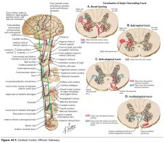
The Anterior Corticospinal Cord only extends as far as the upper thoracic cord.
The Lateral goes down all the way. |
|
|

What is the bone indicated by the red arrow?
|

The (body of) the Sphenoid Bone.
|
|
|
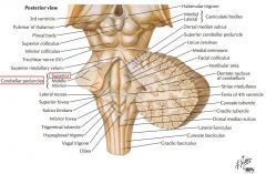
What happens to the fibres of the Superior Cerebellar Peduncle at the level of the rostral Pons and the caudal Mesencephalon?
|
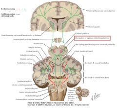
They undergo a decussation.
|
|
|

White arrow.
|

Nucleus of the Abducens Nerve.
|
|
|

What structure is indicated by the white arrow?
|

The Corpora Quadrigemina (well, a part of it)
|
|
|

What bone is indicated by the red arrow?
|
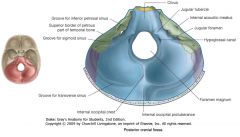
The Occipital Bone
|
|
|
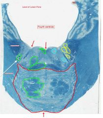
White arrow.
|

Spinal Tract of the Trigeminal Nerve.
|
|
|
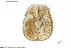
What structure is indicated by the white arrow?
|

The Cerebral Aqueduct.
|
|
|

What part of the Temporal Bone is indicated by the red arrow?
|

The Petrous Part of the Temporal Bone.
|
|
|

White arrow.
|

Spinal Nucleus of the Trigeminal Nerve.
|
|
|

What structure is indicated by the white arrow?
|

The Cerebral Aqueduct.
|
|
|
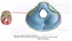
What part of the Temporal Bone is indicated by the red arrow?
|

The Mastoid Part of the Temporal Bone.
|
|
|

White arrow.
|

Medial Longitudinal Fasciculus
|
|
|
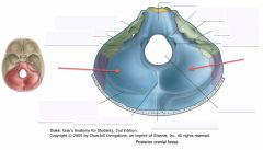
What part of the Occipital Bone is indicated by the red arrows?
|

The Cerebellar Fossae
|
|
|

What is the function of the Posterior Commissure?
|

It consists of axons which mediate reflexes associated with pupillary constriction.
|
|
|

White arrows.
|

The Pontine Nuclei
|
|
|

What foramen is indicated by the red arrow?
What passes through it? |

The Internal Acoustic Meatus.
CN VII and VIII |
|
|

What portion of the Fornix is indicated by the white arrow?
|

The Body.
|
|
|

White arrow.
|

Longitudinal Pontine Fibers
|
|
|

What foramen is indicated by the red arrow?
What passes through it? |

Jugular Foramen
CN IX, X and XI, and Internal Jugular Vein |
|
|
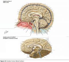
What portion of the Fornix is indicated by the white arrow?
|

One of the Columns.
|
|
|

White arrow.
|

Transverse Fibers of Pons.
|
|
|

What foramen is indicated by the red arrow?
What passes through it? |

The Foramen Magnum
The Spinal Cord, Vertebral Arteries, and Spinal Accessory Nerve (CN XI) |
|
|

What septum is indicated by the white arrow?
|

The Septum Pellucidum.
|
|
|
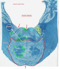
White arrow.
|

Inferior Cerebellar Peduncle
|
|
|

What foramen is indicated by the red arrow?
What passes through it? |

Hypoglossal Canal
CN XII |
|
|

What structure is indicated by the white arrow?
|

The Thalamus.
|
|
|

White arrow.
|

Middle Cerebellar Peduncle
|
|
|

What feature of a vertebra is indicated by the red arrow?
|

Vertebral Body
|
|
|

What structure is indicated by the orange arrow?
|

The Hypothalamus.
|
|
|

White Arrow
|

Medial Lemniscus
|
|
|
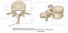
What feature of a vertebra is indicated by the red arrow?
|
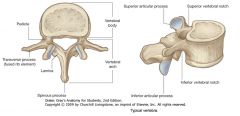
Spinous Process
|
|
|
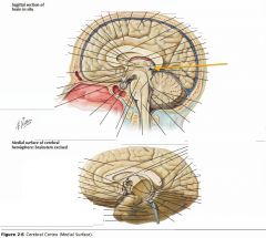
What structure is indicated by the orange arrow?
|

The Pineal Body.
|
|
|
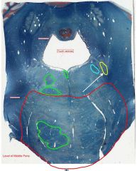
White arrow.
|

Principal Sensory Nucleus of the Trigeminal Nerve
|
|
|
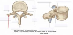
What feature of a vertebra is indicated by the red arrow?
|

Transverse Process
|
|
|

What is the function of the Thalamus?
|

Its nuclei process sensory, motor, and limbic information, along with a variety of other kinds of information which are then sent to the cerebral cortex.
|
|
|

White arrow.
|

Motor Nucleus of the Trigeminal Nerve
|
|
|
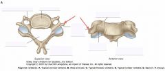
What feature of a vertebra is indicated by the red arrows?
|

The Transverse Process
|
|
|

What is the function of the Hypothalamus?
|

It's intimately involved in the control of visceral activity and in the maintenance of homeostasis.
Certain regions are also involved with the limbic system. |
It acts both with neurologic control and with hormonal control (as an endocrine organ).
|
|

White arrow.
|

Medial Longitudinal Fasciculus.
|
|
|

What is the function of the Pineal Body?
|

It secretes melatonin.
|
|
|
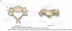
What feature of a vertebra is indicated by the red arrow?
|
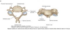
The Foramen Transversarium
|
|
|
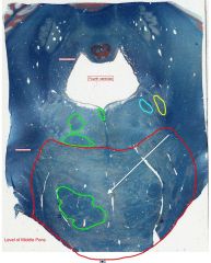
White arrow.
|

Pontine Nuclei
|
|
|

What gyrus is indicated by the blue arrow?
|

The Gyrus Rectus (or Straight Gyrus)
|
|
|
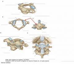
What is the name of the specially-shaped vertebra indicated by the red arrows?
What is a unique feature of this vertebra? |

The Atlas Vertebra (C1)
It doesn't have a vertebral body. |
|
|

White arrow.
|

Middle Cerebellar Peduncle
|
|
|

What structure is indicated by the blue arrow?
|

The Olfactory Bulb
|
|
|

What is the name of the specially-shaped vertebra indicated by the red arrows?
What is a unique feature of this vertebra? |

The Atlas Vertebra (C1)
It doesn't have a vertebral body. |
|
|
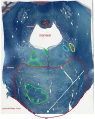
White arrow.
|

Transverse Fibers of Pons
|
|
|
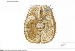
What tract is indicated by the blue arrow?
|

The Olfactory Tract.
|
|
|
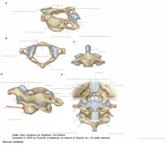
What is the name of the vertebra indicated by the red arrow?
What special joint does it form with its neighbour? |

The Axis Vertebra (C2)
The Atlanto-Axial Joint |
|
|

White arrow.
|
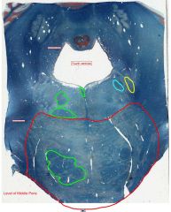
Longitudinal Pontine Fibers
|
LPF include Corticopontine and Corticospinal fibers, among others)
|
|

What sulcus is indicated by the blue arrow?
|

The Olfactory Sulcus
|
|
|
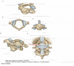
What is the name of the joint indicated by the red arrow?
|
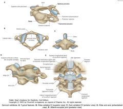
The Atlanto-Occipital Joint.
|
|
|

White arrow.
|

Superior Cerebellar Peduncle
|
|
|

What structure is indicated by the blue arrow?
|

The Medial Olfactory Stria
|
|
|

What feature of the mandible is indicated by the red arrow?
|

The Angle
|
|
|

White arrow.
|

Medial Lemniscus
|
|
|

What structure is indicated by the blue arrow?
|

The Lateral Olfactory Stria.
|
|
|

White arrow.
|

Medial Longitudinal Fasciculus.
|
|
|

What structure is indicated by the blue arrow?
|
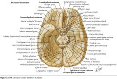
The Anterior Perforated Substance
|
|
|

What feature of the mandible is indicated by the red arrow?
|

The Angle
|
|
|

White arrow.
|

Mesencephalic Tract of the Trigeminal Nerve
|
|
|
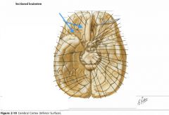
What gyri are indicated by the blue arrows?
|

The Orbital Gyri
|
|
|
|
What are the borders of the Anterior Triangle of the neck?
|
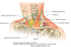
Posteriorly: the anterior border of the sternocleidomastoid muscle.
Superiorly: the inferior border of the Mandible. Anteriorly, the anterior midline of the neck. |
|
|
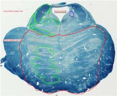
White arrow.
|
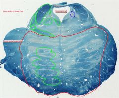
Longitudinal Pontine Fibers.
(Including Corticospinal and Corticopontine Fibers) |
|
|
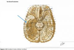
What gyrus is indicated by the blue arrow?
|

The Inferior Temporal Gyrus.
|
|
|
|
What are the boundaries of the Posterior Triangle of the neck?
|

Anteriorly: the sternocleidomastoid muscle.
Posteriorly: the superior fibres of the Trapezius muscle. Inferiorly: the medial half of the clavicle. |
|
|

White arrow.
|

Pontine Nuclei
|
|
|
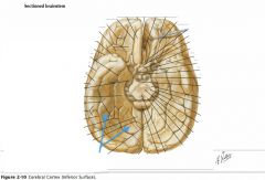
What gyri are indicated by the blue arrows?
|

The Occipitotemporal Gyri (lateral and medial)
|
|
|
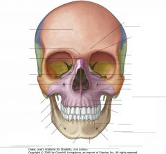
Identify the location of the Symphysis Menti.
|
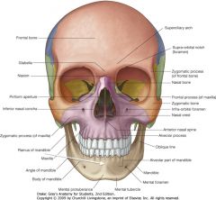
It is the medial ridge above the mental protuberance, indicated by the white arrow.
|
|
|

White arrow.
|
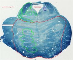
Middle Cerebellar Peduncle
|
|
|

What gyrus is indicated by the blue arrow?
|

The Parahippocampal Gyrus.
|
|
|
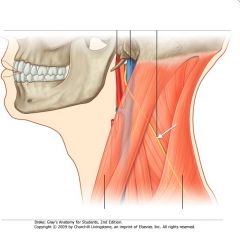
What nerve is indicated by the white arrow?
|
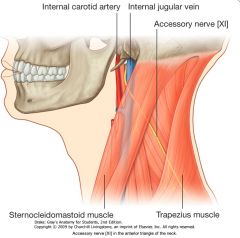
The Spinal Accessory Nerve (Cranial Nerve XI)
|
|
|

White arrow.
|

Superior Cerebellar Peduncle
|
|
|

What structure is indicated by the blue arrow?
|

The Uncus
(a prominent bump on the Parahyppocampal Gyrus) |
|
|
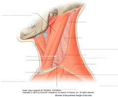
What muscle is indicated by the white arrow?
|

The Scalenus Anterior (or Anterior Scalene) muscle
|
|
|

White arrow.
|

Medial Lemniscus
|
|
|

What (dissected out) nerve is indicated by the blue arrow?
|

The Optic Nerve.
|
|
|

What muscle is indicated by the white arrow?
|
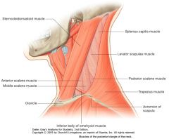
The Scalenus Medius (or Middle Scalene) Muscle
|
|
|
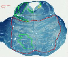
White arrow.
|

Spinal Lemniscus
|
|
|

What structure is indicated by the blue arrow?
|

The Optic Chiasm (or chiasma)
|
|
|
|
What is the function of the scalenus (scalene) muscles?
|

They assist with flexion and lateral flexion of the neck.
|
|
|

White arrow.
|

Mesencephalic Nucleus and Tract of CN V.
|
|
|

What happens at the Optic Chiasma?
|

There is a partial crossing of the Optic Nerves.
|
|
|
|
Describe the innervation of the scalenus muscles.
|
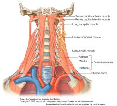
They are innervated by anterior rami of cervical spinal nerves.
|
|
|
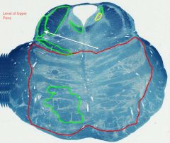
White arrow.
|

Lateral Lemniscus
|
|
|
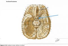
What structure is indicated by the blue arrow?
|

An Optic Tract.
|
|
|

What nerves are indicated by the white arrow?
|
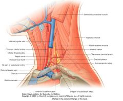
The Roots of the Brachial Plexus
|
|
|

White arrow.
|

Medial Longitudinal Fasciculus.
|
|
|

Where do the Optic Tracts terminate?
|
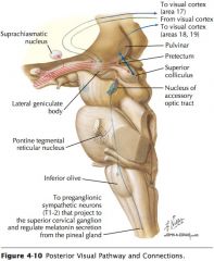
Mainly in the lateral geniculate bodies of the thalamus.
|
|
|

What is the artery indicated by the white arrow?
|

The Subclavian Artery
|
|
|

White arrow.
|
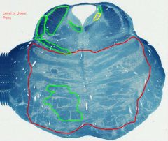
Superior Cerebellar Peduncle
|
|
|

What structure is indicated by the blue arrow?
|

A Mamillary Body
|
|
|

What is the vein indicated by the white arrow?
|

The Subclavian Vein
|
|
|

White arrow.
|

Pontine Nuclei
|
|
|

What are the Mamillary Bodies?
|
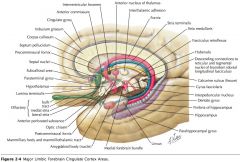
Two hypothalamic nuclei that receive input from the limbic lobe via the fornix.
|
|
|

What muscles in the neck does the Subclavian Artery pass between?
|

It passes laterally between the Anterior and Middle Scalene Muscles (Scaleni Anterior and Medius)
|
|
|
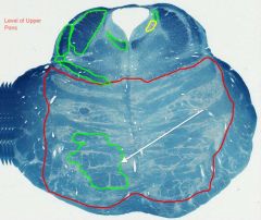
White arrow.
|

Corticospinal and Corticopontine Tracts.
|
|
|

Which division of the brainstem is indicated by the blue arrow?
|

The Medulla Oblongata.
|
|
|

What bones does the Subclavian Vein pass by?
|

It passes posteriorly to the Clavicle, and anteriorly to the First Rib (and the Anterior Scalene muscle).
|
|
|
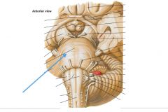
Which division of the brainstem is indicated by the blue arrow?
|

The (Basilar) Pons
|
|
|

What nerve is indicated by the white arrow?
|

The Phrenic Nerve
|
|
|

What is the source of the Phrenic Nerve?
|

It arises from the 3rd, 4th and 5th cervical spinal nerves.
|
|
|
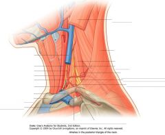
What does the Phrenic Nerve innervate?
|

It supplies motor fibres to the diaphragm.
|
|
|

What is the artery indicated by the white arrow?
|

The Common Carotid Artery
|
|
|

Which division of the brainstem is indicated by the blue arrow?
|

The (Basilar) Pons
|
|
|

What is the artery indicated by the white arrow?
|

The External Carotid Artery.
|
|
|

What is the artery indicated by the white arrow?
|

The Internal Carotid Artery.
|
|
|
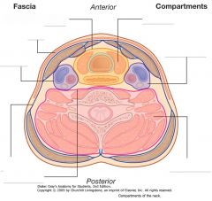
What are the contents of the Carotid Sheath?
|
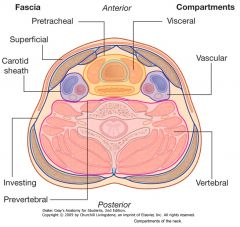
The Carotid Arteries
The Internal Jugular Vein The Vagus Nerve (CN X) |
|
|

Which structure is indicated by the blue arrow?
|

The Olive of the Medulla Oblongata.
|
|
|
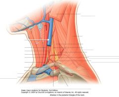
What vessel is indicated by the white arrow?
|

The Internal Jugular Vein
|
|
|
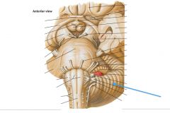
Which region of the brain is indicated by the blue arrow?
|

The Cerebellum
|
|
|

What nerve is indicated by the white arrow?
|

The Vagus Nerve (CN X)
|
|
|

What is the branching white matter indicated by the blue arrow?
|

The Arbor Vitae (the Tree of Life)
|
|
|
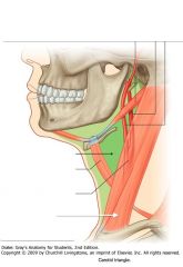
What muscle is indicated by the white arrow?
|
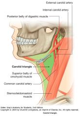
The Sternocleidomastoid
|
|
|
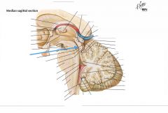
Which space is indicated by the blue arrow?
What does it do? |

The Cerebral Aqueduct, connecting the Third and Fourth Ventricles.
|
|
|
|
What are the subdivisions of the Anterior Triangle of the neck?
|

The Carotid Triangle
The Submandibular Triangle (and the submental and muscular triangles) |
|
|

What structure is indicated by the blue arrow?
|

The Corpora Quadrigemina (Quadrigeminal Plate)
|
|
|
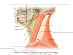
What subdivision of the Anterior Triangle of the neck is indicated by the white arrow?
|
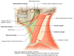
The Carotid Triangle
|
|
|

What structure is indicated by the blue arrow?
|

A Superior Colliculus of the Quadrigeminal Plate
(there are two superior colliculi) |
|
|

What subdivision of the Anterior Triangle of the neck is indicated by the white arrow?
|

The Submandibular Triangle
|
|
|
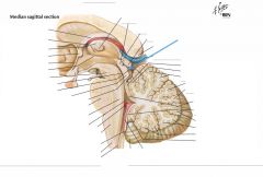
What is the function of the Superior Colliculi of the Corpora Quadrigemina?
|

They are involved with visual functions.
|
|
|
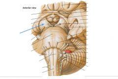
What structure is indicated by the blue arrow?
|

The Basis Pedunculi
(The Cerebral Crus is the anterior surface of the Cerebral Peduncle, another name for Basis Pedunculi) |
|
|

What are the borders of the Submandibular Triangle?
|

Superiorly: the inferior border of the mandible.
Inferiorly: the anterior and posterior bellies of the Digastric Muscle. |
|
|

What surface is indicated by the blue arrow?
|

The Posterior Perforated Substance.
|
|
|
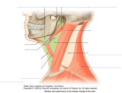
What are the borders of the Carotid Triangle?
|

Anteroinferiorly: the superior belly of the Omohyoid Muscle.
Superiorly: the Stylohyoid Muscle and the posterior belly of the Digastric Muscle. Posteriorly: the anterior border of the Sternocleidomastoid. |
|
|

What is the space located anterior to the Posterior Perforated Surface?
|

The Interpeduncular Fossa.
|
|
|

What muscle is indicated by the blue arrow?
|

The posterior belly of the Digastric Muscle
|
|
|
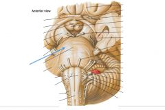
Describe the fibers of the Basilar Pons.
|

They are Transverse Fibers, and they cross over the anterior surface of the brainstem and continue into the Cerebellum.
|
|
|

What muscle is indicated by the white arrow?
|

The superior belly of the Omohyoid Muscle
|
|
|
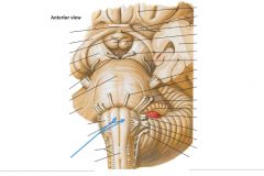
What tracts are indicated by the blue arrows?
|

The Pyramidal Tracts of the medulla.
|
|
|

What vessel is indicated by the white arrow?
|

The Superior Thyroid Artery.
|
|
|
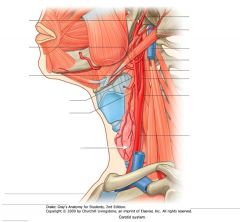
What structure is indicated by the white arrow?
|
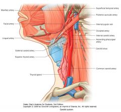
The Thyroid Gland.
|
|
|
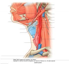
What artery is indicated by the white arrow?
|

The Lingual Artery
|
|
|

What course does the Lingual Artery characteristically take by the Hyoid Bone?
|

The Lingual Artery often has a characteristic upward bend just above the greater horn of the Hyoid Bone.
|
|
|

What artery is indicated by the white arrow?
|

The Facial Artery.
|
|
|
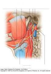
What nerve is indicated by the white arrow?
|
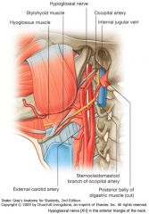
The Hypoglossal Nerve (CN XII)
|
|
|

What does the Hypoglossal Nerve innervate?
|

The Hypoglossal Nerve (CN XII) provides motor innervation to the tongue muscles.
|
|
|

What nerve is indicated by the white arrow?
|

The Internal Laryngeal Nerve,
a branch of the Superior Laryngeal Nerve, off the Vagus (CN X). |
|
|
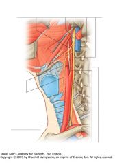
What does the Internal Laryngeal Nerve innervate?
|
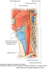
The Internal Laryngeal Nerve provides sensory and parasympathetic fibres to the mucous membrane of the larynx above the level of the vocal cords.
|
|
|

What gland are indicated by the white arrows?
|

The Parathyroid Glands
|
|
|
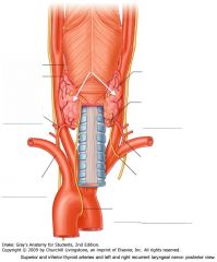
What structure is indicated by the white arrows?
|

The Thyroid Gland
|
|
|
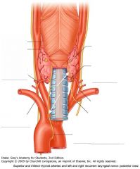
What nerves are indicated by the white arrows?
|

The right and left Recurrent Laryngeal Nerves.
|
|
|

Why are the Recurrent Laryngeal Nerves important to remember for Thyroidectomies?
|

They need to be protected during surgery to prevent paralysis of the vocal cords.
|
|
|
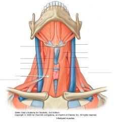
What muscle is indicated by the white arrow?
|
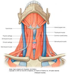
The Sternohyoid Muscle.
|
|
|

What muscle is indicated by the white arrow?
|

The Sternothyroid Muscle
|
|
|

What muscle is indicated by the blue arrow?
|

The Thyrohyoid Muscle
|
|
|
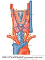
What feature of the Thyroid is indicated by the white arrows?
|
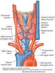
The Lateral Lobes.
|
|
|
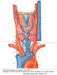
What feature of the Thyroid is indicated by the white arrow?
|

The Isthmus
|
|
|
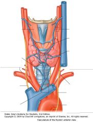
What feature is immediately posterior to the Isthmus of the Thyroid Gland?
|

The 2nd-4th Tracheal Rings.
|
|
|
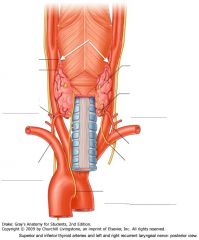
What vessels are indicated by the white arrows?
|

Superior Thyroid Arteries
|
|
|

What vessels are indicated by the white arrows?
|

Inferior Thyroid Arteries
|
|
|

What does the Inferior Thyroid Artery branch from?
|

It is a major branch of the Thyrocervical Trunk, off of the Subclavian Artery.
|
|
|

Describe the course of the right Recurrent Laryngeal Nerve.
|

It branches from the Vagus Nerve, wraps around the Subclavian Artery, and re-ascends to its sites of innervation up to the larynx.
|
|
|
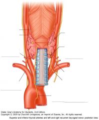
Describe the course of the left Recurrent Laryngeal Nerve.
|

It branches from the Vagus Nerve, wraps around the Arch of the Aorta, and re-ascends to its sites of innervation up to the larynx.
|
|
|

What muscle is indicated by the white arrows?
|
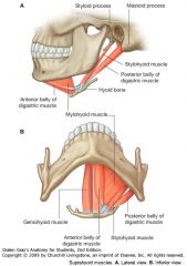
The Anterior belly of the Digastric Muscle
|
|
|

What muscle is indicated by the white arrows?
|

The Posterior Belly of the Digastric Muscle.
|
|
|
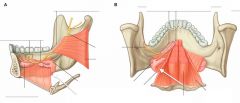
What structure is indicated by the white arrows?
|
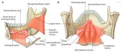
The Superficial Part of the Submandibular Gland
|
|
|

What muscle is indicated by the blue arrows?
|

The Mylohyoid Muscle
|
|