![]()
![]()
![]()
Use LEFT and RIGHT arrow keys to navigate between flashcards;
Use UP and DOWN arrow keys to flip the card;
H to show hint;
A reads text to speech;
23 Cards in this Set
- Front
- Back
|
"Stroke" Clinical DX |
80-85% Ischemic Infarct 15-20% Hemorrhage |
|
|
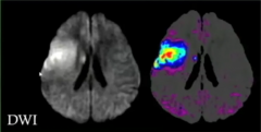
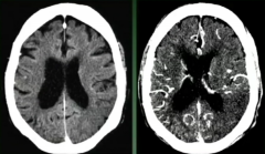
Differing Degrees of Restriction Diffusion Can Bring This Out with Wider Window Levels Central Area- Definite Infarct Peripheral Areas May Not Be Infarct Diffusion Not All or None There is a Spectrum |
|
|
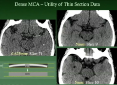
Dense Vessel Thrombus Seen Better on Thin Slices! |
|
|
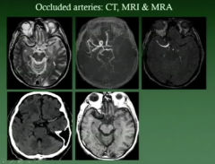
T2 Axial Would Miss Occluded T2 MCA Vessel. Better To Look at Other Images! |
|
|
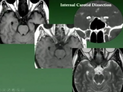
Intracranial Carotid Dissection Seen Subtlely on ax T2.
See on ax T1 easily.,
See on for CTA |
|
|
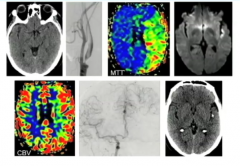
CTA Source DATA poor man's perfusion map |
|
|
MTT Shows Left MCA Decreased Perfusion
CBV shows some increased MCA perfusion.
This indicates collateral vessels supplying MCA needs.
F/U Patient Did Well |
|
|
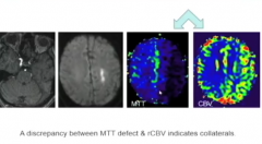
MRI source occluded left ICA Small diffusion restriction ( Must be collaterals ) Decrease in left MCA MTT Increase in left MCA CBV (Indicating good collaterals) |
|
|
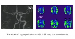
Paradoxical Hyperperfusion on ASL CBF map due to collaterals. Slower flow on right side, so recover more signal.
|
|
|
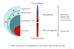
Tissue Saved in Penumbra may Appear Normal-- But may not be normal- micorcystic areas of infarction- red dots |
|
|
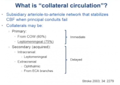
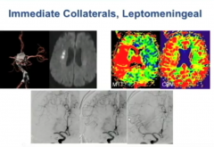
Collateral Circulation Immediate: Most Important Cow 60% Leptomeningeal 75% Delayed Intracanial Leptomeningeal Extracranial Ophthalmic / From ECA Branches |
|
|
Occluded MCA small infarct core on DWI Large Penumbra on MTT Increase Perfusion on CBV Due to Collaterals Angio-- Occluded MCA with good collaterals After MCA Opened, get disappearance of leptomeningeal collaterals Dynamic Leptomeninges collateralCome andGo |
|
|
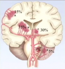
Hypertensive Hemorrhage 2001 NEJM 35% Basal Ganglia 30% Medial Thalamus 15% Cortical/Lobar 5% Medial Cerebellum-Deep Gray 1% Pons |
|
|
|
|
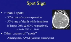
May Indicate Expanding Hematoma |
Other Causes Spot Sign - Aneurysm <<<AVM |
|
|
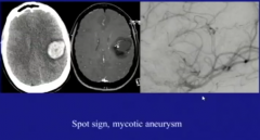
Another Cause Spot Sign Mycotic Aneurysm |
|
|
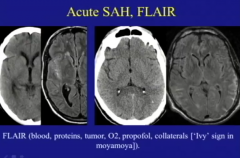
Acute SAH on Flair 1. Blood 2. Proteins- Meningitis 3. Tumor- Leptomeningeal tumor 4. O2 Administration (SAS- Not in Ventricle) 5. Propofol 6. Collaterals [Ivy sign moyamoya] |
|
|
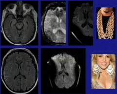
Fake Out on Flair Images Can Mistake for SAH
Bright Signal SAS with metal artifacts 1. Dental amalgam/braces frontal lobe 2. Neck chain 3. Ear Rings |
|
|
Flair Best for Supratentorial SAH
SWI Better for Intratenorial SAH |
|
|
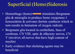
Superficial Siderosis Due to Bergmann Glia & Microglia >Produces Ferritin Only Need One Bleed |
|
|
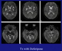
Levy AJNR 2012 Patients May Improve with Chelating Agents One Yr Later with Deferipone |
|
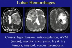
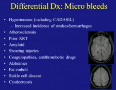
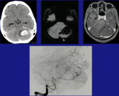
Hypertension, GBM, AVM |
Lobar Hemorrhages 1. Hypertension 2. Amyloid >70 Y/O 3. Anticoagulation 4. AVM 5. Mycotic Aneurysms 6. 1 and 2 Tumors 7. Venous Thrombosis |
|
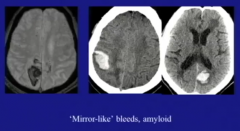
Amyloid "Mirror Like Bleeds" |
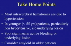
Amyloid "Mirror Like Bleeds" |