Reading...
![]()
Play button
![]()
Play button
![]()
Use LEFT and RIGHT arrow keys to navigate between flashcards;
Use UP and DOWN arrow keys to flip the card;
H to show hint;
A reads text to speech;
66 Cards in this Set
- Front
- Back
|
Eye
3 layers outer to inner |
sclera
choroid retina |
|
|
Eye
Sclera (characteristics) |
tough, white tissue expect anterior-most region (cornea) which is transparent
|
|
|
Eye
Choroid layer (highly _, includes _ _) |
highly vascularized
includes ciliary body (a muscle) and iris |
|
|
Eye
Retina (includes _ _) |
includes the photoreceptors and the pigmented epithelium
|
|
|
Cornea
(function) |
coarse focus, not adjustable
|
|
|
Anterior chamber
(space b/w) |
space b/w iris & cornea
|
|
|
Posterior chamber
(space b/w) |
space b/w lens & iris
|
|
|
Aqueous humor
(function) |
supplies nutrients to cornea and lens (fills anterior and posterior chambers)
|
|
|
Vitreous humor
(function) |
maintains eye shape
also removes blood and cellular debris from the eye |
|
|
Iris
(function) |
controls amount of light that enters the eye
|
|
|
Lens
(function) |
fine focus
adjustable |
|
|
Retina
(function) |
actual site of photoreception and transmission to brain
|
|
|
Pigmented epithelium
(function) |
absorbs light, thereby increasing acuity
supplies nutrients to & removes debris from the retinal layer |
|
|
Glaucoma
(caused by, results in) |
caused by increase in pressure in anterior and posterior chambers due to accumulation of aqueous humor in these two chambers
any swelling is then transmitted to vitreous body resulting in increase in pressure throughout eye this reduces blood supply to eye, causing damage to retina |
|
|
in the center of the retina is a region called _
|
macula lutea
|
|
|
in the center of the macula is a depression called the _
|
fovea
|
|
|
fovea
(depression due to, region of _, thinning of the layers does what, also absence of _) |
due to thinning of layers of eye that are over this part of the retina
region of highest visual acuity in retina due to exclusive presence of cones in the region thinning of the retinal layers minimizes the distortion of light before it reaches the photoreceptors also absence of blood vessels around the fovea for same reason |
|
|
macula lutea
|
region in center of retina
|
|
|
macula/fovea specialized for _ vision, while regions outside the macula are responsible for _ vision
|
central
peripheral |
|
|
pale, circular region next to macula
|
optic disc
|
|
|
optic disc
(opening in eye through which _ enter and _ exit to form _, no _ in this region resulting in _) |
blood vessels enter eye
axons from retinal ganglion cells exit to form optic nerve no photoreceptors = blind spot in visual field |
|
|
Papilledema
(what, indicative of, causes) |
swelling of the optic disc
indicative of swelling in the brain due to trauma, infection, or tumor swelling due to these causes will cause increase in intracranial pressure that will be transmitted throughout CSF, which includes the junction where optic nerve enters the eye |
|
|
Macular Degeneration
(loss of _, what compromised) |
involves loss of photoreceptor cells in retina limited to macula
central-vision dependent tasks compromised, peripheral unaffected blurred vision or darkened areas in central vision total blindness not observed but reading, driving severely disrupted distance vision unaffected 2 categories: "dry" and "wet" |
|
|
Focusing apparatus of eye are _ _ _
|
cornea, lens, iris
|
|
|
Light that enters eye must be refracted in order to focus image on retina
Refraction carried out by _ but _ responsible for _ It does this by changing shape: far vision requires _ near vision requires _ |
lens and cornea
lens = fine adjustments in focus far vision - relatively flat and thin lens near vision - thicker and rounder lens |
|
|
Lens shape controlled by _ and _
|
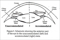
ciliary muscles and zonule fibers
(see pg. 5 for how they work) |
|
|
accommodation
|
contraction of ciliary muscles is associated with focusing the lens for near vision
|
|
|
Decreases in pupil size _ the level of visual resolution or clarity
|
increase
|
|
|
changes in _ also contribute to level of resolution of a visual image
|
pupil size
|
|
|
pupil size controlled by _
|
dilation and constriction of iris
|
|
|
Cataracts
|
clouding of lens that disrupts the passage of light through the lens resulting in blurring of vision
cataracts occur when the proteins that make up the lens (lens is mostly water and protein fibers) start to degenerate resulting fragments clump together and form the cataract untreated, can cause blindness |
|
|
Retina
(part of the eye where _, damage = ?) |
where actual sensing of light occurs
part of CNS and damage leads to permanent loss of vision since these neurons can't regenerate |
|
|
Retinal layers
(name - starting with outermost) |
pigment epithelium layer
layer of photoreceptor outer segments external limiting membrane layer outer nuclear layer outer plexiform layer inner nuclear layer inner plexiform layer ganglion cell layer optic nerve layer |
|
|
Retina
Pigment epithelial layer |
cuboidal cells containing melanin
this black pigment absorbs any light that is not captured by photosensitive cells in the retina, preventing light from reflecting back to retina which would: (1) lead to degeneration of visual signal on retina (2) potentially allow damaging levels of light onto the retina photoreceptors this layer also provides nutritional support to photoreceptors |
|
|
retinal detachment
|
connection b/w pigment epithelium and rest of the retina is not very strong and there is potential for separation = retinal detachment
leads to damage/loss of photoreceptors due to loss of nutritional support |
|
|
layer of photoreceptor outer segments
|
consists of outer segments of the rods and cones
|
|
|
external limiting membrane layer
|
simply a membrane that the rods and cones pass through
|
|
|
outer nuclear layer
|
contains cell bodies of rods and cones
|
|
|
outer plexiform layer
|
contains synaptic connections made by the rods & cones onto the bipolar cells
|
|
|
inner nuclear layer
|
contains cell bodies of the bipolar cells
|
|
|
inner plexiform layer
|
contains synaptic connections made by the bipolar cells onto the ganglion cells
|
|
|
ganglion cell layer
|
contains cell bodies of ganglion cells
|
|
|
optic nerve layer
|
contains axons of the ganglion cells on their way to form the optic nerve
|
|
|
Retinitis Pigmentosa (RP)
|
inherited visual disorder
characterized by progressive vision loss due to degeneration of photoreceptors early symptoms: night blindness, reduction of visual field, thinning of retinal blood vessels, formation of clumps of pigment within the retina pigment is from pigmented epithelial layer disrupted by the disease night blindness = rods lost first, cones doing all photoreception as disease progresses, cones also lost = blind RP really several diseases with similar symptoms |
|
|
photoreception
(what, carried out by) |
conversion of photostimuli (light) into a neurosignal
rods and cones |
|
|
Cones vs. Rods
sensitivity to light |
Cones:
low (100 times less sensitive) Rods: high (one rod can respond to single photon of light) |
|
|
Cones vs. Rods
Distribution |
Cones:
high in fovea very low in rest of retina Rods: absent in fovea high in rest of retina |
|
|
Cones vs. Rods
Color vision |
Cones:
Yes Rods: No (achromatic) |
|
|
Cones vs. Rods
Visual Acuity |
Cones: high
Rods: low |
|
|
Cones vs. Rods
Relative abundance |
Cones:
fewer cones than rods Rods: 20 times more |
|
|
Cones vs. Rods
Specialized for |
Cones:
day vision Rods: night vision (rod responses saturate in daylight) |
|
|
Cones vs. Rods
Effects of damage |
Cones:
loss of cones = blindness Rods: loss causes night-blindness and loss of peripheral vision |
|
|
Rods are _ to light, but have _ spatial resolution
|
extremely sensitive
low |
|
|
Cones are _ to light, but have _ spatial resolution
|
relatively insensitive to light
high |
|
|
Rods use _ photopigment(s)
Cones use _ photopigment(s) |
rods - one:
**rhodopsin** cones - three: S (short) = violet M (middle) = green L (long) = yellow A single cone uses only one type (length refers to the wavelength of light that each pigment has the shortest absorbance to) |
|
|
How do we detect so many colors with only 3 photopigments?
|
visual system compares the relative activities of cones with different photopigments to determine what color (what wavelength) the light is
|
|
|
Color Blindness
|
caused by lack of cones that are sensitive to a particular color
most common form is red-green (6% of population) which is due to loss of the middle cone photopigment green or blue color blindness exist also (blue is uncommon) red-green blindness is inherited via recessive allele on X-chromosome and is more common in males than females |
|
|
Phototransduction
(how do rods and cones respond to light, the response results in _) |
*rods and cones do not respond to light by firing action potentials, but instead respond with graded changes in membrane potential
this response to light results in hyperpolarization of rod/cone membrane potential |
|
|
Flow of info thru retina
|
light strikes the rods/cones>
hyperpolarizes rods/cones decreasing neurotransmitter release > bipolar cells are depolarized increasing their neurotransmitter release > Ganglion cells are depolarized & action potentials are initiated |
|
|
light adaptation
|
process of adapting to overall levels of illumination (like going from inside to outside)
|
|
|
Refractive errors = ametropia
(name) |
Myopia
Hypermetropia Emmetropia Astigmatism Presbyopia |
|
|
Myopia
|
cannot bring distant objects into focus (nearsighted)
caused by the cornea being too curved or the eyeball being too long |
|
|
Hypermetropia
|
cannot bring near objects into focus (farsighted)
caused by eyeball being too short |
|
|
Emmetropia
|
normal vision
|
|
|
Astigmatism
|
either the lens or cornea do not have a uniform curvature, therefore the light rays do not all get focused onto the same point
|
|
|
Presbyopia
|
aging leads to decreases in elasticity of lens,
making it increasingly difficult for lens to obtain appropriate curvature when the ciliary muscle contract to focus on near objects the minimal distance at which we can focus upon near objects gets farther away, impairing detailed near vision tasks (reading) |