Reading...
![]()
Play button
![]()
Play button
![]()
Use LEFT and RIGHT arrow keys to navigate between flashcards;
Use UP and DOWN arrow keys to flip the card;
H to show hint;
A reads text to speech;
375 Cards in this Set
- Front
- Back
|
What does the prosencephalon further subdivided into?
|
telencephalon
diencephalon |
|
|
What does the rhombencephalon further subdivide into"?
|
metencephalon
myelencephalon |
|
|
What does the telencephalon form?
|
lateral ventricles
cerebral cortex basal ganglia rhinencephalon semioval cent |
|
|
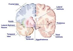
What does the diencephalon form?
|
third ventricle
thalamus hypothalamus epithalamus subthalamus |
|
|
What does the metencephalon form?
|
forth ventricle
pons cerebellum |
|
|
what does the myelencephalon form?
|
medulla oblongata
|
|
|
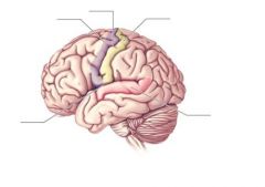
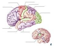
What are the 5 lobes of the telencephalon?
|
frontal
parietal occipital temporal insula |
|
|
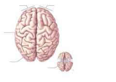
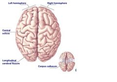
What divides the left and right hemispheres of the brain?
|
longitudinal fissure
|
|
|
Where is the frontal lobe located?
|
occurs between frontal poles to the central sulcus posterior, and the lateral fissures laterally
|
|
|
Where is the parietal lobe located?
|
extends from central sulcus to parieto-occipital sulcus posteriorly
|
|
|
Where is the occipital lobe located?
|
posterior to pareito-occipital sulcus
|
|
|
Where is the temporal lobe located?
|
inferior to the lateral fissure and extends back to the level of the parieto-occipital sulcus
|
|
|
Where is the insula located?
|
lies deep to the lateral fissure and is comprised of 4-5 gyri
|
|
|
DEFINE white matter
|
the cellular processes (axons) of cell bodies
|
|
|
DEFINE grey matter
|
the cell bodies/dendrites of neurons (unmyelinated=grey)
makes up cerebral cortex |
|
|
What is the thickness of grey matter inside the brain dependent on?
|
size of gyrus
4.5mm in precentral gyrus 1.5mm in calcarine gyrus |
|
|
What is the hystological layers of the cortex from superficial to deep?
|
molecular
external granular external pyramidal internal granular internal pyramidal polymorphic |
|
|
How are the hystological layers organized?
|
named by type of neuron presend
density of cells arrangement of cells |
|
|
if the cytoarchitecture of the hystological layers is disrupted, what occurs?
|
cortex dysfunction
|
|
|
What is functional cortical mapping based on?
|
pathological data
electrostimulation data blood flow data metabolic data |
|
|
DEFINE pathological data
|
crude approach, made assumptions based on area of the brain that is injured and the behavior ensued
|
|
|
DEFINE electrostimulation data
|
tap areas of the brain with current and see what happens; often used in neurosurgery to make sure the right spot is operated on
|
|
|
DEFINE blood flow data
|
functional MRI (fMRI); there is an increase or decrease in blood flow as a result of a task
|
|
|
What is the difference between a fMRI and a MRI?
|
during fMRI, pt does a task while in MRI tube and physicians observe which area of the brain has a "hot spot"
|
|
|
What are some limitations of fMRI?
|
the lighting up jdoes not occur in real time
do not know if area is excitatory or inhibitory |
|
|
what are 2 scans to detect metabolic data of the brain?
|
PET scan
SPECT |
|
|
DEFINE: PET scan
|
pt is injected w/ radioactive isotope with a short half-life that binds to oxygen and glucose; pt performs a task and the area active has a greater emission of photons
|
|
|
DEFINE: SPECT
|
Single photon emitted CT scan
|
|
|
DEFINE historical maps
|
based on pathological and electrostimulation data
|
|
|
What are the 3 main historical maps?
|
Campbell (1905) mapped 20 areas
Broadmann (1909) mapped 47 areas Economo (1929) mapped 97 areas |
|
|
Which historical map is still used as a reference today?
|
Economo's map
|
|
|
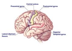
LOCATION primary motor cortex
|
located in precentral gyrus of frontal lobe, in front of central sulcus
|
|
|
What is the area anterior to the precentral gyrus?
|
an association area to the primary motor cortex
|
|
|
FUNCTION primary motor cortex
|
controls voluntary skeletal mm contraction on the contralateral side through UMN
|
|
|
What is the system that the primary motor cortex is considered a part of?
|
pyramidal system
|
|
|
How is the precentral gyrus organized?
|
somatotopically (specific part of gyrus controls certain part of body)
|
|
|
DEFINE motor unit
|
ratio of LMN to number of mm fibers being innervated
|
|
|
LOCATION primary sensory cortex
|
occurs in the postcentral gyrus (behind central sulcus) of the parietal lobe
|
|
|
FUNCTION primary sensory cortex
|
allows discrimination and location of sensory stimuli on contralateral side
|
|
|
What happens in sensory info does not reach this cortex?
|
person is not conciously aware of sensory stimuli; therefore, cannot respond to it
|
|
|
can a pt still perceive pain if the post central gyrus is damaged/removed?
|
yes, through the thalamus
|
|
|
LOCATION primary visual cortex
|
in occipital lobe, adjacent to calcarine fissure
|
|
|
What is another name for the primary visual cortex?
|
area #17
|
|
|
FUNCTION primary visual cortex
|
images are received inverted, then the visual cortex flips them righ side up to make sense of them
|
|
|
What will damage to the primary visual cortex cause?
|
blindness; despite intact eyes, nerves, aqueous humor, etc.
|
|
|
LOCATION primary auditory cortex
|
superior temporal gyrus of the temporal lobe
|
|
|
FUNCTION primary auditory cortex
|
helps us to perceive sound
|
|
|
what will damage to the primary auditory cortex cause?
|
deafness
|
|
|
LOCATION personality/intellect cortex
|
anterior portion of the frontal lobe
|
|
|
FUNCTION personality/intellect cortex
|
forms personality, intellect, psychic, abstract, self-control, cognition
|
|
|
What will damage to the personality cortex cause?
|
malfunction of all functions
see-Phineas Gage |
|
|
FUNCTION olfactory cortex
|
perceive smell
|
|
|
What are the 2 areas of the olfactory cortex?
|
lateral olfactory stria
medial olfactory stria |
|
|
LOCATION lateral olfactory stria
|
lateral ot optic chiasm and terminates at the uncus in the parahippocampal gyrus.
|
|
|
FUNCTION lateral olfactory stria
|
conscious olfactory response
|
|
|
LOCATION medial olfactory stria
|
medial and under the optic chiasm
|
|
|
FUNCTION gustatory cortex
|
perceive taste
|
|
|
What are the 3 areas of the gustatory cortex?
|
1. most ventral portion of postcentral gyrus (parietal lobe)
2. insular cortex (insula) 3. frontal operculum (frontal lobe) |
|
|
FUNCTION association areas (general)
|
refinement and interpretation of cortexes
|
|
|
LOCATION association areas (general)
|
adjacent to or surround major cortical areas
|
|
|
What occurs when an association area is damaged?
|
intact cortex (sensory info) but lesion in association area can cause senses to be less refined/duller
|
|
|
DEFINE motor speech cortex
|
allows a person to initiate speech by influencing portions of precentral gyrus that control skeletal mm for speech (larynx, pharynx, tongue, face, mouth)
|
|
|
LOCATION Broca's area
|
located around the inferior frontal gyrus in the frontal lobe
|
|
|
FUNCTION Broca's area
|
primary cortex for motor speech, but technically an assocaition area of the frontal lobe that finesses the precentral gyrus
|
|
|
What does damage to Broca's area cause?
|
aphasia
|
|
|
DEFINE aphasia (general)
|
a general term for language disorders to include reading, writing, speaking, or comprehension of written and spoken words due generally to cerebral cortex or conduction dysfunction
|
|
|
DEFINE Broca's aphasia
|
caused by damage to Broca's area, generally in stroke of middle cerebral artery; pt cannot or has difficulty forming words even though vocal cordes and innervations are normal
|
|
|
What area's of language are compromised in Broca's aphasia?
|
Verbally and Graphically (writting) compromised
|
|
|
DEFINE non-fluent
|
words do not flow, despite the ability to perceive language and organize thought processes
|
|
|
Are pts with broca's aphasia aware that they cannot get words out?
|
yes, can cause them to get very frustrated
|
|
|
What other physiological symptoms can occur with Broca's aphasia?
|
menianopsia (loss of 1/2 a visual field) and paralysis of facial mm on rt bc optic pathway and internal capsule are close to Broca's area
|
|
|
FUNCTION wernicke's area
|
language cortex
|
|
|
LOCATION wernicke's area
|
90% located in the posterior part of the superior temporal gyrus
10% extends into parietal lobe (still considered functional part of temporal) |
|
|
FUNCTION wernicke's area
|
controls comprehension of spoken words and written and auditory language
|
|
|
In which hemisphere is wernicke and broca's area more dominant in 90% of people?
|
left
|
|
|
DEFINE wernicke's aphasia
|
involved w/ comprehension of spoken and written language. able to speak and write words but the sequence is not normal, so they don't make sense.
|
|
|
how will pt respond to their wernicke's aphasia
|
will be aware that they do not make sense and become frustrated
|
|
|
What is compromised if there is a large lesion in wernicke's area?
|
visual (cannot read words) and linguistic ability is compromised
|
|
|
DEFINE empty speech
|
speaking but it does not make sense
|
|
|
DEFINE paraphasia
|
substitute one word for another
|
|
|
DEFINE neologisms
|
create new and meaningless words and put them into sentences
|
|
|
DEFINE jargon aphasia
|
words and phrases are strung together w/ no meaning but seems logical to patient
|
|
|
What artery feeds both wernicke's and broca's area (and therefore affects both if site of stroke)?
|
middle cerebral artery
|
|
|
DEFINE conduction aphasia
|
occurs when something impairs the conduction from Wernicke's to Broca's area
|
|
|
Where is the lesion/damage in conduction aphasia?
|
lesion destroys the arcuate fasciculus (efferent connection from wernicke's to broca's)
|
|
|
What are characteristics of conduction aphasia?
|
`1. less fluent in language than pts with wernicke's aphasia
2. may make paraphasic errors 3. naming is impaired 4. reading aloud is impaired, but pt can read silently w/ good comprehension 5. writing (Broca's) is abnormal with misspelled and omitted words |
|
|
DEFINE global aphasia
|
most sever form of aphasia; inability to use language in any form due to extensive damage to Broca's, Wernicke's, and arcuate fasciculus; LINGUISTICALLY and VERBALLY compromised (unable to read/write well, unable to comprehend speech, unable to produce jintelligible speech)
**stroke that causes this has low survival rate** |
|
|
LOCATION memory cortex
|
2 areas w/in temporal lobe
|
|
|
what are the 2 areas that make up the memory cortex?
|
hippocampus of temporal lobe
Amygdala nuclear complex |
|
|
LOCATION hippocampus of temporal lobe
|
deep to the parahippocampus
|
|
|
what does damage to the hippocampus cause?
|
anterograde amnesia (would remeber events before accident but no events after)
|
|
|
FUNCTION amygdala nuclear complex
|
processing and retaining memory; if it doesn't pass through here, it doesn't get remembered.
|
|
|
if you're going to remember something consciously what parts of the brain must process this info?
|
hippocampus and amygdala nuclear complex
|
|
|
What causes UMN paralysis to the contralateral side of the body?
|
damage to the precentral gyrus; contains UMN that feed LMN on the opposite side of the body--as well as teh association motor cortex areas
|
|
|
DEFINE spastic
|
wiped out UMN but LMN is still intact; mm are still innervated due to sensory input; reflexes still occur, but there is no control bc the umn is not working to influence LMN
*due to damage in internal capsule that contains UMN axons |
|
|
What can cause loss of general sensation to contralateral side of the body?
|
damage to postcentral gyrus of the parietal lobe
|
|
|
Where is instantaneous spatial coordination of all parts of the body integrated at?
|
posterior parietal lobe
|
|
|
Which parietal lobe is more dominant and involved in spatial organization?
|
right
|
|
|
What can damage to the posterior part of the right parietal lobe cause?
|
deficit in perception of person and spatial relationships (L-sided neglect)
|
|
|
DEFINE hemi-innattention
|
ignoring due to a lack of integration of senses with the rest of the body
-non-movement of L extremitiy -lack of awareness of sensory stimuli -lack of personal hygiene and grooming -pt plans movement around the rt side of the body |
|
|
DEFINE bilateral spatial neglect
|
cuased by damage to R parietal lobe. lack of understanding of spatial relationships; pt does no include the left side in anything (paint only R side of face)
|
|
|
Can R sided neglect occur?
|
yes, if severe damage to left parietal lobe--must be SUBSTANTIAL damage.
**survival rate is low** |
|
|
DEFINE apraxia
|
inability to carry out or regulate a complex or skilled movement when there is no LMN paralysis, no ataxia (loss of coordination), loss of sensory input, and the pt is not confused
|
|
|
When does apraxia occur?
|
when there is a lesion in the premotor and supplemental motor cortexes.
|
|
|
DEFINE agraphia
|
inability to write, not due to LMN paralysis, ataxia,or sensory input
**may be able to type** |
|
|
DEFINE transmissive apraxia
|
inability to carry out a sequence of skilled motor movements
|
|
|
What part of the brain is effected in transmissive apraxia?
|
supramarginal gyrus in the parietal lobe
|
|
|
DEFINE agnosia (general)
|
inability to perceive sensations through otherwise normally functioning sensory pathways
**sensory info is not consciously integrated** |
|
|
DEFINE tactile agnosia
|
inability to recognize familiar object through touch and proprioception; due to a lesion in the posterior parietal lobe of the dominant hemisphere
|
|
|
DEFINE disturbance of body image
|
due to a parietal lobe lesion; pt may not recognize their thumb from their pinky finger, they can confuse left and right sides.
|
|
|
DEFINE depth agnosia
|
inability to appreciate depth and thickness of objects due to a lesion in the occipital lobe
|
|
|
DEFINE movement agnosia
|
inability to recognize stationary and moving objects due to a lesion in the occipital lobe
|
|
|
DEFINE prosopagnosia
|
inability to recognize faces due to a temporal lobe lesion; pt can see the face, but not know who it belongs to until they hear the voice of the face
|
|
|
Which side of the brain is dominant in 90% of humans? Why?
|
left because that is the hemisphere that controls language in 90% of population
|
|
|
what are left hemisphere dominance characteristics?
|
*contains Broca's and Wernicke's areas
*logical and analytical abilities *general math ability *processing large volumes of info *ability to be rational and pragmatic *what we think vs what we feel *doing-consciousness |
|
|
what are right hemisphere dominance characteristics?
|
*geometric spatial orientation
*musical perception and skills *artistic talent *formation of ideas (non-verbal ideation) *perception and processing of emotions *coordination of sensory info *what we feel vs what we think *being conscious of our environment and emotions |
|
|
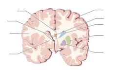
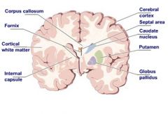
DEFINE semioval center of telencephalon
|
white matter within cerebral hemispheres; made of myelinated axons and dendrites; extends between the cerebral cortex, basal ganglia, and ventricular system
|
|
|
What are the 3 types of fibers of the semioval center?
|
commissural, association, and projection fibers
|
|
|
DEFINE commissural fibers
|
connect corresponding cortical regions of the two hemispheres
|
|
|
what are the 3 main commissural fibers?
|
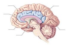
corpus callosum
anterior commissure posterior commissure |
|
|
DEFINE corpus callosum
|
largest of the 3 commisures; millions of fibers that are a primary means of communication between hemispheres.
|
|
|
What are the 4 parts of the corpus callosum?
|
rostrom (connect frontal lobe)
genu (frontal lobes) body (frontal & parietal lobes) splenium (temporal & occipital lobes) |
|
|
DEFINE anterior commissure
|
(telencephalon) cjrosses midline rostrally through the fornix to connect portions of the temporal lobe; functionally involved in olfactory pathways
|
|
|
DEFINE posterior commisure
|
(diencephalon) visual reflexes that rely on optical info; connects superior calliculi and pretectum of the midbrain (reciprocal pathways)
|
|
|
DEFINE association fibers
|
connect cortical regions in the SAME hemisphere
|
|
|
DEFINE short association fibers
|
(arcuate) not specifically named; arch the floor of each sulcus to connect adjacent gyri
|
|
|
DEFINE long association fibers
|
cables, always reciprocal; connect cortical regions in different lobes within the same hemisphere
|
|
|
DEFINE fasciculus
|
long association fibers that form bundles
|
|
|
what does the uncinate fasciculus connect?
|
connects frontal lobe to temporal lobe
|
|
|
what does the arcuate fasciculus connect?
|
frontal, temporal, and occipital lobes; major fasiculus that connects Broca's and Wernicke's areas
|
|
|
what does the cingulum fasciculus connect?
|
primary association bundle on medial side of the hemisphere of the brain; connects parietal, temporal, and frontal lobes; can be surgically removed to decrease pain
|
|
|
What are the 2 association areas that are deep to the insula?
|
external and extreme capsules.
|
|
|
What are the layers of the brain (from superficial to deep) when coming from the lateral side in a horizontal cross section of the brain?
|
insula-extreme capsule-claustrum-external capsule-corpus striatum-internal capsule
|
|
|
DEFINE projection fibers
|
mostly axons that converge on the brainstem; connect one specific part of the cerebral cortex and another part of the CNS (reciprocal);
|
|
|
DEFINE corona radiata
|
radiating mass bc everything has to narrow into the brain stem
|
|
|
DEFINE internal capsule
|
compact band of projection fibers formed rostrally. composed of an anterior limb, genu, posterior limb
|
|
|
DEFINE anterior limb of internal capsule
|
afferent sensory; partially separates the caudate nucleus (medially) and the putamen (laterally)
|
|
|
Which part of the internal capsule is 90% afferent?
|
anterior limb
|
|
|
What specific fibers is the internal capsule composed of?
|
thalamocortical projection fibers (afferent)
frontopontine (efferent) |
|
|
If the anterior limb of the internal capsule is damaged what is effected?
|
sensory
|
|
|
What are the boundaries of the anterior limb of the internal capsule?
|
medially by the head of the caudate nucelus
laterally by the putamen |
|
|
Which part of the internal capsule is 90% efferent?
|
posterior limb
|
|
|
What are the boundaries of the posterior limb of the internal capsule?
|
medially by the thalamus
laterally by the globus pallidus |
|
|
What sort of fibers make up the posterior limb of the internal capsule?
|
thalamocortical (afferent)
corticofugal (efferent) |
|
|
What are the 4 corticofugal fibers found in the posterior limb of the internal capsule?
|
corticothalamic
corticopontine corticobulbar (to cranial nn) corticospinal |
|
|
DEFINE genu of the internal capsule
|
tratransition area between limbs that contains both afferent and efferent fibers
|
|
|
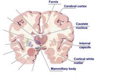
DEFINE fornix
|
made up of commissural and projection fibers; connects telencephalon and diencephalon with reciprocal movement of into
|
|
|
DEFINE fimbria of the fornix
|
formed from the axons of the pyramidal neuronsof the hippocampus
|
|
|
how far to the fimbria of the fornix reach? what do they become?
|
axons proceed forward until they reach the posterior end of the hippocampus. they then arch beneath the splenium of the corpus callosum where they become the crura of the fornix
|
|
|
DEFINE crura of the fornix
|
a bilateral structure connected by the fornical commissure; converge and form the body of the fornix
|
|
|
DEFINE body of the fornix
|
runs forward under the corpus callosum to the rostral margins of the thalamus where it bifurcates and forms the 2 anterior column of the fornix
|
|
|
What does the anterior columns of the fornix divide into?
|
postcommisural fibers
precommissural fibers |
|
|
What do the postcommissural fibers of the fornix terminate in?
|
thalamus and mammillary bodies of the hypothalamus
|
|
|
What do the precommissural fibers terminate in?
|
thalamus
|
|
|
FUNCTION fornix
|
major input/output structure associated with the limbic system
|
|
|
DEFINE limbic system
|
coritcal and subcortical structures which are active with emotions and the visceral and behavioral responses associated with those emotions
|
|
|
DEFINE limbic lobe
|
collection of specific structures in the telencephalon/diencephalon
|
|
|
What are the 3 specific gyri associated with the hippocampal formation?
|
dentate gyrus
hippocampal gyrus parahippocampal gyrus (all of in temporal lobe) |
|
|
Whate are the specific structures in the telencephalon/diencephalon that make up the limbic lobe?
|
hippocampal formation (Temporal)
amygdaloid nuclear complex (Temporal) anterior nucleus of thalamus (Diencephalon) hypothalamus (Diencephalon) |
|
|
What are the 3 connecting pathways of the limbic system?
|
fornix
stria terminalis mammilothalmic tract |
|
|
FUNCTION stria terminalis
|
reciprocal connection between amygdala and hypothalamus
|
|
|
DEFINE mammilothalamic tract
|
connects mammillary nuclei of hypothalamus and thalamus
|
|
|
What anatomically makes up the basal ganglia?
|
corpus striatum
amygdaloid nuclear complex claustrum |
|
|
What makes up the corpus striatum?
|
caudate nucleus
lenticular nucleus |
|
|
DEFINE caudate nucleus
|
elongated gray mass that follows the curvature of the lateral ventrical; lies medial to the putamen
|
|
|
What separates the body of the caudate nucleus from the thalamus?
|
stria terminalis
|
|
|
what does the tail of the caudate nucleus terminate at?
|
amygdaloid nuclear complex
|
|
|
DEFINE lenticular nucleus
|
buried in the semioval center and surrounded by white matter; in close contact w/ internal capsule (which separates it from the thalamus)
|
|
|
What makes up the lenticular nucleus?
|
putamen
globus pellidus |
|
|
DEFINE putamen
|
larger of the 2 halves that make up the lenticular nucleus; lies between external capsule and the lateral medullary lamina
|
|
|
DEFINE globus pallidus
|
medial and smaller of 2 halves of lenticular nucleus; lateral boundary is lateral medullary lamina and medial boundary is posterior limb of the internal capsule
|
|
|
DEFINE amygdaloid nuclear complex
|
oldest (one of the first parts of brain to develop evolutionary wise); internal to the uncus; nuclear structure divided into subnuclear structures
|
|
|
FUNCTION amygdaloid nuclear complex
|
*makes afferent and efferent connections: hypothalamus, thalamus, olfactory regions
*involved w/ limbic system; process and stores memory |
|
|
DEFINE claustrum
|
small; gray matter; thin plate that lies between lenticular nucleus and insular cortex
|
|
|
What anatomical structures of the basal ganglia are not part of the extrapyramidal system?
|
amygdaloid nuclear complex
claustrum |
|
|
FUNCTION basal ganglia
|
principal component of extrapyramidal system responsible for involuntary skeletal muscle contractions
|
|
|
What makes up the extrapyramidal system?
|
corpus striatum (telencephalon)
subthalamus (diencephalon) substantia nigra (mesencephalon) red nucleus (mesencephalon) |
|
|
FUNCTION extrapyramidal system
|
*instinctive or involuntary movement
*inhibits involuntary movement that gets in the way of desired voluntary movement *helps inhibit cocontraction of antagonistic mm *adjusts body position appropriately for a given task |
|
|
Where do nuerons of the basal ganglia project into?
|
structures within the brainstem, NOT spinal cord
|
|
|
DEFINE dyskinesisas
|
abnormal involuntary movement (ie-parkinson's tremors) due to damage of basal ganglia
|
|
|
DEFINE disturbance of muscle tone
|
rigidity (hypertonicity) due to damage of basal ganglia
|
|
|
What part of the brain is the rhinencephalon associated with?
|
telencephalon
|
|
|
what system is the rhinencephalon associated with?
|
olfactory system
|
|
|
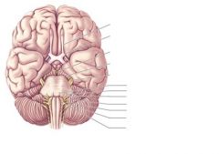
DEFINE olfactory bulbs
|
part of CN 1; outgrowth of the brain that lie on the cribiform plate that is part of the ethmoid bone
|
|
|
FUNCTION olfactory bulbs
|
receives neurons from nasal mucosa (1st order sensory) that synapse w/ 2nd order sensory neurons w/in the olfactory bulb and travel through the olfactory N
|
|
|
What occurs after the olfactory N enters the rhinencephalon?
|
info can go either right or left
right: go into medial stria (ant. perforated substance) left: go into lateral stria that terminates in the temporal lobe to the uncus of the parahippocampus) |
|
|
how can olfactory info cross over from other side after entering the rhinencephalon?
|
through the anterior commissure
|
|
|
How is the sense of smell unique?
|
only sensation that does not have to go to the thalamus before going to the cortex to be perceived. GOES DIRECTLY TO CORTEX
|
|
|
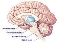
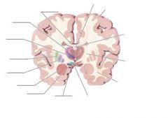
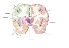
DEFINE diencephalon
|
contains 4 thalami in an area rostral to the brainstem, and accounts for about 2% of the brain mass
|
|
|
What are the 4 parts of the diencephalon?
|
Epithalamus
thalamus hypothalamus subthalamus |
|
|
What is contained within the epithalamus?
|
habenular trigone
stria medullaris pineal body roof of 3rd ventricle |
|
|
DEFINE habenular trigone
|
traingle shaped nuclei that belong to the diencephalon; have both afferent and efferent connections to other parts of the brain (ie-hypothalamus, thalamus, basal ganglia)
|
|
|
DEFINE stria medullaris
|
fibers connecting to hypothalamic, thalamic, afferent and efferent to basal ganglia; rostral to habenula
|
|
|
FUNCTION epithalaums
|
plays minor role in limbic system
|
|
|
DEFINE pineal body
|
attached to roof of 3rd ventricle that helps to determine the onset of puberty by producing melatonin
|
|
|
How does the pineal body determine the onset of puberty?
|
the pineal body stops producing melatonin and triggers the onset of puberty at a predetermined age.
The pineal body quits functioning once puberty is triggered and begins to calcify around age 40 |
|
|
What makes up the largest portion of the diencephalon?
|
thalamus
|
|
|
What connects the right and left halves of the thalamus?`
|
massa intermedia which crosses the 3rd ventricle
|
|
|
what are the thalami internally composed of?
|
numerous functional nuclei
|
|
|
DEFINE specific relay nuclei (r nuclei)
|
project to and receive fibers (ascending tracts such as the spinothalamic tract and dorsal column) from well-defined cortical areas related to specific functions
|
|
|
DEFINE association nuclei (a nuclei)
|
DO NOT receive fibers from ascending systems, but project to association areas of the cerebral cortex
|
|
|
DEFINE subcortical nuclei (sc nuclei)
|
project to subcortical areas (ie-basal ganglia, red nucleus)
|
|
|
DEFINE diffuse cortical connection nuclei (dc nuclei)
|
relay to various areas of the cortex but their destinations are not as specific as r nuclei
|
|
|
Which is the largest nuclei in the thalamus?
|
pulvinar nuclei
|
|
|
FUNCTION pulvinar nuclei
|
is a specific r nucleus that contains 2 smaller nuclei:
medial geniculate body lateral geniculate body |
|
|
DEFINE medial geniculate body
|
r type nucleus; involved with auditory pathway
|
|
|
DEFINE lateral geniculate body
|
r-type nucleus; involved with optic pathway
|
|
|
FUNCTION thalamus (general)
|
*great relay station of the brain of ALL sensory info (minus smell)
-processes sensory info -sends it to appropriate areas *plays dominant role in maintenance and regulation of consciousness, alertness, and attention *involved w/ subcortical perception of pain and temp *some nuclei serve as integrative areas for motor function (reflexes) *NOT purely sensory |
|
|
Where in the brain do you discriminate where pain is coming from and what kind of pain it is? where in the brain does the actual appreciation of pain occur?
|
postcentral gyrus
thalamus |
|
|
DEFINE hypothalamus
|
part of the diencephalon which is involved with visceral, autonomic, and endocrine functions
|
|
|
What are the 4 functional areas (nuclei) of the hypothalamus?
|
supraoptic region
preoptic region tuberal region mammillary region |
|
|
What are the 2 distinct nuclei of the supraoptic region of the hypothalamus and where do they project to?
|
paraventricular nucleus
supraoptic nucleus project to the posterior lobe (neurohypophysis) of the pituitary gland |
|
|
FUNCTION supraoptic lobe of hypothalamus
|
involved with PARASYMPATHETIC nervous system
receives direct projections from the retina and responsible for motion sickness |
|
|
DEFINE preoptic region
|
contains 3 nuclei
involved with parasympathetic ns |
|
|
DEFINE tuberal region
|
largest region (contains 4 nuclei)
involved with sympathetic ns |
|
|
DEFINE mammillary region
|
contains 2 prominent mammillary bodies (where fornix terminates)
|
|
|
Where does the hypothalamus have extensive afferent and efferent connection with other parts of the brain?
|
reticular formation
amygdaloid nuclear complex hippocampal formation fornix retina olfactory regions |
|
|
FUNCTION hypothalamus (general)
|
*synthesize RELEASING FACTORS that stimulate pituitary glands to secrete hormones
*involved with supraspinal control of the Autonomic NS -sympathetic: tuberal region -parasympathetic: preoptic & supraoptic regions *thirst/water uptake *temp regulation *hunger *emotional states *libido |
|
|
how does the hypothalamus influence the anterior lobe of the pituitary gland (adenohypophysis)?
|
*hypothalamus synthesizes Releasing Factors (RF)
*RF enter blood stream in infundibulum and carried to adenohypophysis *RF leaves blood stream and stimulates synthesis and secretion of hormones (CHEMICAL CONTROL) |
|
|
How many major hormones are synthesized and released from the anterior lobe of the pituitary?
|
8 hormones
|
|
|
how does the hypothalamus influence the posterior lobe of the pituitary (neurohypophysis)
|
*HORMONES are synthesized in the supraoptic and preoptic regions of the hypothalamus
*transported by neuron through infundibulum *when stimulated, neurons release hormones into posterior pituitary (NEUROLOGICAL CONTROL) |
|
|
what are the 2 main hormones associated with the neurohypophysis? what do they do?
|
oxytocin: causes smooth mm contraction during labor
vasopressin: vasoconstrictor and antidiuretic |
|
|
Which is the largest nuclei that makes up the subthalamic region?
|
subthalamic nucleus
|
|
|
DEFINE subthalamic nucleus
|
has extensive efferent and afferent connection; most important is the pallidosubthalamic fribers from the globus pallidus (afferent)
|
|
|
FUNCTION subthalamic nucleus
|
involved with motor integration activities (part of extrapyramidal system)
|
|
|
CLINICAL SIGNIFICANCE subthalamic nucleus
|
destruction of subthalamic nucleus will result in hemiballism (usually following stroke)
|
|
|
DEFINE hemiballism
|
violent, forceful, involuntary movements of the extremities on the contralateral side of the lesion to the subthalamic nucleus
|
|
|
What part of the brain are the optic nerves embryonically an outgrowth of?
|
diencephalon
|
|
|
What makes up the tectum of the mesencephalon?
|
superior colliculi
inferior colliculi |
|
|
FUNCTION superior colliculi
|
involved with visual pathway and visual reflexes
relay visual info from the superior colliculi to the lateral geniculate bodies of the thalamus |
|
|
DEFINE brachium of superior colliculi
|
axons that jconnect the superior colliculi to the lateral geniculate bodies of the thalamus
|
|
|
FUNCTION inferior colliculi
|
involved with auditory pathway and auditory reflexes
|
|
|
FUNCTION brachium of the inferior colliculi
|
axons which connect inferior colliculi to the medial geniculate body
|
|
|
What does the dorsal surface of the mesencephalon collectively make up?
|
roof of the cerebral aquaduct
|
|
|
DEFINE sensory nuclei
|
contains the cell bodies of 2nd order sensory neurons in the brainstem
also where 1st and 2nd order neurons synapse |
|
|
DEFINE motor nuclei
|
contain cell bodies of the LMC which are going to exit via cranial nerves and innervate skeletal muscle fibers
|
|
|
Which cranial nerves are associated with the midbrain?
|
3 and 4 (oculomotor and trochlear nn)
|
|
|
which cranial nerves are associated with the pons?
|
5-8
|
|
|
Which cranial nerves are associated with the medulla oblongata?
|
9-12
|
|
|
What are the 3 main parts that make up the cerebral peduncles?
|
crus cerebri
substantia nigra tegmentum |
|
|
DEFINE tegmentum (general)
|
continuous elongated mass of gray matter that extends the full length of the brain stem
|
|
|
What does the tegmentum contain?
|
*motor nucleus of the Trochlear n (IV)
*Tegmental nuclei of mesencephalic nucleus (CN V) *Mesencephalic nucleus (CN V) *Oculomotr Nuclear complex (CN III) *Edinger-Westphal nucleus (CN III) *Red nucleus *reticular formation *Decussation of Cerebellar peduncles *Lateral lemniscus *trigeminal lemniscus *medial lemniscus *spinal lemniscus *medial longitudinal fasciculus (MLF) |
|
|
FUNCTION motor nucleus of Trochlear n (IV)
|
contain cell bodies of LMN of trochlear n
INN superior oblique extrensic eye m |
|
|
FUNCTION tegmental nuclei of mesencephalic nucleus
|
involved with limbic system
|
|
|
FUNCTION mesencephalic nucleus
|
deals with proprioception of mm of mastication
|
|
|
What makes the mesencephalic nucleus "an exception to the rule"?
|
This nucleus contains the CELL BODIES of 1st order sensory neurons (usually contains in ganglia OUTSIDE CNS)
|
|
|
FUNCTION oculomotor nuclear complex
|
INN inferior oblique, medial rectus, superior rectus, inferior rectus mm (each mm has own nuclei)
**ALSO INN: levator palpebra superioris** |
|
|
DEFINE Edinger-Westphal nucleus
|
parasympathetic motor nucleus that contains cell bodies of preganglionic parasympathetic neurons
|
|
|
FUNCTION Edinger-Westphal nucleus
|
constricts pupilla sphincter m (iris of eye) to make pupil smaller (miosis)
INN cilliary body for accomodation |
|
|
DEFINE miosis
|
when pupil gets smaller (caused when pupilla sphincter m contracts)
|
|
|
DEFINE cilliary body
|
smooth mm surrounding the lens of the eye to change shape for accomodation
|
|
|
DEFINE red nucleus
|
most comprehensive, encapsulated nuclear structure.
Oval columns of cells that runs the full length of the midbrain |
|
|
FUNCTION red nucleus
|
part of the extrapyramidal sysem
communicates with other areas of extrapyramidal system (basal ganglia) and areas outside of system (cerebellum, cerebral cortex, SC) |
|
|
DEFINE reticular formation
|
made up of a diffused network of small nuclei that run the full lenght of tegmentum (brain stem)
|
|
|
FUNCTION reticular formation
|
*communicates with ascending, sensory pathways and descending motor pathways
*sleep *conciousness *arousal *pain modulation/inhibition *limbic system *refinement of motor activity |
|
|
Where do decussations of the cerebellar peduncles occur?
|
in the tegmentum
|
|
|
DEFINE decussation of cerebellar peduncles
|
occur in tegmentum
Made of 3: superior cerebellar peduncles (cerebellum to midbrain) inferior cerebellar peduncles (cerebellum to medulla) middle cerebellar peduncles (cerebellum to pons) |
|
|
FUNCTION decussation of cerebellar peduncles
|
where info crosses over from the cerebellum to the midbrain
|
|
|
DEFINE lateral lemniscus
|
contain 2nd order sensory neurons of the auditory pathway that cross to opposite hemisphere
|
|
|
DEFINE trigeminal lemniscus
|
2nd order sensory neurons
|
|
|
FUNCTION trigeminal lemniscus
|
primary nerve to head/neck for general sensation
part of the general sensation pathway for the head and neck |
|
|
DEFINE medial lemniscus
|
part of the dorsal column pathway
contains 2nd order sensory neruons originate in nucleus cuneatus and nuclear gracillis, both in the tegmentum of the medulla |
|
|
FUNCTION medial lemniscus
|
conscious proprioception
fine tough 2-pt discrimination |
|
|
DEFINE lemniscus
|
Pathways (axons/fibers)
|
|

|

|
|
|
|
|

|

|
|
|

|
|

|

|
|
|

|
|

|

|
|

|

|
|
|
|
|

|

|
|

|
|
|

right side only
|
|
|

|

|
|

|

|
|

|

|
|
|

|
|

|
|
|

|

|
|

right 1/2 only
|

|
|
top 1/2 only
|
|
|
top 1/2 only
|

|
|

bottom 1/2 only
|

|
|

|

|
|

|

|
|

|

|
|

|
|
|

|

|
|
|
|
|
left 1/2 only
|

|
|

|

|
|

|
|
|

bottom 1/2 only
|

|
|

left 1/2 only
|
|
|
|

|
|

|

|
|
|
What 3 tracts are contained w/in the medial longitudinal fasciculus?
|
tectospinal tract
reticulospinal tract medial vestibulospinal tract |
|
|
FUNCTION medial longitudinal fasciculus
|
*ascending tracts involved w/ control of the eyeball movements (both eyes need to coordinate)
*descending tracts involved w/ equilibrium reflexes |
|
|
FUNCTION spinal lemniscus
|
part of spinothalamic tract
associated with pain, crude touch, temp |
|
|
DEFINE substantia nigra
|
separates crus cerebri from the tegmentum and extends the full length of the brain stem
|
|
|
FUNCTION substantia nigra (general)
|
part of extrapyramidal system
(if substantia nigra malfunctions, basal ganglia malfunctions) |
|
|
DEFINE crus cerebri
|
most ventral portion of cerebral peduncles
contains corticofugal fibers (UMN) to provide LMN w/ motor functions |
|
|
What are the 3 divisions of the crus cerebri?
|
lateral 1/3
Middle 1/3 (pyramidal tracts) medial 1/3 |
|
|
What are the 3 divisions of the middle 1/3 of the crus cerebri?
|
lateral 1/3 (corticospinal tract-LE)
middle 1/3 (corticospinal tract-UE) medial 1/3 (corticobulbar tract--mm of head, neck, face) |
|
|
What fibers are found in the lateral 1/3 of the crus cerebri?
|
occipitotemporopontine fibers
|
|
|
what fibers are found in the medial 1/3 of the crus cerebri?
|
frontopontine fibers
|
|
|
What are the 2 divisions of the pons?
|
dorsal (tegmentum-cont. w/ that of midbrain and medulla)
ventral (pons proper--larger) |
|
|
What ascending sensory pathways are found in both the midbrain and dorsal pons?
|
medial lemniscus
lateral lemniscus trigeminal lemniscus spinal lemniscus medial longitudianl fasciculus |
|
|
What is the trapezoid body of the pons?
|
where fibers from the lateral lemniscus cross over and form a trapezoidal shape
|
|
|
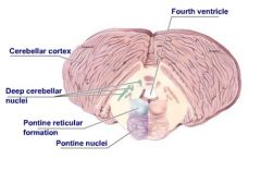
DEFINE pontine reticular nuclei
|
contains diffuse, small, nuclear structures (same as midbrain)
|
|
|
FUNCTION pontine reticular nuclei
|
arousal, sleep
|
|
|
What cranial nuclei are associated with the pons?
|
v, vi, vii, viii
|
|
|
What are the nuclei associated with CN in the pons?
|
*facial motor nucelus
*salivatory nuclei *solitary nucleus *spinal nucleus of V *motor nucleus of the trigeminal nerve *mesencephalic nucleus of V *chief sensory nucleus of V *abducent motor nucleus *dorsal and ventral cochlear nuclei *vestibular nuclei |
|
|
FUNCTION facial motor nuclei
|
inn mm of facial expression (VII)
|
|
|
what CN are associated with the salivatory nuclei?
|
VII, IX
|
|
|
FUNCTION salivatory nuclei
|
parasympathetic fx to CN VII
inn sublingual, mucus, lacrimal, and submandibular glands *contains cell bodies of preganglionic parasympathetic nn |
|
|
what CN are associated with the solitary nucleus?
|
VII, IX, X
|
|
|
FUNCTION solitary nucleus
|
involved with sensory function: taste inn for anterior 2/3 of the tongue (CN VII)
|
|
|
what CN are associated with the spinal nucleus of V?
|
V, VII, IX, X
|
|
|
FUNCTION spinal nucleus of V
|
sensory V: involved w/ pain, temp, pressure and crude touch
sensory VII: pain, temp, pressure and crude touch in a small area behind the external ear |
|
|
FUNCTION motor nucleus of the trigeminal n
|
perceive general sensations from the head and neck
inn mm of mastication |
|
|
What 3 nn come off of the trigeminal n?
|
opthalmic (V1), maxillary (V2), mandibular (V3)
|
|
|
which CN have parasympathetic functions (ie-have preganglionic parasympathetic nn)?
|
III, VII, IX, X
|
|
|
FUNCTION mesencephalic nucleus of V
|
sensory: receives proprioceptive info of head and neck
|
|
|
what CN are associated the chief sensory nucleus of V?
|
V, VII, IX, X
|
|
|
FUNCTION chief sensory nucleus of V
|
discrimination of fine touch
|
|
|
FUNCTION abducent motor nucleus
|
inn lateral rectus extrinsic eye m
|
|
|
FUNCTION dorsal and ventral cochlear nuclei
|
perception of hearing, equilibrium
*originate in cochlea of inner ear |
|
|
FUNCTION vestibular nuclei
|
equilibrium
*originate in semicircular canals of inner ear |
|
|
What structures are located within the pons proper?
|
pontine nuclei
longitudinal fibers |
|
|
FUNCTION transverse fiber in pons proper
|
take info into the cerebellum from the pons.
connects collateral processes of UMN to cerebellum |
|
|
what fibers give rise to the middle cerebellar peduncles?
|
superficial transverse fibers (thicker)
deep transverse fibers |
|
|
What fibers are located between the superficial and depp transverse fibers of the pons proper?
|
longitudinal fibers
|
|
|
What are the 2 major tracts w/in the longitudinal fibers
|
corticospinal tract
coricobulbar tract |
|
|
FUNCTION pons (general)
|
*relay station btw the midbrain na dthe medulla
*allows cerebellum to communicate with the brain stem *very important w/ fx of reticular formation (sleep, arousal, circadian rhythms, consciousness) *involved w/ cranial nerve activity *secondary respiratory system *primary sleep center |
|
|
is the superior peduncle a predominately efferent or afferent structure?
|
efferent
|
|
|
Are the middle and inferior peduncles predominately efferent or afferent?
|
afferent
|
|
|
What are the 3 lobes of the cerebellum?
|
anterior, posterior, flocculonodular lobes
|
|
|
FUNCTION anterior lobe of the cerebellum
|
muscle tone maintenance
maintenance of posture gross voluntary movements |
|
|
FUNCTION posterior lobe of the cerebellum
|
coordination of fine voluntary movements (largest)
|
|
|
FUNCTION flocculonodular lobe
|
maintenance of equilibrium (inferior and smallest)
|
|
|
what will damage to the posterior lobe of the cerebellum cause?
|
ataxia and intentional tremor
|
|
|
What are the 3 layers of cells located w/in the gray matter of the cerebellum from outer to inner?
|
molecular
perkinje layer granular layer |
|
|
What cells are found in the molecular level of the cerebellum?
|
baskel cells, stellate cells, golgi cells
|
|
|
What is contained w/in the white matter of the cerebellum?
|
deep cerebellar nuclei (1 in each hemisphere)
|
|
|
What are the layers of the deep cerebellar nuclei from medial to lateral?
|
fastigal (equilibrium)
globose emboliform dentate |
|
|
What types of info are processed w/in the cerebellar cortex?
|
constant proprioceptive info so body can adjust
equilibrium info muscle tone skeletal muscle activity (from corticospinal and corticobulbar fibers) |
|
|
Where does the cerebellum get most of its info from?
|
pons and medullar (via peduncles)
|
|
|
embryonically, what does the medullar oblongata originate from?
|
myelencephalon and rhombencephalon
|
|
|
what divides the medulla oblongata into columns?
|
anterior and posterior median fissures
|
|
|
DEFINE obex of medulla
|
on dorsal aspect it is a v-shaped structure formed where the 4th ventricle narrows into the central canal of the spinal cord
|
|
|
DEFINE anterolateral sulcus
|
marks the lateral limits of the pyramids
|
|
|
What are the 4 CN associated with the medulla?
|
IX, X, XI, XII
|
|
|
What can be seen in the anterolateral sulcus?
|
the 4 CN associated w/ the medulla
|
|
|
what are the contents of the medulla?
|
cranial nerve nuclei
nucleus gracilis and nucleus cuneatus ascending sensory tracts descending motor tracts corticospinal decussation decussation of the medial lemniscus medullary reticular formation inferior cerebellar peduncle inferior olivary nuclear complex |
|
|
What are the CN nuclei of the medulla?
|
spinal nucleus of V
inferior salivatory nucleus nucleus ambiguus dorsal motor nucleus hypoglossal motor nucleus solitary nucleus |
|
|
What CN are associated with the spinal nucleus of V
|
V, VII, IX, X
|
|
|
FUNCTION spinal nucleus of V
|
general sensation (nucleus descedns from the pons into the medulla)
|
|
|
FUNCTION inferior salivatory nucleus
|
motor nucleus of the parasympathetic system of CN VII, IX
inn salivary and mucous glands |
|
|
what CN are associated with the nucleus ambiguus?
|
IX, X, XI
|
|
|
FUNCTION nucleus ambiguus
|
motor nucleus which contains cell bodies of LMC involved w/ inn of the pharynx, larynx, and soft palate
|
|
|
what CN are associated with the dorsal motor nucleus
|
X
|
|
|
FUNCTION dorsal motor nucleus
|
parasympathetically inn the viscera of the thorax, abdomen, and pelvis
|
|
|
FUNCTION hypoglossal motor nucleus
|
inn the intrinsic and extrinsic mm of the tongue
|
|
|
what CN are associated with the solitary nucleus?
|
VII, IX, X
|
|
|
FUNCTION solitary nucleus
|
taste
|
|
|
what do the nucleus gracilis and nucleus cuneatus form?
|
medial lemnisci
|
|
|
What are the nucleus gracilis and nucleus cuneatus part of?
|
the conscious proprioception pathway (dorsal collumn)
|
|
|
DEFINE corticospinal decussation
|
where corticospinal tract decussates in the pyramids of the medulla
|
|
|
DEFINE decussation of the medial lemniscus
|
where 2nd order sensory neurons originating from the nuclei cuneatus and nuclei gracilis decussate and form the medial lemnisci
|
|
|
DEFINE inferior olivary nuclear complex
|
a large nuclear complex involved w/ cerebellar coordination activities (both afferent and efferent)
|
|
|
FUNCTION medulla oblongata (general)
|
respiration (primary)
heart rate blood pressure vomitting |