![]()
![]()
![]()
Use LEFT and RIGHT arrow keys to navigate between flashcards;
Use UP and DOWN arrow keys to flip the card;
H to show hint;
A reads text to speech;
44 Cards in this Set
- Front
- Back
|
What Dural Fold Attaches to the Crista Galli |
Faux Cerebri Longitudinally Separates the Left and Right Sides (runs down midline) |
|
|
Broken Cribiform Plate of Ethimoid bone can lead to lost in what sensation? |
Lost of smell *Damage to Olfactory Nerve |
|
|
Subdural Space |
Below the Dura Matter and Where Veins of CNS are found |
|
|
Contains CSF and where Arteries are found |
Sub Arachnoid Space |
|
|
Sub-Dural Hematoma occurs as a result of rupturing of? |
Veins Present in the Sub-Dural Space |
|
|
Rupture of one of the arteries that forms the circle of Willis could lead to? |
Sub Arachnoid Hematoma * Sudden Onset Headache *Stiff Neck *Photophobia *Nausea/Vomiting *Transient loss of Consciousness |
|
|
Patient Presents with headaches and dizziness after receiving a blow to their temple with a 5 iron golf club after he was caught cheating with the maid. Which arteries are most likely damaged and where is his bleeding localized |
Middle Meningeal arteries Extra Dural (epidural Space) Space |
|
|
3 different types of Cortex ; Telencephalon |
Archicortex Neocortex Paleocortex |
|
|
Fleeting memory Lost of Temporary Memory is associated with which part of the brain? |
Cingulum a Gyri in close Proximity to the Corpus Callusom |
|
|
Difference between Primary Brodman's Areas & Associated Areas |
Primary = Receive Impulses Associated = Process these impulses and makes sense of them. |
|
|
This has a really crappy job with pispoor income Things can get really messy if he doesn't work. |
Para Central Lobule |
|
|
Hemisphere to Hemisphere Communication Mediated by what Fibers? |
Commisural Fibers Corpus Callosum |
|
|
Area to Area Communication within Hemisphere |
Association Fibers |
|
|
Brain to spinal Chord Communication |
Projection Fibers (Internal Capsule) |
|
|
The Input to the Basal Ganglion |
Cortex (higher center) Thalamus Substantia Nigra (part of midbrain) |
|
|
The Output of Basal Ganglion is from the? |
Pallidum |
|
|
Motor Actions are initiated via |
Direct Pathway |
|
|
Indirect Pathway for Motor Actions |
1) Cortex 2) Striatum 3) External Segment of Pallidum 4) Subthalamus 5) Globus Pallidus Interna 6) Thalamus 7) Cortex |
|
|
Direct Pathway for Initiation of Motor Actions |
1) Cortex 2) Striatum 3) Globus Pallidum Interna 4) Thalamus 5) Cortex |
|
|
Communication Between the Substantia nigra and the Striatum |
Striato Nigral Nigral Striatal |
|
|
The Neurotransmitters of the Striatum are? |
1) GABA 2) Substance P 3) Enkephalin |
|
|
The Neurotransmitter of the Substantia Nigra is |
Dopamine |
|
|
Dopamine D1 Receptors are involved in which Pathway? |
Direct Pathway |
|
|
Dopamine D2 Receptors are involved in which Pathway? |
Indirect Pathway |
|
|
Striato =========> Nigra |
GABA |
|
|
Nigra =============> Striato |
Dopaminergic |
|
|
Patient Received damage to Substantia Nigra Where would you implant stem cells to regrow dopaminergic Neurons? |
Striatum You can't grow axons from the Substantia Nigra back to the Striatum |
|
|
A Knocked out Sub thalamus can lead to? |
Hemiballism |
|
|
Direct Pathway after Striatum goes to? |
Goes to Globus Pallidus Internal Segment |
|
|
Indirect Pathway after Striatum |
Goes to Globus Pallidus External Segment |
|
|
All sensations except what are relayed in the Thalamus? |
Olfaction (SMELL) |
|
|
Pain and Temperature sensation from the body passes through the Spinal Lemniscus. What part of body is this an exception for? |
Face |
|
|
diamond shaped fossa at back pryamid of medulla |
4th ventricle |
|
|
Arteries that arise off the Internal Carotid Arteries |
MAAPO Middle Cerebral Artery Anterior Choroid Artery Anterior Cerebral Artery Posterior Communicating Artery |
|
|
Arteries that arise off the Vertebral Arteries |
Superior Cerebellar Anterior Spinal AICA PICA Posterior Spinal Posterior Cerebral Pontine |
|
|
The portions of the brain are supplied by which arteries |

|
|
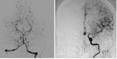
These arteries are found at which level of the Meninges |
SubArachnoid Space |
|
|
•Astroke in the cortical distribution of one ACA results in sensorimotor deficitin the opposite foot and leg. Urinary incontinence and contralateral frontallobe signs may also be observed.•Astroke in the cortical distribution of the MCA results in a severe sensorimotordeficit in the contralateral face and upper limb. With dominant hemisphereinvolvement, global aphasia also results; with nondominanthemisphere involvement, the neglect syndrome or amorphosynthesisresults. •Clinically,the lenticulostriatevessels are the most common site of spontaneous hypertensive hemorrhage inindividuals with long-standing hypertension.•Astroke in the distribution of the vertebral artery (or the PICA) results in an ipsilateralloss of pain and temperature sensations in the face; contralateral loss of painand temperature sensation in the limbs, trunk, and neck; an ipsilateralHorner syndrome; hoarseness; dysphagia; nystagmus;vertigo;diplopia; ipsilateral ataxia; and ipsilateralloss of taste. This combination of signs is the lateral medullary or Wallenberg syndrome.•Astroke in the cortical distribution of the PCA results in a contralateralhomonymous hemianopsia.With dominant (usually left) hemisphere involvement, reading and writingabnormalities also result. •Thelargest radicular artery is the so-called artery of Adamkiewicz,which generally enters the spinal cord in the lower thoracic or upper lumbararea. Clinically, this area of the spinal cord is susceptible to vascularinsult should this radicular artery be compromised.•Astroke in the distribution of the anterior spinal artery results in thedevelopment of total motor paralysis and dissociated sensory loss below thelevel of the lesion. The sensory loss, if dissociated (loss of pain andtemperature but no involvement of position and vibration sense), is caused bysparing of the dorsal columns supplied by the posterior spinal arteries.
|
Rakesh Foot Note Clinical Correlations |
|
|
Abnormal accumulation of CSF in an individual causing: increased inter cranial pressure tunnel vision Progressive head enlargement in childhood (Megolocephaly) |
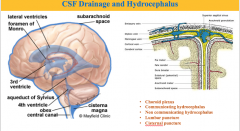
Hydrocephalus |
|
|
Pain and Temperature Sensation |
.p.p.p. |
|
|
Solitary Tract carries what sensation from Trigminal Meniscus |
Taste Recall: VPM |
|
|
Anterior Nucleus & Medial Nucleus of the Thalamus is associated with? |
Papez Circuit Look out for question about Amnesia or fleeting memory |
|
|
Pre-optic Nucleus helps with? |
Sexual Regulation of human beings |
|
|
Folich Syndrome Damage to Feeding Center |

|