Reading...
![]()
Play button
![]()
Play button
![]()
Use LEFT and RIGHT arrow keys to navigate between flashcards;
Use UP and DOWN arrow keys to flip the card;
H to show hint;
A reads text to speech;
47 Cards in this Set
- Front
- Back
|
How is the diagnosis of Alzheimer's disease made when alive?
Is it always correct? |
Clinically.
No not always correct. |
|
|
What is pseudodementia?
|
dementia caused by depression and hypomania.
|
|
|
What is the most common primary dementia? second most common?
|
Alzheimer's disease: most common
Dementia with Lewy Bodies (DLB) - second most common |
|
|
What is sundowning?
In what disorder do you observed this? |
Sundowning is episodes of delirium at night.
It is observed in Alzheimer's disease. |
|
|
List 3 most prediposing factors in development of Alzheimer's disease.
|
Age, genetic influences, apolipoprotein E status.
|
|
|
Is myoclonus late or early neurologic sign of Alzheimer's?
|
Late
|
|
|
What is snout reflex? In what dementia disorder do you observe this?
Is this an early or late sign? |
Snout reflex - frontal release sign
observed in Alzheimer's Early neurologic sign |
|
|
What 2 regions are atrophied in Pick's disease?
|
Frontal and temporal lobes
|
|
|
What abnormal protein is observed in lewy bodies?
|
alpha synuclein
|
|
|
Name 3 associated clinical features/signs of Lewy body dementia.
|
1. parkinsonism
2. fluctuating level of consciousness 3. visual hallucinations |
|
|
What 2 neurodegenerative disorders have lewy bodies?
|
Dementia with lewy bodies
Parkinson's disease |
|
|
What is Percheron's artery?
What happens with occlusion of this artery? |
one artery supplying both thalami
vascular dementia |
|
|
List the features of NPH(normal pressure hydrocephalus) triad.
|
gait ataxia, dementia, incontinence
|
|
|
What is the cause of CJD (Creuztfeldt Jacob disease)?
List 2 features of CJD. |
Prion
spongiform encephalopathy rapidly progressive dementia |
|
|
How do you diagnose CJD (Crueztfeldt Jacob disease)?
List 3 |
a) Radiology: bright cortical signal in clinically affected areas, thalami, or basal ganglia
b) CSF 14-3-3 protein – not specific c) EEG: periodic sharp waves |
|
|
List neurodegenerative disorders that are associated with following protein abnormalities.
1. Taupathies 2. alpha-synuclein 3. Polyglutamate (CAG repeat) |
1. Alzheimer's, Pick's diseaes (FTD), and progressive supranuclear palsy (PSP)
2. Dementia with Lewy bodies (DLB) and Parkinson's disease 3. Huntington's and spinocerebellar ataxia |
|
|
What 2 enzymes cut APP to generate Abeta-amyloid proteins?
What enzyme cut APP to make normal soluble APP? |
producing abnormal Abeta protein - beta and gamma secretase
normal - alpha secretase |
|
|
Name 4 genes involved in Alzheimer's disease and list the chromosome numbers for each gene.
|
1. APP, chrom 21
2. ApoE4, chrom 19 3, Presenilin 1 (PS-1), chrom 14 4. Presenilin 2 (PS-2) , chrom 1 |
|
|
What are the early clinical signs of FTD? late?
|
early - personality/behavioral disturbance and language problems
Late - memory loss |
|
|
What part of the temporal lobe is saved in FTD?
|
posterior 2/3 of the superior gyrus in the temporal lobe
|
|
|
What 2 areas lose their pigment in Parkinson's pts?
|
substantia nigra
Locus ceruleus |
|
|
Name the disease with combined symptoms of parkinsonism and vertical gaze palsy.
|
progressive supranuclear palsy (PSP)
|
|
|
What is the genetic problem with Huntington's disease? chrom?
What genetic quality does this mutation show down the generations? |
large CAG repeats on chrom 4.
shows anticipation |
|
|
What gene encodes normal prion protein?
What is the abnormal prion called? |
normal gene - PrPc
Abnormal - PrPsc |
|
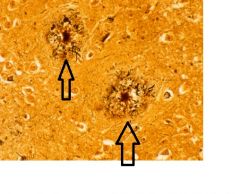
this is a biopsy of Alzheimer's brain.
What protein is in the middle? |
this is a neuritic plaque.
It has Abeta amyloid protein core. |
|
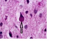
What protein is in the above structure pointed by the arrow.
|
It's a pick body with TAU protien
|
|
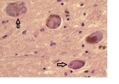
This is a section of the midbrain.
What 3 diseases do you suspect? |
Parkinson's
Dementia with lewy bodies Progressive supranuclear palsy |
|
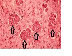
What protein would be found in the structures indicated by the arrows?
|
Alpha synuclein
These are lewy bodies |
|
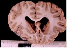
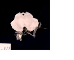
This is a brain of a pt who showed symptoms of chorea and demetia.
What disease do you suspect? What is the enlarged cavity called? |
Huntington's disease (atrophy of caudate and putamen)
Ex vacuo (enlarged ventricles due to atrophied caudate) |
|
|
How is the demyelinization process different b/t MS and leukodystrophy?
|
Leukodystrophy is a dysmyelinating disease in which a metabolic inborn error causes little or no formation of meylin.
MS is a demyelinating disease in which a normally formed myelin is destroyed by a disease process. |
|
|
ID the type of leukodystrophy with the following accumulated proteins:
1. Sulphatide 2. Psychosine 3. Very long chains of fatty acid |
1. Metachromic leukodystrophy
2. Krabbe (globoid) leukodystrophy 3. adrenoleukodystrophy |
|
|
What is the most common leukodystrophy?
|
metachromic leukodystrophy.
|
|
|
Which leukodystorphy cause inflammation?
|
Adrenoleukodystrophy
|
|
|
What type of demyelinating or dysmyelinating disorder is this?
presence of rosenthal fibers and diffuse demyelinazation |
Alexander's disease
|
|
|
Which functional fibers are affected in leukodystrophy? Is it bilateral or unilateral?
|
both sensory and motor.
bilateral |
|
|
Describe the pathogenesis of demyelination process in MS.
|
Cellular immune response activates CD4+ Th1 cells against myelin self-antigen. T cells secrete INF-gamma, which activates macrophages.
Activated macrophages and their toxic products cause demyelination. |
|
|
Where are the lesions usually found in MS brain?
|
Optic nerve, paraventricular white matter and juxtacortical white matter.
|
|
|
What are shadow plaques and inactive plaques in MS.
|
Shadow plaques - show thin myelin sheath due to either remyelination or incomplete loss of myelin; somewhere b/t normal and inactive plaque
Inactive plaques - there is total myelin loss and loss of oligodendrocytes. Completely healed. |
|
|
Name the disorder:
Positive antibody for aquaporin-4 channels. vision loss and paraplegia. |
Neuromyelitis optica (NMO) or Devic disease
|
|
|
Describe tumefactive MS.
|
Single large lesion in white matter with edema.
Biopsy reveals acute myelin loss, axonal sparing, lymphocyte, foamy macrophages, and gliosis |
|
|
What are predisposing factors for ADEM (acute disseminated encephalomyelitis)? (2)
|
post viral infection or post vaccination
|
|
|
Which subtype of MS is most common in early stage of MS? late?
|
early - relapsing-remitting
Late - secondary progressive |
|
|
On expanded disability status scale (EDDS), what does 3.5 indicate? 7?
|
3.5 - highest score at which walking assistance is not needed
7 - can't walk even with assistance. |
|
|
Waht is another name for first MS attack?
|
clinically isolated syndrome.
|
|
The arrows indicate accumulation of what protein?
What are they? What disease? |
psychosine
The arrows indicate globoid cells Krabbe leukodystrophy |
|
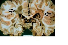
What do the arrows indicate?
What disease do you suspect? |
inactive plaques paraventricularly located
MS |
|
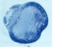
the above picture is a cross section of pons, stained against myelin.
This was obtained from a pt with Hx of chronic alcoholism. What disorder do you suspect? Will the progression of disease slow or rapid? What is the most likely and direct cause of this disorder? |
Central pontine myeliolysis
Rapid progression due to rapid correction of hyponatremia to correct electrolyte imbalance. |