Reading...
![]()
Play button
![]()
Play button
![]()
Use LEFT and RIGHT arrow keys to navigate between flashcards;
Use UP and DOWN arrow keys to flip the card;
H to show hint;
A reads text to speech;
60 Cards in this Set
- Front
- Back
|
What is the defining characteristic that differentiates a Glioblastoma from an anaplastic astrocytoma?
|
Necrosis (serpentine pseudopallisading)
Vascular Proliferation |
|
|
what is the frequency of brain tumors in the US? And what percentage are primary?
|
1-7/10,000
1/2-3/4 are primary mets |
|
|
what are the four major classes of intracranial tumors?
|
1) Gliomas
2) Neuronal Tumors 3) Poorly differentiated neoplasms 4) Meningiomas |
|
|
what are the three factors a physcian must consider when diagnosing brain tumors?
|
Location, Age of Patient, Imaging Characteristics
|
|
|
what is the most common form of glioma? ~80%
|
fibrillary astrocytoma
|
|
|
at what age are fibrillary astrocytomas usually found?
|
40-60
|
|
|
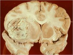
what do fibrillary astrocytomas look like microscopically and marcro?
|
marco: is grey infiltrative, and distorts the brain
micro: increase in the number of glial cell nuclei, variable pleomorphism, and GFAP+ (which are astrcytic cell processes which give a fibrillary background appearance. |
|
|
what do anaplastic astrocytomas look like micro and macro?
|
and more densely cellular, greater nuclear pleomorphism and mitotically active cells
|
|
|
what is characteristic of an astrocytoma WHO grade 2
|
it does not show mitosis or necrosis but you will see the fibrillary appearance
|
|
|
what is the factor that is thought to be contributing to the vascular proliferation in Glioblastomas?
|
VEGF which is vascular endothelial cell growth factor...possible as a result from hypoxia
|
|
|
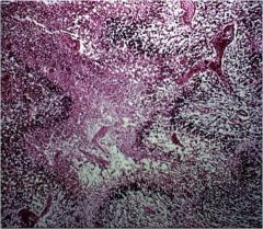
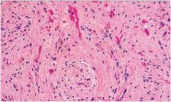
it is showing necrosis and diasnostic of glioblastoma...only glioblastoma has necrosis
|
what is this photo showing and why is this diagnostic?
|
|
|
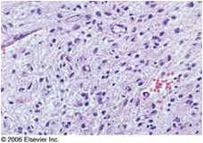
poorly differentiated cells which are huge tumor cells
|
what should you take note about this picture of glioblastoma?
|
|
|
what age group are pilocytic astrocytomas found? and where?
|
children?
usually in the cerebellum |
|
|
what appearance is commonly found in pilocytic astrocytoma
|
1) cystic mass with enhancing mural nodule
2) bipolar cells with long thin "hairlike" GFAP+ projections 3) ROSENTHAL FIBERS |
|
|
When are brainstem tumors usually formed?
|
0-20 Y.O.
|
|
|
which brainstem gliomas are most common, aggressive and have a short survival?
|
pontine gliomas
|
|
|
which age group are oligodendrogliomas common? and where are they usually found?
|
40-50 Y.O.
cerebral hemispheres |
|
|
A WHO grade 2 oligodendroglioma will usually have what appearance?
|
1) delicate netwoek of anastomosing capillaries
2) calficication 3) LITTLE MITOTIC activity |
|
|
what is characteristic of rosenthal fibers?
|
is a thick, elongated, worm-like eosinophilic (pink) bundle that is found on H&E staining of the brain in the presence of long standing gliosis
|
|
|
what is the difference between grade 3 and 2 oligodendrogliomas?
|
grade three has mitotic activity, increased cell density, nuclear anaplasia, and NECROSIS.
|
|
|
which has a better prognosis, astrocytomas or oligodendrogliomas?
|
oligo
|
|
|
what age group and where are ependymomas found?
|
0-20 Y.O.
found in the 4th ventricle in adults the central canal of the spinal cord is most commonly effected (NF II) |
|
|
why is resection of the epydymoma difficult or not possible?
|
b/c of their relation to the medullo/pontine nuclei
|
|
|
what do the tumor cells sometimes form in ependymomas?
|
rosettes
perivascular pseudorossetes=these are tumor cells surrounding vessels with an intervening zone made up of thin ependymal processes directed toward the wall |
|
|
what do the different rosettes look like?
|
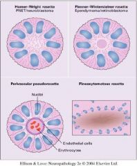
like this bitch
|
|
|
what genetic disorder is spinal cord ependymomas associated with? and where are these tumors found (called what)?
|
neurofibromatosis 2
filum terminale called myxopapillary ependymoma |
|
|
what tumor is commonly found in children in their lateral ventricals or found in the 4th ventrical in adults which results in hydrocephalus to due to obstruction of ventriuclar system
|
choroid plexus papilloma
|
|
|
what are neuronal tumors that are made up of entirely ganglion cells?
|
gangliocytoma
|
|
|
what is the typical features of gangliocytoma?
|
neoplastic ganglion cells are present as clumps of cells separated by relatively ACELLULAR STROMA
|
|
|
what neuronal tumors have a cyctic component, irregularly clustered and have random orientation of neurites?
|
gangliogliomas
|
|
|
dysembryoplastic neuroepithlial tumors usually present how?age?
|
childhood, seizures, growth in superficial temporal lobe, and a good prognosis
|
|
|
what are the typical features of the dysembroplastic neuroepithlial tumors?
|
1) blue/small cells
2) glial-neuronal element 3) multiple intacortical nodules with a myxoid background 4) "floating neurons" of the myxoid background |
|
|
the most common malignant CNS tumor in children is?
|
medulloblastoma
|
|
|
where do medulloblatomas usually form?
|
inferior vermis
|
|
|
what should you see with medulloblastomas if they are neuroepithelial?
|
small/blue cells
|
|
|
what rosettes may be found in medulloblastomas
|
homer-wright
|
|
|
what tumor is a grade 4 and involves small round blue cells tumors/commonly presented in young children and infants
|
PNET-primitive neuroectodermal tumor
|
|
|
what age group does Atypical teratoid/rhabdoid tumor present in?
|
usually under 5
|
|
|
what is the typical micro morphology of an AT/RT...where are they commonly presented?
|
rhabdoid cells with eosinophilic cytoplasm and eccentric nuclei
abundant mitosis grade 4 commonly found in the cerebellum/ or posteroir cerebrum |
|
|
what is the most common CNS neoplasm in immunosuppressed patients such as AIDS patients?
|
primary CNS lymphomas
|
|
|
The malignant lymphoma cells infiltrate the brain parenchyma and accumulate around blood vessels
|
yes
|
|
|
where are germ cell tumors such as germinoma in the CNS commonly found?
|
midline almost always around the 3rd ventrical
and pineal region |
|
|
what is seen in teratomas?
what nationality has a strong relation to teratomas? |
these are the crazy looking tumors with hair and teeth..they usually present with hydrocephalus, visual problems, pituitary failure or precocious puberty
japanese |
|
|
what tumors are described as rounded masses with a well defined dural bas and compresses the underlying brain with possible focal deficits?
|
meingioma=mainly benign
|
|
|
the mass of meningiomas may be polypoid (sheet like tumor spread along the surface of dura which is associated with hyperostotic reactive changes in overlying bone with what kind of bodies?
|
psammoma bodies
|
|
|
a histological pattern of meningiomas is described as whorled clusters of cells which sit in groups without a visible cell membrane...what is this pattern called?
|
syntial
|
|
|
what histological pattern is described as forming from calcification of syncytial nests of meningothelial cells?
|
psammomatous bodies
|
|
|
what is the criteria for a grade 2 atypical meningioma?
|
-greater then 4 mitoses/10power
OR (3 or more atypical things) 1) increased cellularity 2) small cells with high nuclear/cytoplasmic ratio 3) patternless growth 4) necrosis 5) prominent nucleoli |
|
|
how many mitosis/10power field is considered the anaplastic meningioma?
|
20/hpf
|
|
|
what are the most common metastatic tumors to the brain? 5 are included
|
lung, breast, kidney, GI, skin (melanoma)
|
|
|
which tumors have micro patterns of "nuclear free zones" that lie between areas of nuclear palisading called VEROCAY BODIES?
|
Peripheral Nerve Sheath Tumors: Antoni A pattern
|
|
|
which nerve are peripheral nerve sheath tumors attached to if they occur in the cranium?
|
vestibular branch of 8th CN..called an acoustic neuroma
|
|
|
what pattern in peripheral nerve sheath tumors is considered less dense cellular areas with loose meshwork of cells with microcyts and myxoid changes?
|
Antoni B
|
|
|
neurofibromas are usually formed with what genetic disorder?
|
neurofibromatosis type 1
|
|
|
what is the difference between a schwannoma and the neurofibroma?
|
you can not seperate the lesion from the nerve in with a neurofibroma. there are fingers of the tumor that insert themselves into the nerve fibers
|
|
|
in neurofibromatosis type 1 which kind of tumor is likely to undergo malignant degeration at a higher rate then in the general population?
|
plexiform type neurofibromas
|
|
|
what are the common tumors seen in neurofibromatosis type 2
|
bilateral VIII nerve schwannoma and multiple meningiomas, and ependymomas of the spinal cord
|
|
|
what is the triad of tuberous sclerosis? In addition with hamartomas and other benign neoplasms
|
mental retardation
seizures adenoma sebaceum (facial rash) |
|
|
what astrocytomas are unique to tuberous sclerosis and are a WHO grade 1 but they may obstruct CSF of the ventricles?
|
SEGA: subependymal giant cell astrocytoma
|
|
|
what are the microscopic features of oligoddendrogliomas?
|
halo surronding the nuclei (fried egg cells)
anastomosing capillaries (chicken wire fencing) 90% show calcification |