Reading...
![]()
Play button
![]()
Play button
![]()
Use LEFT and RIGHT arrow keys to navigate between flashcards;
Use UP and DOWN arrow keys to flip the card;
H to show hint;
A reads text to speech;
144 Cards in this Set
- Front
- Back
|
Sensory input
|
Gathered information occuring inside and outside of the body relayed to brain via nerves
|
|
|
Integration
|
Nervous system processes and interprets sensory input and decides what should be done at each moment
|
|
|
Motoroutput
|
response to stimuli
|
|
|
Central Nervous System
|
Brain and spinal chord
integration command center |
|
|
Peripheral nervous system
|
Consists of Spinal Nerves
Cranial Nerves everything that isn't CNS |
|
|
Spinal Nerves
|
Carry impulses to and from the spinal chord
|
|
|
Cranial nerves
|
carry information two and from brain
|
|
|
Functional Subdivisions of Cranial Nerves
|
Sensory afferent
Motor Efferent |
|
|
Sensory Afferent division
|
Division of PNS consisting of nerve fibers that convey impulses to the CNS from specific sensory receptors throughout the body
|
|
|
Neurons
|
principle cells of nervous system
excitable cells tat transmit electrical signals |
|
|
Nuroglia AKA Glial Cells
|
- Supporting cells
- 6 types four in CNS and two in PNS - most have branching porcesses and a central cell body - make up half the mass of the brain - provide supportive scaffolding for neurons - segregate and insulate neuron -guide young neurons to proper connections -promote health and growth |
|
|
Neuroglia in CNS
|
Astrocytes
Microglia Ependymal cells Ogliodendrocytes |
|
|
Astrocytes
|
- Nuroglia cell in CNS
- Shaped like delicate branching sea anemone - Are most abundant and versatile glial cell -support and brace neurons - anchor neurons to their nutrient supplies - guide migration of young neurons - control chemical environment cleaning leaked potassium ions and recapturing neuro-transmitters - control chemical environment around neurons |
|
|
Microglia
|
Small ovoid cells with spiny processes
-neuroglia cells in CNS -Deffensive glial cells in CNS - Phagocytes that montinor health of neurons -Phagocytizes microorganisms or neuronal debris - important because cells of immune system have limited access to CNS |
|
|
Ependymal Cells
|
- glial cells in CNS
- range in shape from squamous to columnar many are ciliated - Line central cavities of the brain and spinal columns - Form permeable barriers between the cerebralspinal fluid and tissue fluid -beating cilia helps to circulate cerebrospinal fluid |
|
|
Ogliodendrocytes
|
- Neuroglia in CNS
- have fewer processes thn astrocytes - wrap around nerve cell when fully formed to produce make up myelin sheath - Can grab and mylinate multiple CNS cells at once |
|
|
Neuroglia in PNS
|
Satellite Cells
Schwann Cells |
|
|
Satellite cells
|
Neuroglia in PNS
- Surround neuron cell bodies in PNS have similar support and brace neurons - anchor neurons to their nutrient supplies - guide migration of young neurons - control chemical environment cleaning leaked potassium ions and recapturing neuro-transmitters - control chemical environment around neurons |
|
|
Schwann Cells
|
Neuroglia Cells in PNS
-Surround all nerve fibers in PNS and form myelin sheaths around the thicker nerve fibers -similar to oglodendrocytes - vital to regeneration of damaged peripheral nerve fibers -can grab up to 15 PNS cells nerve fibers but can only mylinate one |
|
|
Neurons Structure
|
Structural units of Nervous system
- All have cell body and slender process to be considered a neuron - Composed of body(containing nucleus) - Dendrites which receive information from presynaptic cell -axon terminal which transmit information to postsynaptic cell |
|
|
Neuron characteristic
|
- conducts nerve impulses
- extreme longevity - Amototic: when assume role as communicating links lose ability to divide - High metabolic rate and require continuous and abundant supplies of oxygen and glucose |
|
|
Plasma Membrane
|
Help with electrical signalling as well as cell to cell signaling during development
|
|
|
Neuron Cell Body
|
AKA soma or parykaryon
-consists of spherical nucleus with nucleolus surrounded by cytoplasm -holds a majority if not all normal cell organelles -focal point for outgrowth of neuronal processes -no centrioles: amototic Contains axon hillock |
|
|
Axon Hillock
|
is a specialized part of the cell body (or soma) of a neuron that connects to the axon. As a result, the axon hillock is the last site in the soma where membrane potentials propagated from synaptic inputs are summated before being transmitted to the axon
|
|
|
axon
|
An axon also known as a nerve fibre; is a long, slender projection of a nerve cell, or neuron, that typically conducts electrical impulses away from the neuron's cell body.
|
|
|
Nissal Bodies
|
AKA Chromatophilic substance
ER in neuron that is more developed than any other in body |
|
|
Nuclei
|
Clusters of cell bodies in CNS
|
|
|
Ganglia
|
Clusters of cell bodies in PNS
|
|
|
Neuron Processes
|
Armlike processes extend from the cell body of all Neurons
|
|
|
Tracts
|
bundle of neuron proceses in CNS
|
|
|
Nerves
|
Bundles of Neuron processes in PNS
|
|
|
Dendrites
|
- Short tapering diffusely branching extensions of neuron Cell body
- typically hundreds branch off neuron close to the cell body -contain virtually all organelles present in the cell body that occur in dendrites -convey incoming messages toward cell body -typically graded potentials not action potentials |
|
|
Dendritic spine
|
Area on dendrite that branches out to create closeness between other neurons lessening synapse space
|
|
|
Input/Receptive regions on dendrite
|
Provide an enormous surface area for receiving signals from other neurons
|
|
|
Axon structure
|
Each neuron has a single axon arising from axon hillock and narrows to form a slender process that is uniform in diameter for the rest of the length
-Some neurons don't have axon hillock while in others |
|
|
Nerve Fiber
|
a long axon
|
|
|
Axolemma
|
Plasma membrane of axon
|
|
|
Axon Collaterals
|
branches that extend from the axon of a neuron in right angles
|
|
|
Terminal Branches
|
AKA terminal Arborizations
Branches at the end of an axon |
|
|
Trigger Zone
|
The area of the axon hillock and axon junction where nerve impulses are generated
|
|
|
Axon terminals
|
AKA Terminal Boutons
knob like distal ending of terminal branches that secrete neurotransmitters into synapse |
|
|
Secretory Region
|
area on terminal boutons where neurotransmitters are secreted
|
|
|
Axon Functional characteristics
|
-act as conducting region of neuron
- generates nerve impulses and transmits away from cell body along Axolemma -Secrete neurotransmitters from axonal terminals "Secretory regions" |
|
|
Transport along the axon
|
Consistently occurring as a result of motor neurons
Movement along axon occurs in two ways o Anterograde – toward axonal terminal o Retrograde – away from axonal terminal |
|
|
Anterograde motion along axon
|
toward axonal terminal
|
|
|
Retrograde motion along axon
|
away from axonal terminal
|
|
|
Myelin Sheaths
|
Cover nerve fibers particularly those that are long or large in diameter
formed by Schwan cells PNS or Oligodendrocytes CNS Whittish Fatty protein lipid protects and electrically insulates fibers |
|
|
Myolanated fibers
|
axons bearing a myelin sheath conduct impulses rapidly
|
|
|
Unmylonated fibers
|
conduct impulses slowly
will be surrounded by Schwann cell not coiled thus not insulated |
|
|
Mylenation in PNS process
|
Formed by schwann cells
1) Schwann cells will indent and then will envelope an axon 2) Schwann cell rotates around axon wrapping its plasma membrane loosely around it in sucesive layers 3) Schwann cell cytoplasm (former neurolemma) is forced from between the membranes (Outer collar of perinuclear cytoplasm) -these tight membrane wrappings form myelin sheath |
|
|
Nodes of Ranvier
|
Gaps between mylenation allowing for chemical signalling to occur
axon collaterals can emerge from these sites |
|
|
Mylenation in CNS
|
• Oligodendrocyte form myolin sheaths
• Oligodendrocyte has multiple flat processes that can mylenate multiple axons at one time • No neurolemma exists (plasma membrane) |
|
|
White matter
|
myelanated fibers in CNS
|
|
|
Gray Matter
|
contains mostly nerve cell bodies and nonmyelinated fibers
in CNS |
|
|
Structural classification of neurons
|
Multipolar
Bipolar Unipolar |
|
|
Multipolar Neurons
|
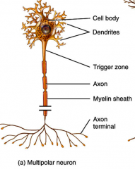
is a type of neuron that possesses a single (usually long) axon and many dendrites, allowing for the integration of a great deal of information from other neurons. These dendritic branches can also emerge from the nerve cell body. Multipolar neurons constitute the majority of neurons in the brain and include motor neurons and interneurons.
|
|
|
Unipolar Neuron
|

Have a single short process that emerges from the cell body and divides t-like into proximal and distal branches
Most are sensory neurons that conduct along afferent pathway |
|
|
Bipolar Neuron
|

- All sensory neurons located in special sense organs
- Have two processes, axon and dendrites that extend from opposite sides of the cell body Peripheral process: Associate with a sensory receptor Central process: enters CNS Ex olfactory |
|
|
Functional classification of neurons
|
- Sensory (afferent) Neurons
- Motor Neurons |
|
|
Sensory Neurons
|
- Afferent Neurons that transmit impulses from sensory receptors in the skin or internal organs towards or into the CNS
- Virtually all unipolar - Only most distal parts Unipolar cells act as impule receptor sites |
|
|
Motor Neurons
|
- Efferent Neurons that carry impulses way from CNS to the Effector organs of the body periphery.
- Motor Neurons are multipoalr - Cell bodies located in CNS |
|
|
Interneurons/Association Neurons
|
- Lie between motro and sensory neurons and shuttle signals through CNS pathway where integration occurs
- Most confined within CNS - Make up 99% of neurons of the body |
|
|
Action potentials
|
• Electrical impulses carried along length of axon
• Always same regardless of stimulus • Underlying functional feature of nervous system |
|
|
Membrane Potentials
|
Established by difference in either/both Concentration gradients and Electrical gradients on opposite sides of membrane
o Ions move from higher to lower concentration gradients o Ions move towards of opposite electrical charge electrical gradient o Electrochemical gradient: the electrical and chemical gradients taken together o Inherent difference in voltage created by membrane creates PE |
|
|
Voltage
|
Measure of potential energy created by membrane
|
|
|
Electrochemical Gradient
|
electrical and chemical gradients taken together
|
|
|
Current
|
The flow or movement of elecrical charge
|
|
|
Resistance
|
Resistance to charge flow
|
|
|
Types of Plasma membrane channels
|
Passive/leakage channels
Chemically Gated Channels Voltage Gated Channels Mechanically Gated Channels |
|
|
Passive/Leakage Channels
|
channels in membrane that are always open and allow for consistent passage of certain molecules
|
|
|
Chemically Gated Channels
|
• Classification of membrane channel
• Open when appropriate chemical binds • Example Na K ligand gated channel |
|
|
Voltage gated channels
|
• Classification of membrane channel
• Open and close in response to changes in membrane potential |
|
|
Mechanically gated Channels
|
• Membrane channel
• Open in response to physical deformation of the receptor (ie touch) |
|
|
Gated Channels
|
When opened allow for the quick moving or passing of ions across their electrochemical gradients
-results in generation of current and a change in voltage across membrane |
|
|
Resting Membrane Potential
|
Typical Potential difference across membrane
- Cytoplasm inside cell is negatively charged relative to outside -results in polarization of membrane - typically -70 mv -established based on ionic composition between intra and extra cellular fluids as well as the permeability difference to those specific ions |
|
|
Neurolemma
|
the plasma membrane surrounding a Schwann cell of a myelinated nerve fiber and separating layers of myelin
|
|
|
Difference in ionic composition inside v outside cell membrane
|
• K+ is higher in higher concentration inside cell where as Na + higher concentration outside K+ playing essential role in generating ion potentials
• Negative protein anions balance charge inside molecule |
|
|
Plasma Membrane permeability
|
• Membrane more permeable to potassium ions than sodium
• Potassium is consistently lost through leakage channels • Loss of potassium increases negative charge of Neuron • However dragged back in because of negative charge inside cell generating Resting potential • Resting potential is due to this difference in permeability |
|
|
Sodium potassium pump
|
Stabilizes resting membrane potential by taking in two K and rejecting three Na Molecules
|
|
|
Membrane potentials used?
|
- Used to integrate send and receive information
- Changes in membrane potential produced by - change in membrane permeability to ions - alteration of ion concentration across membrane |
|
|
Classification of changes that occur to membrane potentials
|
• Depolarization: a decrease in membrane potential where membrane becomes less negative than resting potential
• Repolarization: the membrane returns to its resting membrane potential • Hyperpolarization: increase in membrane potential where membrane is more negative than resting potential |
|
|
Graded potentials
|
- Short lived localized changes in membrane potential
-Short lived localized changes in membrane potential that can be either depolarizations or hyperpolarizations -Occur in cell body - Can cause current flow that decreases in magnitude with distance due to leaky plasma membrane of Cell body - The stronger the stimulus the farther down it will travel |
|
|
Steps of Action Potentials
|
Resting
Depolarization Repolarization Hyperpolarization |
|
|
Resting state of membrane
|
a. All gated Na and K channels closed membrane potential stable
i. NOTE: Na+ channel has two gates a 1. voltage sensitive activation channel 2. inactivation gate that blocks channel once opened (both gates must be open in order for Na+ to enter but closing either gate effectively closes channel ii. K+ only has single voltage sensitive gate that is closed in resting state and opens slowly in response to depolarization |
|
|
Depolarization
|
a. Local currents depolarize axon membrane causing Na+ ion channels to open
b. depolarization of stimulation site causes more sodium pumps to open c. When depolarization reaches Threshold -50 or -55 depolarization becomes self generating cascades down axon resulting in opening of all sodium channels d. Resulting cascade brings membrane potential to +30 MV |
|
|
Threshold
|
o Not all depolarization produce action potentials must reach threshold of cell to cause action potential to occur
o Typically 15-20 mv from resting value o Strong stimuli depolarize membrane o Weaker stimuli must be applied for longer to provide crucial amount of current flow |
|
|
Repolarization
|
a. Sodium inactivation gates close decreasing sodium permeability to resting levels
b. Votage gated K+ open c. K+ exits cell restoring negativity of resting neuron |
|
|
Hyperpolarization
|
a. Potassium gates remain open causing excessive efflux of K+
b. Causes hyper polarization of membrane c. Neuron is insensitive to stimulus and at this time d. Repolarization restores resting electrical conditions but not ionic conditions e. ionic condition corrected through sodium potassium pump |
|
|
All or None Phenomenon
|
Action potentials either occur completely or not at all
|
|
|
How is action potential propogated
|
o Influx of Na across membrane leads to depolarization of regions further down the axon as Na+ ions go towards negative regions
• Opens voltage gated channels and triggers action potentials • Because area where AP originated just generated AP cannot generate again • Propagation occurs in one direction away from point of origin • AP is self propagating continuing down axon at constant velocity • After depolarization membrane repolarizes restoring resting membrane potential • AP is regenerated a new at each patch |
|
|
Coding for stimulus intensity
|
o Strong stimuli generate nerve impulses more often in a given time than weak
o Stimulus intensity coded for by number of impulses per second |
|
|
Absolute Refractory period
|
• When a neuron cannot respond to another stimulus no matter how strong
• Period occurs from the opening of na channels to they reset • Ensures all or none event • Enforces one way transmission of nurve impulse |
|
|
Relative refractory period
|
• Interval following the absolute refractory period
• Most Na channels have returned to resting state • Some K channels still open • Repolarization occurring • Threshold for AP generation is substantially elevated • Exceptionally strong stimulus can reopen sodium gates |
|
|
Conduction Velocity depends on two factors
|
Axon diameter
Degree of Myelination |
|
|
Axon Diameters affects on conduction velocity
|
a. The Larger the axon’s diameter the faster it conducts impulses because offer less resitance to flow of current
b. Bring adjacent areas of membrane to threshold more quickly |
|
|
Degree of Myelination's affects on conduction velocity
|
a. Presence of myelin sheath dramatically increases rate of propagation because its insulation does not allow for leakage of charge
b. Current can only pass through membrane at myaline sheet gaps |
|
|
Sultatory conduction
|
o Electrical signal appears to jump from gap to gap along axon due to mylenation
o 30 times faster than continuous conduction |
|
|
Continuous Conduction
|
o AP propagation involving nonmyelinated axons
o Channels are immediately adjacent to each other o Relatively slow |
|
|
Multiple Sclerosis
|
o Autoimmune disease that destroys myelin sheets
o Loss of myelin resulting in slowing down of successive gaps o Eventually impulse conduction ceases o More Na Channels appear where myelin lost • Compensation for loss of myelin • May account for symptom free periods and relapse |
|
|
Synapse
|
• A space at junction between neurons that mediates information transfer from one neuron to the next
|
|
|
Axondentric synapse
|
Synapse between the axon endings of one neuron and the dendrites of other neuron
|
|
|
Axosomatic Synapses
|
Synapses between axon endings of one neuron and cell bodies of other neurons
|
|
|
Axoaxonic
|
axon-axon synapse
|
|
|
Electrical Synapse
|
o Synchronize activity of interconnected neurons
o Consist of gap junctions o Less common then chemical synapses o Contain protein channels called conenexons that • Connect cytoplasm of adjacent neurons • Allow ions and small molecules to flow directly from one neuron to the next • Neurons are electrically coupled • Transmission across synapse rapid • Communication may be unidirection or bidirectional o Important in CNS for • Arousal from sleep • Mental attention • Emotions and memory • Ion and water homeostasis |
|
|
Chemical Synapse
|
o Specialized to allow the release and reception of chemical neurotransmitters
o Current from presynaptic membrane dissipates in fluid filled cleft thus preventing nerve impulses from being directly transmitted o Transmission across Synaptic cleft is chemical event not electrical Ensuring unidirectional communication btwn neurons |
|
|
What two parts are Chemical Synapses composed of?
|
• Axon Terminal
• receptor region |
|
|
Axon Terminal
|
Part of presynaptic neuron
important in Chemical synapses b/c Contains Synaptic vesicles: membrane bounded sacs containing neurotransmitter molecules |
|
|
Synaptic Cleft
|
A fluid filled space in chemical synapses that separates pre and postsynaptic membrane
important because ensures current from presynaptic membrane dissipates preventing impulse being directly transmitted |
|
|
Process of Signal Transfer across Chemical Synapses
|
1. Action potential arrives at presynaptic axon terminal causing opening of Ca and Na channels on presynaptic neuron
2. Entering Ca Binds to Synaptotagmin communicating to snare proteins to releases neurotransmitters through exocitosis 3. Ca Removed from presynaptic Cell and cell repolarizes 4. Neurotransmitter crosses synaptic cleft binding to receptors on postsynaptic neuron 5 postsynaptic membrane permeability changes causing excitatory or inhibitory effect 6. Neurotransmitter effects are terminated |
|
|
As long as neurotransmitter binds to receptor
|
i. continues to have affect
ii. blocks reception of additional messages iii. must be removed from its receptor |
|
|
Effects of neurotransmitter terminated in what different ways?
|
Reuptake
Degredatin Diffusion away from synapse |
|
|
Reuptake of Neurotransmitters in CNS occurs?
|
1. by astrocytes or
2. By presynaptic terminal or 3. destroyed by enzymes |
|
|
Degredation of Neurotransmitter
|
by enzymes associated with postsynaptic membrane
|
|
|
Synaptic Delay
|
o Neurotransmitter must be released diffuse across membrane and bind ot receptor
o Synaptic delay = time needed to do this o Synaptic delay is rate-limiting step of neural transmission o *Transmission along multisynaptic pathways occurs more slowly |
|
|
What main factors influence the amount of effect neurotransmitters have on the post-synaptic cell
|
• Amount of neurotransmitters released
• Amount of time neurotransmitter bound to receptor |
|
|
Two Types of Postsynaptic Potentials
|
• EPSP- Excitatory postsynaptic potentials
• IPSP – Inhibitory postsynaptic potentials |
|
|
Excitatory Post synaptic potentials
|
• Use only chemically gated channels
• Na+ and K+ channels open • Because Na+ higher concentration outside than K+ inside action results in depolarizing cell • Depolarization lowers makes threshold easier to attain • EPSP are localized at cell body |
|
|
Inhibitory post synaptic potentials (IPSPs)
|
• Neurotransmitter binding to a receptor at inhibitory synapses causes
• Either opens Cl- channels allowing for influx or K+ channels allowing for efflux o Net result in neuron becoming more negative reducing neuron’s ability to produce an action potential |
|
|
Summation
|
• Single EPSP cannot induce AP
• Summate: when EPSPs or IPSPs add together to influence activity of postsynaptic neuron • EPSPs or IPSPs can also summate to cancel each other out |
|
|
Temporal Summation
|
o Occurs when one or more presynaptic neurons transmit impulses in rapid fire bursts producing small EPSP
o Before it dissipates successive impulse trigger more epsps’s o Cause postsynaptic membrane to depolarize/hyperpolarize more than from a single EPSP or IPSP |
|
|
Spatial Summation
|
o When postsynaptic neuron is stimulated by a large number of terminals from presynaptic neurons
o Huge numbers of receptors bind neurotransmitter and initiate EPSP or IPSP o Summate and dramatically enhance depolarization |
|
|
Acetylcholine
|
• First and best understood neurotransmitter identified
• Degraded in synapse by acetylcholinesterase • Released at the neuromuscular junction • Stimulates all skeletal muscle and parts of ANS Example of Neurotransmitter having both excitatory and inhibitory effect Inhibitory in cardiac muscles but excitatory in Skeletal Muscle |
|
|
Classification of Neurotransmitters
|
Excitatory
Inhibitory Direct Indirect NOTE some neurotransmitters can exhibit both classification depending upon site they bind to |
|
|
Excitatatory neurotransmitters
|
Classification of neurotransmitters that Cause Depolarization
|
|
|
Inhibitory Neurotransmitters
|
Classification of neurotransmitters that cause hyperpolarization
|
|
|
Neuron pools
|
• Integrate incoming information received from receptors or different neuronal pools and then forward the processed information to other destinations
• One incoming presynaptic fiber branches out as it enters pool • Excites postsynaptic fibers closest to center (discharge zone) • Bring fibers further from center closer to threshold (Faciliated zone) |
|
|
Discharge Zone of Neuronal pools
|
Neurons that most closely associated with Incoming fiber
will fire when impulse comes down presynaptic fiber |
|
|
Facilitated Zone neuronal pools
|
Neurons farther from incoming fiber will only be slightly depolarized
|
|
|
Divergent Neural Circuit
|
one incoming fiber stimulates ever increasing number of fibers often amplifying circuits
|
|
|
Convergent Neural Circuit
|
Many inputs one output (a concentrating circuit)
|
|
|
Reverberating Neural Circuit
|
Signal travels through a chain of neurons each feeding back to previous neurons
|
|
|
Parallel Neural Circut
|
Signal stimulates neurons arranged in parallel arrays that eventually converge on a single output cell
|
|
|
Serial Processing
|
• Whole system works in a predictable all-or-nothing manner
• One Neuron stimulates the next and so on down the line Ex reflexes |
|
|
Reflexes
|
rapid automatic responses to stimuli which a particular stimulus always causes the same response
example of serial processing |
|
|
Parallel Processing
|
input travels along several pathways
o Pathways are integrated in different CNS systems o One stimulus promotes numerous responses o Eg. Smell |
|
|
Development of Neurons
|
- The nervous system originates from the neural tube and neural crest
- The neural tube becomes the CNS - Cell Differntiation |
|
|
Three phases of neuron cell differentiation
|
Proliferation: cells proliferate to produce appropriate number of cells needed for nervous system development
Migration: potential neurons, become amitotic and move externally to characteristic positions Differentiation: cells differentiate into neuroblasts |
|
|
three parts of simple neuronal pool
|
Input Fiber - presynaptic fiber
Discharge zone - neurons most closely associate with incoming fiber Facilitated zone - neurons farther way from incoming fiber |
|
|
Sensory/Afferent Division of PNS
|
Specific receptor fibers of the sensory afferent division of the PNS
-convey impulses from the skin, skeletal muscles and joints to the CNS |
|
|
visceral afferent fibers
|
Sensory fibers of sensory afferent division of PNS
- Transmit/convey impulses from visceral organs (organs located in ventral body cavity) to the brain |
|
|
Motor/Efferent division and sub divisions
|
Division of the PNS responsible for carrying impulses from teh CNS to effector organs
Is broken down into two parts - Somatic Nervous System (voluntary) - Automatic Nervous System (involuntary) |
|
|
Effector Organs
|
-Muscles or glands that CNS communicates to via motor efferent nerve fibers
-causes glands to secrete or muscles to contract |
|
|
Somatic Nervous System
|
Consists of somatic motor nerve fibers
- conducts impulses from CNS to skeletal muscles Referred to as voluntary nervous system as allows us to consciously control our skeletal muscles |
|
|
Automatic Nervous system
|
-"involuntary nervous system"
-Subdivision of Motor (efferent) Division of PNS -made of visceral afferent nerves - regulates smooth and cardiac muscle and glands - Sub Divided into two regions -sympathetic and parasympathetic work in opposition to each other |