Reading...
![]()
Play button
![]()
Play button
![]()
Use LEFT and RIGHT arrow keys to navigate between flashcards;
Use UP and DOWN arrow keys to flip the card;
H to show hint;
A reads text to speech;
28 Cards in this Set
- Front
- Back
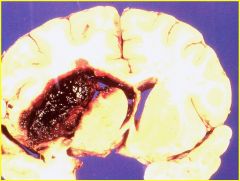
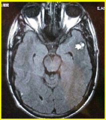
what pathology is shown here? how will pt experience this?
|
hypertensive intracerebral hemorrhage; usually will suddenly rupture and pt with have a sudden headache. then they get homonymous hemi field defect.
|
|
|
what 3 places can you develop intracerebral hemorrhages?
|
BG/thalamus, pons, cerebellum
|
|
|
pt had sudden bad headache and then got ipsilateral ataxia. what may have happened?
|
cerebellar hemorrhage.
|
|
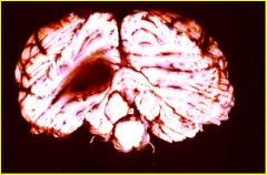
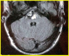
what is shown here? can surgery repair this type of damage? what can surgery NOT repair?
|
cerebellar hemorrhage. pt may get tonsillar herniation very quickly leading to respiratory failrue.
surgery can be done here with good prognosis. however, surgery is not an option when you bleed into your pons or your hemispheres, only hemorrhage in the cerebellum |
|
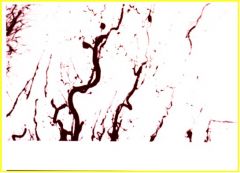
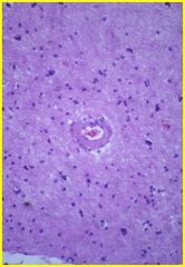
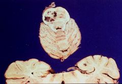
what is this showing? where can this pathology occur?
|
microaneurysms (they can be found in the BG/thalamus, pons, cerebellum). these are the most common sites of hypertensive hemorrhages.
KNOW: hypertensive intracranial hemorrhages are due to microaneuryms!! |
|
|
which is more common cause of stroke, ischemia or hemorrhage?
|
ischemia
|
|
|
what are the 2 types of hemmorhages?
|
intracerebral hematoma (ICH)
subarachnoid hemorrhage (SAH) |
|
|
what are the 4 types of intracerebral hematomas?
|
- high BP-->hypertensive ICH
- amyloid angiopathy --> lobar ICH - hemorrhage into tumors - coagulation defects (either medically induced by warfarin or through pathway defects) |
|
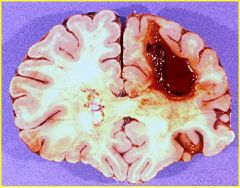
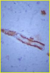
what pathology is shown here? how does it occur? what are its clinical findings?
|
amyloid angiopathy (which is a major cause of LOBAR HEMORRHAGE IN ELDERLY PATIENTS)
DEPOSITION OF AMYLOID IN THE VESSEL WALLS AND THAT WEAKENS STRUCTURAL INTEGRITY OF VESSEL WALL- MORE PRONE TO HEMORRHAGE. FOR SOME REASON, THESE ANEURYSMS OCCUR OUTSIDE OF THE 3 STRUCTURES MENTIONED ABOVE, THESE ARE CALLED LOBAR INTRACEREBRAL HEMORRHAGE. PT SAYS, “MAN I HAVE A HEADACHE!” PT MAY HAVE SOME FRONTAL BEHAVIORAL ABNORMALITIES, CONTRA HEMIPARESEIS, MAYBE A BROCA’S APHASIA (DEPENDING ON THE LOCATION) |
|
what type of hemorrhage is shown here?
|
amyloid angiopathy (structurally unsound vessels with beta amyloid stuck in the vessel wall) ==> lobar ICH
- pateints with alzheimer's are prone to these types of hemorrhages (go figure!) |
|
what is shown here?
|
amyloid angiopathy--> lobar ICH
|
|

what is shown here/
|
multiple hemorrhages in metastatic tumors...this pt had multiple brain tumors thta bled
BUT WHEN YOU SEE A INTRACERREBRAL HEMORRHAGE THAT’S NOT IN THE 3 ABOVE, AND THE PT DOESN’T HAVE HYPERTENSION, OR IF THEY’RE ON WARFARIN FOR DIFFERENT THINGS, THEN YOU HAVE TO THINK OF INTRACEREBRAL HEMORRHAGES LIKE THIS. |
|
|
what is important in the therapy of intracerebral hemorrhage (ICH)?
|
- don't give anti-coagulant (bc you want their blood to remain sticky)
- surgical evacuation of CEREBELLAR hemorrhage, no where else!! - can drain ventricles if hemorrhage goes there - FACTOR VII has been shown to help slow down expansion of ICH |
|
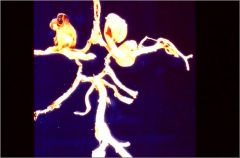
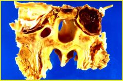
what is being shown here? point to it!
what is the major cause of these kinds of hemorrhages? |
subarachnoid hemorrhage (SAH)
berry aneurysms are major cause CONGENITAL DEFECT OF THE WALL OF THE BIG VESSELS AT BASE OF BRAIN - OVER YEARS OF EXPOSURE WILL RESULT IN GRADUAL DILATTION TO FORM A BERRY ANEURYSM, WHICH ONE DAY WILL RUPTURE. THIS PICTURE SHOWS THAT BERRY ANEURYSM CAN BE MULTIPLE. USUALLY PROXIMAL CIRCLE IN THE ANTERIOR CIRCULATION OF THE CIRCLE OF WILLIS (ACA, A COMM. A, MCA) THERE’S 3 ANEURYMS THAT ARE SHWON ABOVE. THE LEFT TOP IS THE ONE THAT RUPTURED |
|
|
contrast berry aneurysms with mycotic aneurysms
|
berry - on anterior circulation, proximally (can be multiple)
mycotic "infectious" (bacterial/fungal) - on anterior circulation also, but distally IF THERE’S INFECTED HEART VALVE AND SHOWERING BLOOD STREAM WITH INFECTIOUS EMBOLI, MAY GO INTO THE VESSEL WALL OF THIS AREA IN THE HEAD. THEY TEND TO BE VERY DISTAL (IE. ONE OF THE BRANCHES OF THE MCA). |
|
|
what if pt says that they had a sudden onset of THE WORST HEADACHE OF THEIR LIFE?
|
THAT SHOULD RAISE RED FLAG, IT COULD BE A LEAKAGE OF THE ANEURYSM AND THEIR READY TO BLOW ANYTIME. WHEN A PERSON HAS A BLEED FROM BERRY ANEURYSM, 30-40% OF THEM WILL DIE OUTRIGHT. THIS IS A VERY BAD DZ.
|
|
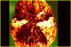
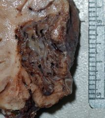
what is shown here? what are 2 ways to keep this from happening again?
|
ruptured berry aneurysm; dark blood in the subarachnoid space
1. neurosurgical (clip the aneuryism and isolate it from circulation so that it will eventually clot) 2. interventional neuroradiology (where you run metal coil into it and then run a current through it. the coil will separate the aneurysm) |
|
what does this do?
|
coil will separate the aneuryism and you build an instant clot
|
|

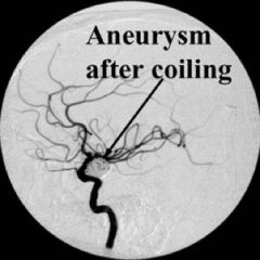
what happened here?
|
pt with a sudden onset of worst headache of her life (from an ACA aneuryism). she was coiled, but after the coiling, the neck ripped and had a subarachnoid hemorrhage
VERY UNCOMMON |
|
What pathology occurred here?what would the clinical findings be?
|
cavernous sinus aneurysm. internal carotid ruptures within cavernous sinus and will bleed into the sinus (better than subarachnoid hem). but you try to shunt this, could mess up CN 3,4,5,6.
|
|
|
what are the similiarities and differences bw these 2 vascular malformations that could cause hemorrhages: arteriovenous malformation (AVM) & cavernous angioma?
|
BOTH CAN CAUSE 3 THINGS: HEMORRHAGE, SEIZURES, AND FOCAL NEURO DEFICITS.
AVM - composed of anastomosed abnormal arterioles/veins; PRESENCE OF INTERVENING PARENCHYMA CA - composed of capillary and small vein sized vessels THAT ARE BACK TO BACK (NO PRESENCE OF INTERVENING PARENCHYMA) |
|
what pathology is imaged forth? how can you treat this?
|
arteriovenous malformation
treat by surgically removing this to prevent hemorrhage/stop seizures. also you can inject crazy glue to harden the vessels and block off all vessels to the AVM |
|
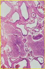
what is the pathology here? how can you tell?
|
arterovenous malformation, as evidenced by the presence of intervening parenchyma
can get 3 things: hemorrhage, seizures, or focal neuro deficit |
|
|
what is the pathology here? how can you tell? what would the clinical findings be?
|
temporal lobe cavernous angioma
you can tell bc of its popcorn appearance. would give you an aura prior to seizure (like smelling burnt rubber) and deja vu. |
|
what is the pathology here? how can you tell? what would the clinical findings be?
|
temporal lobe cavernous angioma
you can tell bc of its popcorn appearance. would give you an aura prior to seizure (like smelling burnt rubber) and deja vu. |
|
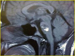
what is this pathology? what is it composed of? name the mutation that could cause this.
|
cavernous sinus in the pons; composed of capillaries and small vein sized vessels that are back to back. "popcorn" appreance on MRI.
"krit" mutation is involved in regulation of vascular growth and involved with CA |
|
what is wrong with this picture?
|
pons showing vascular malformation due to cavernous angioma
|
|
what is this?
|
cavernous angioma found in the pons. If it Oozes too much could give you LT findings, CN findings, encroach on RAS and lead to LOC
|