![]()
![]()
![]()
Use LEFT and RIGHT arrow keys to navigate between flashcards;
Use UP and DOWN arrow keys to flip the card;
H to show hint;
A reads text to speech;
85 Cards in this Set
- Front
- Back
|
in general the nervous system is -------- acting |
fast |
|
|
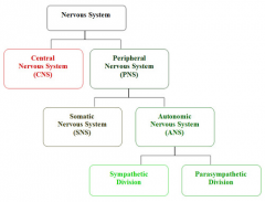
Central nervous system |
brain and spinal cord |
|
|
Peripheral nervous system |
the nervous tissue outside of the brain and spinal cord |
|
|
The main function of the PNS is to |
connect the central nervous system (CNS) to the limbs and organs, essentially serving as a communication relay going back and forth between the brain and the extremities |
|
|
The peripheral nervous system is divided into |
somatic nervous system and the autonomic nervous system |
|
|
Somatic nervous system |
(skeletal muscle) |
|
|
Autonomic nervous system |
(visceral motor system)- provides involuntary regulation of smooth muscle, cardiac muscle, and glandular activity |
|
|
This system is the primary mechanism in control of the fight-or-flight response |
Autonomic nervous system specifically Sympathetic division (adrenaline) |
|
|
The autonomic nervous system has two branches |
Sympathetic division Parasympathetic division |
|
|
Parasympathetic division of the autonomic nervous system |
resting and digesting system (defecation, urination) responsible for stimulation of "rest-and-digest" or "feed and breed" |
|
|
Sympathetic division of the autonomic nervous system |
the fight or flight system (adrenaline) |
|
|
Neurons |
basic units of the nervous system |
|
|
This observation explains why such neurons cannot divide through mitosis |
Most neurons lack centrioles |
|
|
multipolar neuron |
Type of neuron that possesses a single (usually long) axon and many dendrites, allowing for the integration of a great deal of information from other neurons. |
|
|
constitute the majority of neurons in the brain |
multipolar neuron |
|
|
Astrocytes ( astron = star and cyte from = cell) |
characteristic star-shaped glial cells in the brain and spinal cord |
|
|
the most abundant cells of the human brain |
Astrocytes |
|
|
functions of Astrocytes include |
1. support of endothelial cells that form the blood brain barrier
2. provision of nutrients to the nervous tissue
3. role in the repair and scarring process of the brain and spinal cord following traumatic injuries. |
|
|
resting potential of a neuron is |
-70 mV (millivolt) |
|
|
depolarization |
Opening of voltage-gated sodium channels in the membrane of a neuron results in depolarization |
|
|
hyperpolarization |
stimulus that opens gated potassium ion channels, taking the membrane potential away from 0 mV, results in hyperpolarization |
|
|
Generation of an action potential steps |
|
|
|
action potential |
When a stimulus reaches a resting neuron, the neuron transmits the signal as an impulse |
|
|
Generation of an action potential steps (condensed) |
|
|
|
Saltatory conduction (from the Latin saltare, to hop or leap) |
1. have myelinated axons
2.faster than conduction on an unmyelinated axon
3. they skip" from node of Ranvier to node of Ranvier |
|
|
the myelin sheath helps in |
1. increasing the speed of the nerve impulse
2. reducing energy expenditure over the axon membrane as a whole, because the amount of sodium and potassium ions that need to be pumped to bring the concentrations back to the resting state following each action potential is decreased |
|
|
Continuous conduction |
1. No myelin sheath means no nodes of Ranvier, so no "skipping
2. is slower
3. currents depolarize adjacent areas of membrane so that action potentials continue to form along the membrane |
|
|
What Is a Cholinergic Synapse |
a gap where a neuron that produces acetylcholine sends messages to other neurons, or to skeletal muscle cells |
|
|
List the correct order of events that occur at a cholinergic synapse |
|
|
|
Meninges |
The membranes that envelop the brain and spinal cord of the central nervous system.
Primary function of the -------- and of the cerebrospinal fluid is to protect the central nervous system. |
|
|
meninges consist of three layers: |
1. dura mater 2. arachnoid mater 3. pia mater |
|
|
Dura mater [Latin: 'tough mother'] |
1. thick, durable membrane 2. closest to the skull (cranium) |
|
|
Arachnoid mater (spider like ) |
1. middle layer
2. spider web-like appearance
3. This thin, transparent membrane is composed of fibrous tissue and, like the pia mater, is covered by flat cells also thought to be impermeable to fluid. |
|
|
Pia mater [Latin: 'soft mother'] |
layer of the meninges that is in direct contact with the surface of the brain and spinal cord (the gentle layer) |
|
|
Subarachnoid space |
The anatomic space between the arachnoid membrane and pia mater.
|
|
|
Subarachnoid space is occupied by |
Spongy tissue consisting of trabeculae (delicate connective tissue filaments that extend from the arachnoid mater and blend into the pia mater) and intercommunicating channels in which the cerebrospinal fluid is contained. |
|
|
Head injuries that damage cerebral blood vessels are serious conditions because |
these spaces compress and distort the relatively soft tissues of the brain |
|
|
Dorsal root ganglia |
(cell bodies of sensory neurons) |
|
|
If the dorsal root of a spinal nerve is severed |
incoming sensory information would be disrupted |
|
|
Grey matter |
1. Contains numerous cell bodies and relatively few myelinated axons.
2. Projections extend through the white matter toward the outer surface of the spinal cord.
3. Dominated by cell bodies of neurons and glial cells. |
|
|
posterior horns of the spinal cord contain |
sensory nuclei |
|
|
horns |
projections of gray matter extending through the white matter toward the outer surface of the spinal cord |
|
|
White matter |
Columns
Component of the central nervous system, in the brain and superficial spinal cord, and consists mostly of glial cells and myelinated axons that transmit signals from one region of the cerebrum to another and between the cerebrum and lower brain centers. |
|
|
Cerebrum |
region of the brain that is involved in conscious thought and intellectual function as well as processing somatic sensory and motor information |
|
|
The two cerebral hemispheres are separated by |
longitudinal fissure |
|
|
The primary connection between cerebral hemispheres is the |
corpus callosum |
|
|
Association areas of the cerebral hemispheres |
Regions of the brain that are involved in interpreting data or coordinating motor responses |
|
|
Postcentral gyrus |
Contains the primary sensory cortex |
|
|
Neuroglia sometimes called neuroglia or simply glia |
Half the volume of the nervous system
Many types in the cns (4) and pns |
|
|
The visual cortex of the cerebrum is located |
occipital lobe |
|
|
Basal nuclei |
functions in the subconscious control of muscle tone and the coordination of learned movement patterns |
|
|
Diencephalon ("interbrain") |
1. acts as a switching and relay center for integration of conscious and unconscious sensory information and motor commands
2. links cerebrum with brain stem (midbrain, pons and medulla oblongata) |
|
|
Diencephalon is made up of four distinct components: |
1. thalamus 2. subthalamus 3. hypothalamus 4. epithalamus |
|
|
Thalamus |
1. largest portion of the diencephalon
2. functions are the relaying of sensory and motor signals to the cerebral cortex, and the regulation of consciousness, sleep, and alertness. |
|
|
Midbrain |
1. Processes sight, sound, and associated reflexes; maintains consciousness.
2. portion of the central nervous system associated with vision, hearing, motor control, sleep/wake, arousal (alertness), and temperature regulation |
|
|
The midbrain has a slender CSF-filled canal known as the |
cerebral aqueduct |
|
|
Stimulation of the reticular formation results in |
increased attention (retention and altertness) |
|
|
Pons |
control pace and depth of respiration
Sensory nuclei of cranial nerves V-VIII |
|
|
Medulla oblongata |
Major centers concerned with autonomic function, such as heart rate, blood pressure, and respiration |
|
|
Cerebellum |
part of the CNS that adjusts voluntary and involuntary motor activities on the basis of sensory information and stored memory of previous movements; automatic processing center responsible for programming and fine-tuning movements controlled at the conscious and subconscious levels |
|
|
Important role in motor control. Damage produces disorders in fine movement, equilibrium, posture, and motor learning. |
cerebellum (Latin for "little brain") |
|
|
----------- connects the CNS with the body’s external and internal environments |
PNS |
|
|
# of Cranial nerves |
12 |
|
|
: only cranial nerve that is attached to the cerebrum is the |
olfactory nerve |
|
|
cranial nerve that has three branches is the |
trigeminal nerve |
|
|
Damage to the ------- nerve, which is vital for the autonomic control of visceral function, could result in death. |
vagus nerve |
|
|
dermatome |
specific region of the body monitored by each pair of spinal nerves is known as |
|
|
Nerve plexuses |
nerve fibers interwoven together |
|
|
PNS reflexes are |
rapid, automatic responses to stimuli |
|
|
Monosynaptic reflex |
exemplified by the stretch reflex |
|
|
withdrawal reflex |
Pulling away from a painful stimulus |
|
|
Somatic nervous system (SNS) vs Autonomic Nervous System (ANS) |
SNS- under conscious control, control skeletal muscles.
ANS- operates without conscious instruction |
|
|
ganglia neurons |
Nerve cell cluster or a group of nerve cell bodies located in the peripheral nervous system. |
|
|
What are the three functions of the Nervous System? |
1. Sensory Function |
|
|
levels of divisions in the nervous system |
|
|
|
What does the autonomic nervous system control? |
-Smooth muscle (like blood vessels and digestion), cardiac muscle and glands (medulla of the adrenal gland) |
|
|
What are the two major categories of cells found in the nervous system? |
-Glial Cells |
|
|
What are the parts of a neuron? |
-Cell body (aka: soma) |
|
|
What are clusters of cell bodies within the peripheral nervous system called? |
Within the PNS, they are called GANGLIA |
|
|
What are clusters of cell bodies within the central nervous system called? |
Within the CNS, they are called NUCLEI |
|
|
What is the function of the dendrites? |
part of the neuron which RECEIVE electrical impulses (action potentials) |
|
|
What is the function of the dendrites? |
carries impulses AWAY from the cell body (toward another neuron, muscle fiber or gland cell) |
|
|
What is a myelin sheath and what is its function? |
A myelin sheath is a lipid that surrounds the axon |
|
|
Membrane potential |
the difference in electric potential between the interior and the exterior of a biological cell |
|
|
The four classifications of Cranial Nerves |
|