![]()
![]()
![]()
Use LEFT and RIGHT arrow keys to navigate between flashcards;
Use UP and DOWN arrow keys to flip the card;
H to show hint;
A reads text to speech;
97 Cards in this Set
- Front
- Back
|
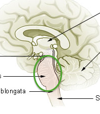
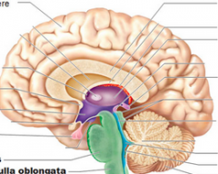
Medulla Oblongata |
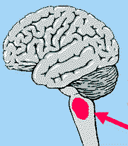
Continuous with spinal cord through foramen magnum (legs of seahorse) |
|
|
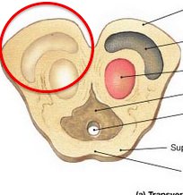
Olives of Medulla |
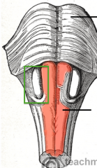
Rounded structures, sides of medulla (green box) |
|
|
Pyramids of Medulla |

In between olives, anterior surface |
|
|
Superior Cerebellar Peduncles |
Posterior of brain, in front of cerebellum |
|
|
Inferior Cerebellar Peduncles |
Posterior of brain, inferior front of cerebellum |
|
|
Middle Cerebellar Peduncles |
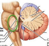
Posterior of brain, middle in front of cerebellum |
|
|
Fourth Ventricle |
Space between peduncles |
|
|
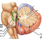
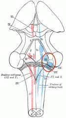
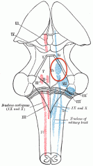
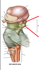
Glossopharyngeal Nerve (CNIX) |

Lateral to olive, superior (on model) |
|
|
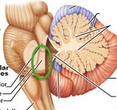
Vagus Nerve CN(X) |
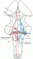
Lateral & inferior to olive (on model) |
|
|
Hypoglossal Nerve CN(XII) |
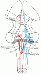
Medial nerve next to olive (on model) |
|
|
Spinal Acessory Nerve |
On bottom of medulla (on model) |
|
|
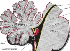
Pons |
Belly of seahorse |
|
|
Rhomboid Fossa |
Anterior Surface |
|
|
Abducens nerve CN(VI) |
Abs of seahorse most medial (on model) |
|
|
Facial Nerve CN (VII) |
More lateral, thin in middle (on model) |
|
|
Vestibulocochlear nerve |
Most lateral (on model) |
|
|
Trigeminal Nerve CN(V) |
Adjacent to middle peduncle |
|
|
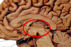
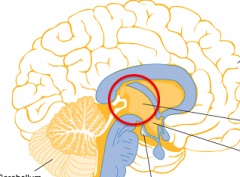
Midbrain |
Neck of seahorse |
|
|
Corpora Quadrigemina |
Back of neck 4 bumps |
|
|
Superior Colliculi |

Top Bumb, 2 sets |
|
|
Inferior Colliculi |
Bottom bump, 2 sets |
|
|
Crus Cerebri |
Mickey mouse's ears |
|
|
Substantia Nigra |
Lines that separate ears from head |
|
|
Cerebral Aqueduct |
Mickeys nose |
|
|
Oculomotor Nerve CN(III) |
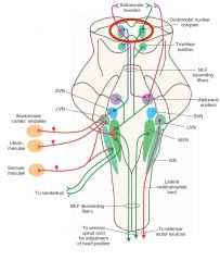
Line on base of neck (on model) |
|
|
Trochlear Nerve CN(IV) |
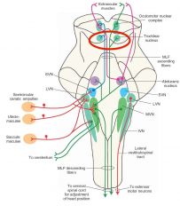
chain on back of necklace, posterior |
|
|
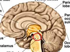
Diencephalon |
Head of seahorse |
|
|
Thalamus |
Mid portion of seahorse head |
|
|
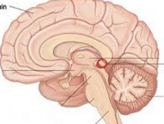
Interthalamic Adhesion |
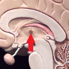
Yellow button in middle of thalamus |
|
|
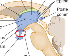
Epithalamus |
Long pink portion, like mullet |
|
|
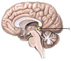
Pineal Gland |
Bulb at end of epithalamus |
|
|
Hypothalamus |
Mouth of seahorse, anterior |
|
|
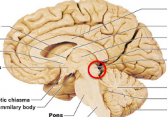
Infundibulum |
Cherry stem in mouth, cherry is pituitary gland |
|
|
Mammilary Bodies |
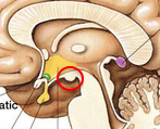
Bump under chin |
|
|
Optic Tracts |
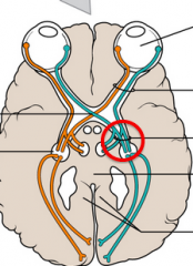
Nerves to brain |
|
|
Optic Chiasm |
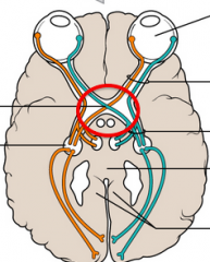
Crossing |
|
|
Optic Nerves |
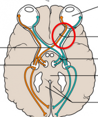
To eyes |
|
|
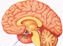
Third Ventricle |
Space between thalamus (purple) |
|
|
Interventricular Foramina |
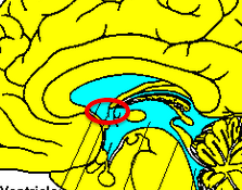
space between 3rd ventricle and lateral ventricle |
|
|
Choroid Plexus |
Fuzzy grapes, in any ventricle |
|
|
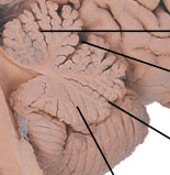
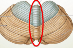
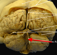
Cerebellum |
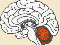
Lower back of brain, posterior |
|
|
Cerebellar Hemispheres |
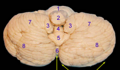
Left + right, biggest part (8) |
|
|
Folia and Fissure |
Folia: ridges, Fissure: space in between |
|
|
Vermis of Cerebellum |
Tissue that connects 2 hemispheres |
|
|
Cerebellar Cortex |
Leaves on tree |
|
|
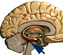
Arbor Vitae |

Brances of tree |
|
|
Cerebellar Nuclei |

Core of the cerebellum |
|
|
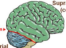
Cerebral Hemispheres |
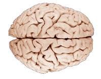
Big portion |
|
|
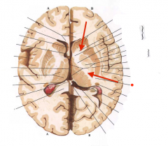
Longitudinal Fissure |
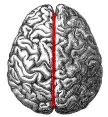
Portion dividing left/right |
|
|
Cerebral Cortex |
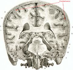
Outer layer, gray matter |
|
|
Cerebral Gyri |
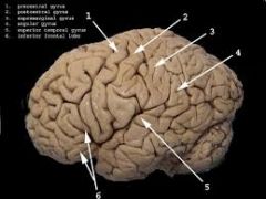
Ridges on top |
|
|
Cerebral Sulci |
Fissures in brain, grooves |
|
|
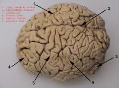
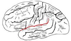
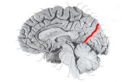
Central Sulcus |
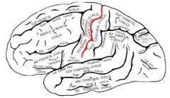
starts at longitudinal fissure -> lateral sulcas |
|
|
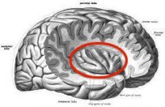
Lateral Sulcus |
Divides parietal and temporal lobe |
|
|
Parieto-Occipital Sulcus |
Divides pareital and occiptal, posterior |
|
|
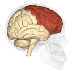
Frontal Lobes |
Frontal Poles: most frontal part |
|
|
Pareital Lobes |

|
|
|
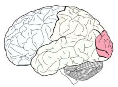
Temporal Lobes |

|
|
|
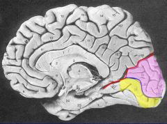
Occipital Lobes |
Posterior portion of brain |
|
|
Insula |
Within lateral sulcus, inside |
|
|
Precentral Gyrus |

Anterior of central sulcus (yellow) |
|
|
Postcentral Gyrus |
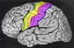
Posterior of central sulcus (purple) |
|
|
Precentral Sulcus |

Divides Frontal and precentral gyrus (Green) |
|
|
Postcentral Sulcus |

Divides postcentral gyrus and parietal lobe (blue) |
|
|
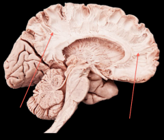
Corpus Callosum |
Sombrero of seahorse |
|
|
Anterior Commissure |
Bumb on seahorse nose (red) |
|
|
Cingulate Gyrus |
Above sombrero |
|
|
Cingulate Sulcus |
Above gyrus |
|
|
Calcarine Sulcus |
Divides horizontally in occipital lobe |
|
|
Fornix |
On brain cross-section, inferior arch |
|
|
Septum Pellucidum |
Hole under flap |
|
|
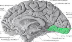
Parahippocampal Gyrus |
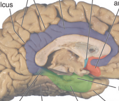
Under brain stem (green) |
|
|
Uncus |
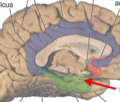
most anterior point of parahippocampal gyrus |
|
|
Lingual Gyrus |
Posteriro to parahippocampal gyrus |
|
|
Limbic Lobe |
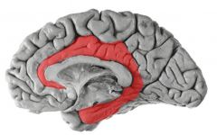
Surrounds brainstem |
|
|
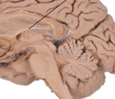
Olfactory Bulbs and Tracts |
Thin shoelace looking thing, transmit smell |
|
|
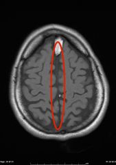
Lateral Ventricles |
space under flap and on top of flap |
|
|
Septal Nuclei |
Gray matter, middle of fornix |
|
|
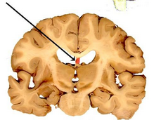
Hippocampus |
Cinnamon roll |
|
|
Basal Nuclei |
Gray matter |
|
|
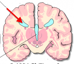
Caudate Nucleus |
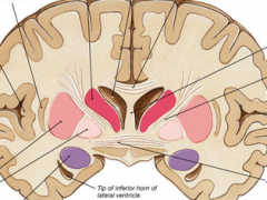
medial smaller (red) |
|
|
Lentiform Nucleus |

Lateral larger (pink) |
|
|
Corona Radiata |
All white matter on top, towards cortex |
|
|
Internal Capsule |
White matter that divides basal nuclei |
|
|
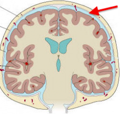
Cranial Dura Mater |

Outer Layer, thicker |
|
|
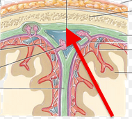
Cranial Arachnoid Mater |
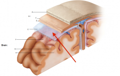
Thin next layer, webbing around brain |
|
|
Cranial Pia Mater |
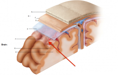
Layer directly on brain |
|
|
Falx Cerebri |
In between hemispheres |
|
|
Tentorium Cerebelli |
Horizontal separating cerebelluma and cerebrum |
|
|
Falx Cerebelli |
Potential space between cerebellar hemispheres |
|
|
Cranial Meningeal |
Potential space |
|
|
Epidural Space |
Potential Space |
|
|
Subdural Space |
Between dura and arachnoid matter |
|
|
Subarachnoid Space |
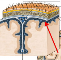
Under arachnoid matter |
|
|
Anterior Median Fissure |
opening at front of spinal cord |
|
|
Conus Medullaris |
Thick fibrous end of spinal cord |
|
|
Central Canal |
Groove for canal |