![]()
![]()
![]()
Use LEFT and RIGHT arrow keys to navigate between flashcards;
Use UP and DOWN arrow keys to flip the card;
H to show hint;
A reads text to speech;
23 Cards in this Set
- Front
- Back
|
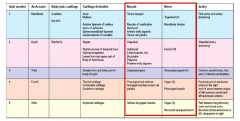
Pharyngeal Arches |
|
|
|
Muscles: Superficial |
(1) Platysma: • IN: CN VII • A: Tenses skin of neck, depresses/wrinkles lower face (2) Sternocleiodmastoid: • IN: spinal portion of CN XI • A: bilaterally extends; unilaterally draws chin up and flexes to same side (3) Trapezius (Superior Portion): • IN: spinal portion of CN XI • A: Draws scapula obliquely upward and rotates glenoid cavity inferiorly (4) Levator Scapulae: • IN: C3-C5 anterior rami • A: scapular movements |
|
|
Muscles: Suprahyoid |
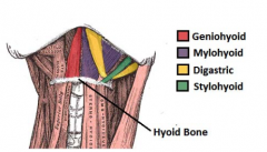
• All insert into hyoid bone (1) Geniohyoid: o O: Inferior mental spine of mandible o IN: Anterior ramus of C1 (2) Mylohyoid: o O: Mylohyoid line o IN: Mylohyoid n. (CN V3) (3) Digastric: o O: Mandible (anterior) + temporal bone (posterior) o IN: Mylohyoid n. (anterior) + CN VII (posterior)(4) Stylohyoid: o O: Styloid process o IN: CN VII |
|
|
Muscles: Infrahyoid |
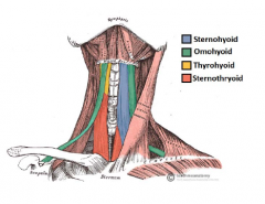
• (1) – (3) innervated via ANSA CERVICALIS • All insert into hyoid unless stated otherwise (1) Omohyoid: o O: intermediate tendon (superior) + superiorborder of scapula (inferior) (2) Sternohyoid: o O: posterior surface of manubrium (3) Sternothyroid: o O: posterior surface of manubrium o I: oblique line of thyroid cartilage (4) Thyrohyoid: o O: oblique of thyroid cartilage o IN: anterior ramus of C1 |
|
|
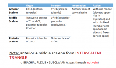
Muscles: Prevertebral |
|
|
|
Thoracic Duct |
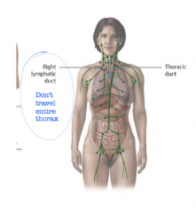
Right lymphatic ductdoesn’t travel entirethorax |
|
|
Arterial Blood Supply |
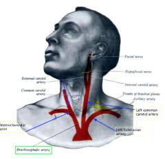
• Brachiocephalic Trunk: suppliesR. side of bodyo R. Subclavian A. o R. Common Carotid: Bifurcates toR. external and internal-Carotid Body + Sinus • L. Common Carotid A. • L. Subclavian A. Carotid Body: chemoreceptors (CN XI & X)Carotid Sinus: baroceptors (CN XI) |
|
|
Carotid body and sinus |
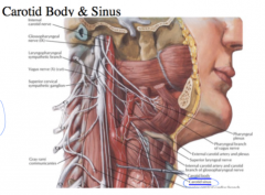
• Both located atbifurcation • Carotid Sinus Swellingextends to ICA* |
|
|
Arterial Blood Supply |
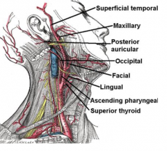
External Carotid • Gives 8 branches to theneck and is OUTSIDEthe skull • Pneumonic: o SALFOPMS |
|
|
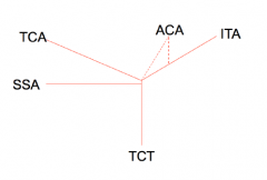
Arterial Branches: R. Subclavian A. |
• Passes throughINTERSCALENE triangle • Mnemonic: ICTV (D) o Vertebral A., InternalThoracic., ThyrocervicalTrunk, Costal Cervical, DorsalScapular A (Sometimespresent*) |
|
|
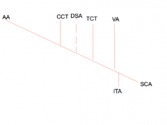
Arterial Blood Supply: Branches of CCT and TCT (shown) |
• Costal Cervical Trunk Branches: o deep cervical a. o supremeintercostal a. • Thyrocercival Trunk: ISAT on the trunk o Inferior Thyroid A. o Suprascapular A. o Ascending Cervical A. Note: travels with Phrenic N. o Transverse Cervical A. |
|
|
Venous Drainage: |
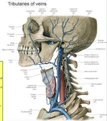
• Maxillary V. + Superficialtemporal = RetromandibularV. • Posterior Auricular V. +Posterior Retromandibular V.= EJV • Facial V. + AnteriorRetromandibular V. =Common Facial (drains intoIJV) • EJV drains to suclavian V.which drains tobrachiocephalic V. |
|
|
Venous Drainage: IJV |
• Drains into Brachiocephalic V. • Mnemonic: Medical School LetsFunny People In o Middle Thyroid V. o Superior Thyroid V. o Lingual V. o Facial V. (joins anterior retromandibular) o Pharygneal V. o Infrapetrosal V. • Note: Inferior thyroid V. drainsdirectly into Brachiocephalic V. |
|
|
Innervation: Cervical Plexus (C1-C4) |
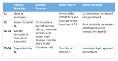
LGTS - Phrenic nerve: C3-C5 shared with brachial plexus; both motor and sensory --> diaphragm |
|
|
Innervation: Cervical Plexus (C1-C4) |
note the phrenic nerve |
|
|
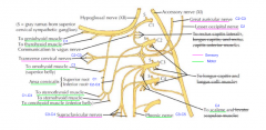
Innervation: Cranial Nerves: Vagus Nerve (X): |
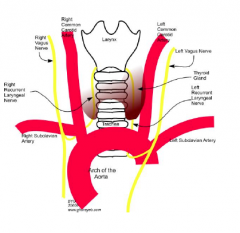
• Sensory: thoracic and abdominal viscera,muscosa of larynx/pharynx, caro=d body • Motor: muscles of pharynx/larnyx • Visceral Motor: regulates HR, BR, digestiveactivity • Superior Laryngeal Br.: o Internal = sensory o External = motor • Recurrent Laryngeal Br.: MIXED o L. Side: loops around arch of aorta o R. Side: loops around subclavian a. |
|
|
Innervation: Cranial Nerves: Accessory Nerve (CN XI) |
MOTOR • 2 components – SPINAL and CRANIAL • Pathway: exits via jugular foramen, descends down carotid sheath, and travelsthrough POSTERIOR triangle • Supplies: SCM and TRAPEZIUS |
|
|
Innervation: Cranial Nerves: Hypoglossal Nerve (CN XII) |
MOTOR • Pathway: exits hypoglossal canal, momentarily in the carotid sheath, wrapsaround external carotid artery and travels to oral cavity • Supplies: 7/8 glossal muscles EXCEPT PLATOGLOSSUS |
|
|
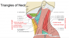
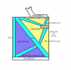
Triangles of Neck |
|
|
|
Triangles diagram |
|
|
|
Triangles of Neck: Anterior: Bounded by SCM, midline of the neck, mandible |
(1) Submandibular (Gastric) Triangle: • Bounded by: mandible, post and ant digastric • Contains: submandibular gland, facial artery, vein and nerve (and branches) (2) Carotid Triangle: • Bounded by: SCM, superior omohyoid, and posterior digastric • Contains: common carotid a, external and internal a, internal jugular, superiorroot of ansa cervicalis, internal laryngeal nerve and nerve to thyrohyoid C1,hypoglossal n, internal jugular vein, facial vein, and superior thyroid artery (3) Muscular Triangle: • Bounded by sternocleidomastoid, superior omohyoid and sternohyoid (4) Submental Triangle: • Bounded by the anterior bellies of the digastric, hyoid bone and mandible |
|
|
Triangles of Neck: Posterior: Bounded by SCM, trapezius, and clavicle |
• Bounded by the sternocleidomastoid, trapezius, and clavicle • Contains: external jugular, cervical plexus, accessory nerve, brachial plexus,transverse cervical artery and vein, phrenic nerve, accessory phrenic nerve,surprascapular artery and vein, subclavian artery and vein • Muscles that form the floor: levator scapulae, scalene (posterior, middleand anterior) (1) Supraclavicular (omoclavicular and subclavian) Triangle: o Bounded by the inferior belly of the omohyoid, clavicle and sternocleidomastoid (2) Occipital Triangle: o Bounded by the inferior belly of the omohyoid, trapezius and sternocleidomastoid |
|
|
Fascia and Spaces |
(1) Prevertebral Fascia: • Splits into ANTERIOR (ALAR) and POSTERIOR layer • DANGER ZONE is located between these regions (2) Pretracheal Fascia: • Muscular portion: holds the infrahyoid muscles• Visceral portion: holds the thyroid gland, trachea, esophagus and pharynx o Continuous with the BUCCOPHARYNGEAL FASCIA (3) Carotid Sheath: • Common carotid artery, internal carotid artery, vagus nerve, internal jugular vein • AT the very base of the skull: contains CN IX, X, XI and XII but they eventuallyleave it |