Reading...
![]()
Play button
![]()
Play button
![]()
Use LEFT and RIGHT arrow keys to navigate between flashcards;
Use UP and DOWN arrow keys to flip the card;
H to show hint;
A reads text to speech;
73 Cards in this Set
- Front
- Back
|
Which areas participate in important feedback loops in which they project back to the cerebral cortex via the thalamus and do not themselves project to lower motor neurons?
|
Basal ganglia and the cerebellum
|
|
|
__________ carry motor system outputs to __________.
|
Upper motor neurons
Lower motor neurons |
|
|
Descending upper motor neurons arise from the ____ and ____. And can be divided into ______ and _______
|
cerebral cortex
brainstem lateral motor systems medial motor systems |
|
|
__________ carry motor system outputs to __________.
|
Upper motor neurons
Lower motor neurons |
|
|
Descending upper motor neurons arise from the ____ and ____. And can be divided into ______ and _______
|
cerebral cortex
brainstem lateral motor systems medial motor systems |
|
|
The lateral corticospinal tracts are essential for ______.
|
Rapid, dextrous movements at individual digits or joints.
|
|
|
How does the lateral corticospinal tract travel down?
|
They cross over from their point of origin and descend to the contralateral lateral spinal cord to control the contralateral extremities.
|
|
|
Over half of the corticospinal tracts originate from the ___. The remainder arise from the ____,_____ or ______.
|
Primary motor cortex.
Premotor supplementary motor parietal lobe areas |
|
|
The two lateral motor systems are:
|
The lateral corticospinal tract and the rubrospinal tract
|
|
|
List the site of origin of the lateral spinocortical tract.
|
Primary motor cortex and other frontal and parietal areas
|
|
|
Where is the site of decussation for the lateral spinocortical tract?
|
Pyramidal decussation at the cervicomedullary junction
|
|
|
Where are the levels of termination in the lateral corticospinal tract?
|
The entire cord (predominantly at the cervical and lumbosacral enlargements)
|
|
|
What is the function of the lateral corticospinal tract?
|
Movements of contralateral limbs.
|
|
|
What are the four medial motor systems?
|
Anterior corticospinal tract, the vestibulospinal tracts, the reticulospinal tracts and the tectospinal tract.
|
|
|
Medial motor systems tend to terminate on ________ that project to _________, controlling movements that involve ________. Thus, unilateral lesions of these systems produce ________.
|
Interneurons
both sides of the spinal cord Multiple bilateral spinal segments No obvious deficits. |
|
|
Lesions of the lateral corticospinal tract produce ______.
|
dramatic deficits
|
|
|
The lateral corticospinal tract is the most clinical important descending motor pathway in the nervous system. Lesions in this pathway _____.
|
Can be precisely localised because they produce characteristic deficits.
|
|
|
The primary motor cortex neurons contributing to the corticospinal tract are located mostly in ____.
|
cortical layer 5
|
|
|
Layer 5 pyramidal cell projections synapse directly onto _______ in the _______ of the _______, as well as onto _______.
|
motor neurons
ventral horn spinal cord spinal interneurons |
|
|
Axons (of the LCT) from the cerebral cortex enter the ______ and descend toward the ______.
|
upper portions of the cerebral white matter (or corona radiata)
internal capsule. |
|
|
In addition to the corticospinal tract, the cerebral white matter conveys __________ between cortex and ________ such as the _______________.
|
bidirectional information
different cortical areas deep structures BG, thalamus and brainstem |
|
|
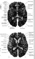
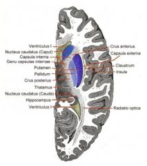
The ______ and ______ are always medial to the internal capsule.
|
thalamus
caudate nucleus |
|
|
The ___ and __ are always lateral to the internal capsule.
|
globus pallidus
putamen |
|
|
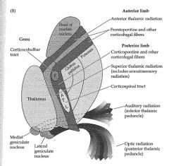
There are three parts to the internal capsule:
|
a) anterior limb
b) posterior limb c) genu |
|
|
The anterior limb of the interior capsule:
|
separates the head of the caudate from the GP and putamen
|
|
|
The posterior limb of the interior capsule:
|
separates the thalamus from the GP and putamen
|
|
|
The genu of the int. capsule
|
The transition between the anterior and posterior limbs at the level of the foramen of monroe.
|
|
|
The corticospinal tract lies in the ______ of the int capsule.
|
posterior limb
|
|
|
What is preserved in the internal capsule?
|
the somatotopic map so motor fibers for the face are most anterior and those for the arm and leg are progressively more posterior.
|
|
|
Fibres projecting from the cortex to the brainstem (including ______) are called ______ because they project from the cortex to the "____" (brainstem).
|
motor fibers for the face
corticobulbar bulb |
|
|
Lesions at the internal capsule cause:
|
weakness of the entire contralateral side because the fibers are compact enough.They occasionally produce more selective motor deficits.
|
|
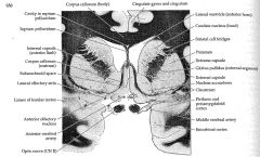
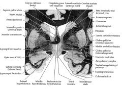
What are all these places?
|
|
|
|
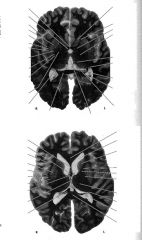
The internal capsules continues into the _____ _____ (Meaning literally the feet of the brain). The corticospinal tract fibers next descend through the ___ ____ where they form somewhat scattered fascicles. These collect on the ventral surface of the _____ to form the ____ _____. For this reason, the corticospinal tract is sometimes known as the ___ ____
|
cerebral peduncles
ventral pons medulla medullary pyramids pyramidal tract |
|
|
The pyramids not only hold fibers of the corticospinal tract but also include:
|
reticulospinal and other brainstem pathways.
|
|
|
The transition from the medulla to spinal cord is called the ____ and occurs at the level of the _____.
|
cervicomedullary junction
foramen magnum |
|
|
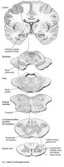
At the cervicomedullary junction, what happens?
|
About 85% of the pyramidal tract fibers decussate (i.e. the pyramidal decussation) to enter the lateral white matter columns of the spinal cord.
|
|
|
Axons of the lateral corticospinal tract enter the spinal cord central grey matter to synapse onto the _____.
|
anterior horn cells.
|
|
|
15% of corticospinal tract fibers do what in the spinal cord?
|
Enter the anterior white matter columns to form the anterior corticospinal tract.
|
|
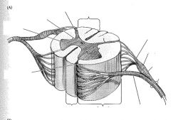
Label these areas
|
|
|
|
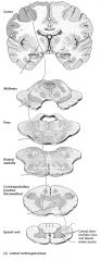
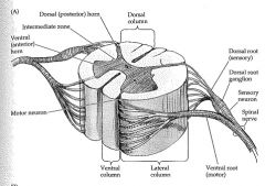
The spinal cord contains a butterfly-shaped _____ surrounded by ascending and descending _____ of ____.
|
central gray matter
white matter columns (funiculi) |
|
|
|
|
|
Motor neurons exit the ____ ___ to form ____ nerves or from segments of the ___ ___ to form ___ nerves.
|
Brain stem
cranial Spinal cord spinal |
|
|
Cranial and spinal segments are called
|
dermatomes (areas on the skin supplied by sensory fibers of the spinal nerves).
|
|
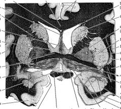
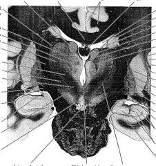
Label these
|
|
|
|
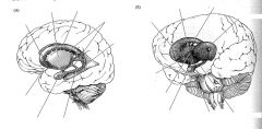
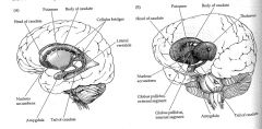
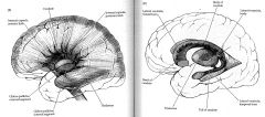
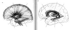
The ___ ___ and the ___ are histologically and embryologically closely related and can be thought of as a single large nucleus called the ___ (or the ___)
|
Caudate nucleus
putamen neostriatum striatum |
|
|
The caudate nucleus is a what shaped structure?
|
C shaped
|
|
|
The CN is divided into three parts:
|
head, body and tail - which do not have a distinct boundary from each other.
|
|
|
The amygdala lies just anterior to the tip of the _____ ____ in the ___ lobe.
|
caudate tail
temporal lobe |
|
|
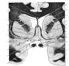
The putamen is a large nucleus forming the ____ portion of the ___ ____.
|
lateral
basal ganglia |
|
|
The putamen fuses with the ___ of the caudate. This region is called ___.
|
anterior and ventral portion of the head
the ventral striatum |
|
|
Most of the ventral striatum consists of the ___ ___.
|
nucleus accumbens
|
|
|
Just medial to the putamen lies the ____ ____.
|
Globus pallidus
|
|
|
What does the globus pallidus stand for?
|
"pale globe" because many myelinated fibers traverse this section.
|
|
|
What is the lentiform nucleus?
|
The putamen and globus pallidus together.
|
|

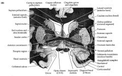
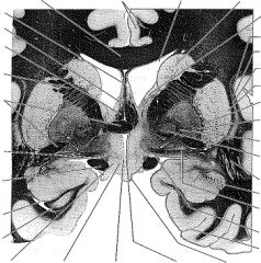
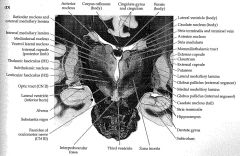
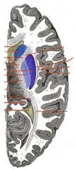
Label these
|
|
|
|
|
|
|
|
|
|
|

|
|

|
|
|
|
|
|
|
|
|
|
|
|
|
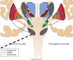
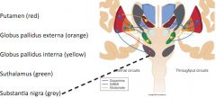
Virtually all inputs to the BG arrive via the ____. Outputs leave the BG via the _____ and the closely related ____.
|
striatum
internal segment of the globus pallidus substantia nigra pars reticula. |
|
|
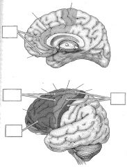
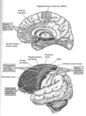
What are the parallel BG pathways for movement, eye movement, cognition and emotion? (or the extrapyramidal motor system-cortical channels). What are the cortical targets of output?
|
Motor channel = supplementary motor area (BA 5), premotor cortex (BA 4), primary motor cortex (BA 6)
Oculomotor channel: Frontal eye fields (BA8) (not primary motor eye movements but it related to cognition) Prefrontal Channel: DLPFC (BA 9/46) Limbic Channel: Anterior cingulate gyrus (BA 24), subcallosal cortex (septal area) (BA 25, 32) |
|
|
|
|
|
The extrapyramidal motor system comprises a system of fibers originating from a variety of cortical regions which then pass through a number integrated ____ to reach the __ and ultimately ____ __ __.
|
Subcortical and brain stem relay centres (basal ganglia, subthalamus, intralaminar thalamic nuclei, midbrain nuclei, reticular formation)
spinal cord lower motor neurons |
|
|

|
|
|
|
|
|
|
|
|
Describe the pathway of the motor channel.
|
Cortical inputs travel mainly to the putamen and outputs arise from the internal segment of the globus pallidus and the substantia nigra pars reticulata to reach the VA and VL of the thalamus. From the thalamus, the motor channel continues to the supplementary motor area, premotor cortex and the primary motor cortex.
|
|
|
Describe the pathway of the oculomotor channel.
|
input is mainly from the body of the caudate nucleus. Output is to the frontal eye fields and supplementary eye fields of the frontal lobes.
|
|
|
Describe the pathway of the prefrontal channel.
|
Input is primarily from the head of the caudate and output reaches the prefrontal cortex.
|
|
|
Describe the pathway of the limbic channel.
|
It is an important ventral pathway through the BG that is involved in limbic regulation of emotions and motivational drives. Inputs come from major areas for the limbic system and project to the nucleus accumbens and other regions of the ventral stream. Outputs arise from the ventral pallidum and go to the thalamus. They then reach the limbic cortex of the anterior cingulate gyrus and subcallosal cortex (septal area).
|