Reading...
![]()
Play button
![]()
Play button
![]()
Use LEFT and RIGHT arrow keys to navigate between flashcards;
Use UP and DOWN arrow keys to flip the card;
H to show hint;
A reads text to speech;
116 Cards in this Set
- Front
- Back
1) Name type of fungi, genus, species.
2) where am I and what temp? |

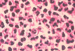
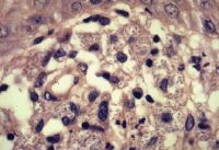
Dimorphic Fungus: Histoplasma capsulatum,
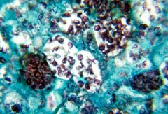
Tissue (37C) Yeast/Other Intracellular yeast in macrophages. Note the histopathologic changes seen in histoplasmosis due to Histoplasma capsulatum using methenamine silver stain. Note the presence of typical yeast cells, some of which are undergoing replication by “budding”. Histoplasmosis can be confined to the lungs, or become systemically disseminated, thereby, producing a fatal outcome. |
|

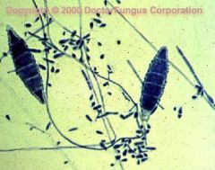
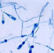
1) Name type of fungi, genus, species.
2) where am I and what temp? |

Dimorphic Fungus: Histoplasma capsulatum,
Nature (25C) Mold phase Description Tuberculate macroconida of the Jamaican isolate of Histoplasma capsulatum PHIL 4023 lores.jpg ID#: 4023 Description: This photomicrograph shows two tuberculate macroconida of the Jamaican isolate of Histoplasma capsulatum. Histoplasma capsulatum grows in soil, and material contaminated with bat or bird droppings. Spores become airborne when contaminated soil is disturbed, and breathing the spores causes histoplasmosis, a disease not transmitted from person to person. |
|

1) Name type of fungi, genus, species.
2) where am I and what temp? |

Dimorphic Fungus: Histoplasma capsulatum,
Tissue (37C) Yeast/Other |
|

1) Name type of fungi, genus, species.
2) where am I and what temp? |
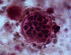
1) Name type of fungi, genus, species.
2) where am I and what temp? Dimorphic Fungus: Histoplasma capsulatum, Tissue (37C) Yeast/Other Intracellular yeast in macrophages. Note the histopathologic changes seen in histoplasmosis due to Histoplasma capsulatum var. duboisii. Note the presence of typical yeast cells, some of which are undergoing replication by “budding”. Histoplasmosis can be confined to the lungs, or become systemically disseminated, thereby, producing a fatal outcome. Methenamine silver stain was used here. |
|

1) Name type of fungi, genus, species.
2) where am I and what temp? |

1) Name type of fungi, genus, species.
2) where am I and what temp? Dimorphic Fungus: Histoplasma capsulatum, Nature (25C) Mold Phase |
|

1) Name type of fungi, genus, species.
2) where am I and what temp? |

Numerous yeast forms of Histoplasma capsulatum in alveolar macrophages. The arrowhead points to a halo formed when there is retraction of the cytoplasm.
1) Name type of fungi, genus, species. 2) where am I and what temp? Dimorphic Fungus: Histoplasma capsulatum, Tissue (37C) Yeast/Other Intracellular yeast in macrophages. |
|

1) Name type of fungi, genus, species.
2) where am I and what temp? |

1) Name type of fungi, genus, species.
2) where am I and what temp? Dimorphic Fungus: Histoplasma capsulatum, Tissue (37C) Yeast/Other Intracellular yeast in macrophages. Note the histopathologic changes seen in histoplasmosis due to Histoplasma capsulatum var. duboisii. Note the presence of typical yeast cells, some of which are undergoing replication by “budding”. Histoplasmosis can be confined to the lungs, or become systemically disseminated, thereby, producing a fatal outcome. Methenamine silver stain was used here. |
|

1) Name type of fungi, genus, species.
|

Dimorphic Fungus
Candidia albicans Candida albicans is a dimorphic fungus, i.e. it can take two forms. Most of the time it exists as oval, single yeast cells, which reproduce by budding. Most yeasts do not produce mycelia (a mass of branching, threadlike hyphal filaments), but Candida has a trick up its sleeve. Normal room temperatures favour the yeast form of the organism, but under physiological conditions (body temperature, pH, and the presence of serum) it may develop into a hyphal form. Pseudohyphae, composed of chains of cells, are also common. |
|
1) Name type of fungi, genus, species.
type of stain |

Candida albicans. Gram-stain of vaginal smear showing C. albicans, epithelial cells, and many Gram-negative rods. (1000X oil)
|
|

1) Name type of fungi, genus, species.
Type of prep |
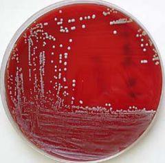
Dimorphic fungus
Candida albicans on blood agar; moist, opaque colonies are characteristic of yeast. |
|

Describe Candida Albicans
|

Candida Species: Direct Examination
•Hyphae show distinct points of constriction resembling sausage links •Budding yeast forms (blastospores) often seen -------------------------------------------------------------------------------- Candida Species •Generally grow at 37°, ferment glucose and may ferment other carbohydrates, and form pseudo- or true hyphae •Harbored by the gastrointestinal tract Candida albicans •Germ tube positive •Creamy colonies, as other yeasts •May display pseudohyphae and true hyphae •Most commonly isolated candidiasis •Virulence factors include rapid germination within tissue, protease production, surface integrin-like molecules for binding extracellular matrix, complement protein binding receptor, phenotypic switching, and surface variation and hydrophobicity |
|
1) Name type of fungi, genus, species.
2) where am I and what temp? |

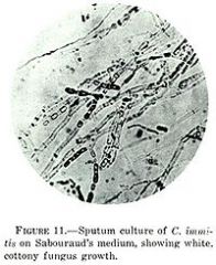
Dimorphic fungi
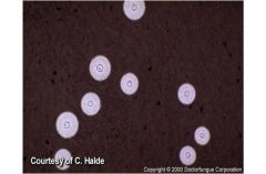
coccidioides immitis barrel -shaped arthroconidia cand be found in nature (25C) as mold phase OR lab ( 37C) yeast/mold |
|
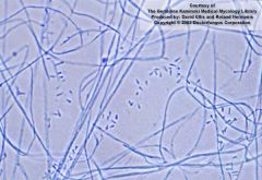
1) Name type of fungi, genus, species.
2) where am I and what temp? |

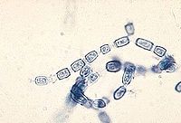
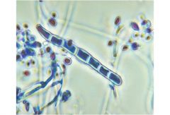
Dimorphic Fungus: Coccidioides immitis
See 'train-car' Septate hyphae of Coccidioides immitis with 90 degree branching and thick walled barrel shaped arthoconidia alternating with empty cells. Nature (25C) mold phase OR Lab (37C) Yeast/mold |
|

1) Name type of fungi, genus, species.
2) where am I and what temp? |
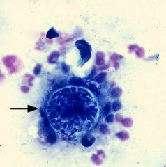
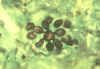
Dimorphic fungi
Coccidioides immitis Spercules with endospore - Tissue (37C) Yeast/other Pleural effusion in a dog with C. immitis infection. A large (60 μm), thick-walled Coccidioides spherule containing numerous endospores is present (arrow). The spherules and endospores of Coccidioides stain blue or purple with the Romanowsky-type stains used in cytology. (© Noah's Arkive, University of Georgia) |
|

1) Name type of fungi, genus, species.
2) where am I and what temp? |
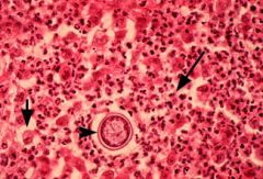
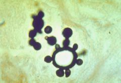
Dimorphic Fungus:
Coccidioides immitis Spherules with endospores, Tissue (37C) Yeast/Other Histologic section of a subcutaneous abscess in a cat. A Coccidioides spherule (arrowhead) is surrounded by numerous degenerate neutrophils (long arrow) and macrophages (short arrow). The spherules and endospores of Coccidioides stain pink with the H&E stain used in histology. (© Noah's Arkive, University of Georgia) |
|

1) Name type of fungi, genus, species.
2) where am I and what temp? |
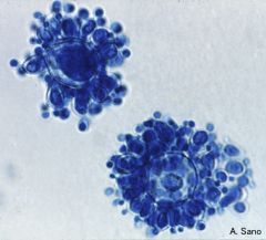
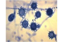
Dimorphic Fungus:
Coccidioides immitis Spherules with endospores (in tissue only) Coccidioides immitis (kok-sid-ee-OID-eez IMM-ih-tiss) is the cause of a nasty fungal disease called coccidioidomycosis (kok-sid-ee-oid-oh-my-KOH-sis). Like the other true-pathogenic, systemic human fungal diseases histoplasmosis, blastomycosis, and paracoccidioidomycosis, Coccidioidomycosis starts out as a lung disease caused by inhalation of the conidia, shown to the left. Most often the disease causes mild flu-like symptoms, but usually is resolved in the lungs. This fungus is a dimorphic pathogen, which means it can change from the room-temperature hyphal form at to the body-temperature spherule form (shown to the right) containing endospores. These endospores can be transported by the bloodstream to other parts of the body, particularly to the brain and central nervous system, where they can germinate and grow to cause even more severe disease. The dimorphism helps the fungus to evade the immune system by the changing of the surface antigens of the fungus. Tissue (37C) Yeast/Other |
|

1) Name type of fungi, genus, species.
2) where am I and what temp? |

Dimorphic Fungus:
Coccidioides immitis Spherules with endospores (in tissue only) Coccidioides immitis (kok-sid-ee-OID-eez IMM-ih-tiss) is the cause of a nasty fungal disease called coccidioidomycosis (kok-sid-ee-oid-oh-my-KOH-sis). Like the other true-pathogenic, systemic human fungal diseases histoplasmosis, blastomycosis, and paracoccidioidomycosis, Coccidioidomycosis starts out as a lung disease caused by inhalation of the conidia, shown to the left. Most often the disease causes mild flu-like symptoms, but usually is resolved in the lungs. This fungus is a dimorphic pathogen, which means it can change from the room-temperature hyphal form at to the body-temperature spherule form (shown to the right) containing endospores. These endospores can be transported by the bloodstream to other parts of the body, particularly to the brain and central nervous system, where they can germinate and grow to cause even more severe disease. The dimorphism helps the fungus to evade the immune system by the changing of the surface antigens of the fungus. Tissue (37C) Yeast/Other |
|

1) Name type of fungi, genus, species.
|
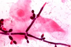
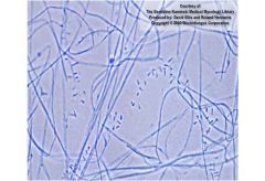
Conidiophores and conidia of the fungus Sporothrix schenckii
|
|

1) Name type of fungi, genus, species.
2) where am I and what temp? |

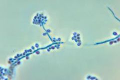
Dimorphic Fungus
Blastomyces dermatitidis Nature (25C) Mold Phase NOT DIAGNOSTIC - looks like paracoccidiodes braziliensis One-celled blastoconidia produced on short conidiophores are typical of Blastomyces dermatitidis at 25°C |
|

1) Name type of fungi, genus, species.
2) where am I and what temp? |
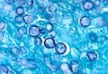
Yeast phase (37C), KOH preparation
DIAGNOSITIC! Broad based single budding yeast that looks like a snowman |
|
1) Name type of fungi, genus, species.
2) where am I and what temp? |

Dimorphich Fungus
Blastymyces dermatidis Broad based single budding yeast, looks like snowman - Tissue (37C) Yeast/Other Smear from foot lesion of blastomycosis showing Blastomyces dermatitidis yeast cell undergoing broad-base budding. ASCP/Atlas of Clinical Mycology II / CDC |
|

1) Name type of fungi, genus, species.
2) where am I and what temp? |

Dimorphich Fungus
Blastymyces dermatidis Broad based single budding yeast, looks like snowman - Tissue (37C) Yeast/Other Histopathology of blastomycosis. Yeast cell of Blastomyces dermatitidis undergoing broad-base budding. Methenamine silver stain. African case. CDC |
|

1) Name type of fungi, genus, species.
2) where am I and what temp? |
Histopathology of blastomycosis of skin. Budding cell of Blastomyces dermatitidis surrounded by neutrophils. Multiple nuclei are visible. CDC
|
|

1) Name type of fungi, genus, species.
2) where am I and what temp? |
Dimorphic Fungus: Paracoccidioides brasiliensis,
Tissue (37C) Yeast/Other DIAGNOSTIC at 37C in tissue OR lab! Multiple budding yeast (mickey mouse) Histopathology of paracoccidioidomycosis. Budding cell of Paracoccidioides brasiliensis. Methenamine silver stain. CDC |
|
1) Name type of fungi, genus, species.
2) where am I and what temp? |

Dimorphic Fungus: Paracoccidioides brasiliensis,
Tissue (37C) Yeast/Other DIAGNOSITIC - at 37C - tissue or lab Histopathology of paracoccidioidomycosis. Budding cells of Paracoccidioides brasiliensis. Methenamine silver stain. CDC/Dr. Lucille K. Georg |
|

1) Name type of fungi, genus, species.
2) where am I and what temp? |

Dimorphic Fungi
Blastomyces dermatitidis Nature (25C) Mold Phase Hyphae with single microconidia (NOT diagnositic) |
|

1) Name type of fungi, genus, species.
2) where am I and what temp? |

Dimorphic Fungus
Histoplasma capsulatum - CULTURE Nature (25C) Mold Phase Tuberculate macroconidia DIAGNOSITC |
|

1) Name type of fungi, genus, species.
2) where am I and what temp? |

Dimorphic Fungus
Paracoccidioides brasiliensis Nature (25C) Mold Phase Hyphae with single microconidia NOT DIAGNOSITC |
|
1) Name type of fungi, genus, species.
2) where am I and what temp? |

Dimorphic Fungus: Paracoccidioides brasiliensis,
Tissue (37C) Yeast/Other Paracoccidioides brasiliensis, yeast form Multiple budding in the yeast form. |
|
|
What are the four dimorphic fungus?
|
histoplasma casulatum
blastomyces dermatidis paracoccidiodes braziliensis coccidioides immitis |
|
|
Which dimorphic fungi has tuberculate macroconidia in the nature (25C) mold phase?
IS it diagnositic? |
histoplasma casulatum
YES |
|
|
which fungus have hyphae with single microconidia?
Is it diagnositic? |
blastomyces dermatidis
paracoccidiodes braziliensis No |
|
|
Which fungus has barrel-shaped arthroconidia?
When do you see this phase? Is it diagnositic? |
coccidioides immitis
Naure (25C) mold phase and Lab (37C) yeast phase - NOT in tissue Yes |
|
|
Which fungus do you see spherules with endosphores?
When do you see this phase? Is it diagnositic? |
coccidioides immitis
Only in TISSUE (37C) yeast phase Yes |
|
|
Which fungus do you see intracellular yeast in macrophages?
Is it diagnostic? |
histoplasma casulatum
ONLY TISSUE (37C) YES |
|
|
Which fungus do you see small single budding yeast?
Is it diagnostic? |
histoplasma casulatum
ONLY LAB (37C) NO! |
|
|
Which fungus do you see broad based single budding yeast?
Is it diagnostic? |
blastomyces dermatidis
At (37C) Yes |
|
|
Which fungus do you see multiple budding yeast?
Is it diagnostic? |
paracoccidiodes braziliensis
At (37C) Yes |
|
|
Which fungus are thermal dimorphs?
|
histoplasma casulatum
blastomyces dermatidis paracoccidiodes braziliensis |
|
|
Which fungus are tissue dimorphs?
|
coccidioides immitis
|
|

1) Name type of fungi, genus, species.
2) where am I and what temp? |
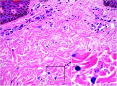
Dimorphic Fungus: histoplasma capsulatum
AIDS PT! Tissue (37C) Yeast/Other Skin biopsy from a profoundly immunosuppressed patient with AIDS-associated histoplasmosis. The dermal host response is negligible despite the presence of numerous Histoplasma capsulatum spores (inset). |
|
|
What is unicellular and reproduce asecually by budding?
|
Yeast
|
|
|
Describe the structure of yeast
|
round unicelluar organisms, in some species the daugher cells remain attached to each other and form a linear structure called psuedohyphae.
|
|
|
Describe the strucure of mold
|
muliticelluar strands of fungi called hyphae.
|
|
|
What is a network of hyphae?
Where do you see them? |
a single colony called mycelium (pl. mycelia)
mold |
|
|
Name the superficial (cutaneous) mycoses
|
Dermatophytes
- Trychonphyton - Microsporum - Epidermophyton |
|
|
What are the subcutaneous Fungi?
|
Sporotrichosis
Chromoblastomycosis |
|
|
What causes sporotrichosis?
|
Sporothrix schenckii
Found in plants, sphagnum moss and vegetation occupational disease of farmers, gardners, florists |
|
|
How do you diagnose Chromoblastomycosis/Chromomycosis?
What do you see? |
direct examination from skin scrapings in 10% KOH demonstrate dark brown sclerotic cells
or H&E stain copper pennies |
|

What is pathongnomonic chromoblastomycosis?
|

Copper pennies - rounded sclerotic bodies
|
|

Name genus and diagnositic finding
|

chromoblastomycosis
H&E stained section showing characteristic dark brown sclerotic cells which divide by binary fission and not by budding. Note all agents of chromoblastomycosis form these sclerotic bodies in tissue. |
|
|
Name the systemic fungi
|
histoplasmosis
blastomycosis |
|
|
Name the opportunistic fungi
|
candida
apergillus |
|
|
What is KOH used for?
|
a potassium hydroxide (KOH) mount of a skin scraping is a common procedure performed to demonstrate the evidence of fungal infection in skin, hairs and nails.
|
|
|
What is the potassium hydroxide mount procedure?
|
Potassium hydroxide 10% is added to the collected material, covered by a cover slip made of fragile glass and gently preheated before examining for fungi.
For thick, hyperkeratotic specimens, leave the potassium hydroxide preparation for 'digestion' and 'clearing' for ½ to 2 h. This clearing time for nails and hairs may extend to 24-48 h. |
|
|
What fungus has a spagetti and meatball appearance?
|
Thick-walled spherical yeast in clusters, often with short filaments resembling'spaghetti and meatballs,' are characteristic of Malassezia furfur in pityriasis versicolor scales.
|
|
|
How do you diagnose dermatophytes?
|
- KOH scrapings/clippings
- culture (LPCB mounts for ID) |
|
|
What is LPCB Stain?
|
•Lactol-phenol cotton blue (LPCB) stain
- primarily used for the microscopic examination of molds |
|
|
How does •Lactol-phenol cotton blue LPCB) stain work?
|
lactic acid enhances the solutions's penetration of the hyphae, the phenol kills the cells and the cotton blue is the dye.
the stain also contains glycerol for a semi-permanent mounting fluid |
|
|
What is Lactol-phenol cotton bue (LPCB) used in combination with?
|
scotch tape - 'quick and dirty' direct smear of a mold colony
|
|
|
What is Lactol-phenol cotton blue (LPCB) stain technique
|
drop of LPCB on slide
firmly touch the surface of the mold with the tape stretch the tape down on the slide |
|
|
What is the india ink staining method?
|
to add to specimens to provide a dark background to highly hyaline yest cells and capsular material (halo effect)
|
|
|
When do you use an india ink procedure
|
to examine cryptococcus neoformans
|
|
|
Which dermatophytes have both macrocondidia and microconidia?
|
microsporum spp
trichophyton spp |
|
|
Which dermatophytes have no microconidia?
|
epidermophyton spp
|
|
|
describe the microscopic features of trichophyton
|
Microscopic Features
Septate, hyaline hyphae, conidiophores, microconidia, macroconidia, and arthroconidia are observed. Chlamydospores may also be produced. Conidiophores are poorly differentiated from the hyphae. Miroconidia (also known as the microaleuriconidia) are one-celled and round or pyriform in shape. They are numerous and are solitary or arranged in clusters. Microconidia are often the predominant type of conidia produced by Trichophyton. Macroconidia (also known as the macroaleuriconidia) are multicellular (2- or more-celled), smooth-, thin- or thick-walled and cylindrical, clavate or cigar-shaped. They are usually not formed or produced in very few numbers. Some species may be sterile and the use of specific media is required to induce sporulation [531, 1295, 2144, 2202]. See above for the specific microscopic features of various species. Histopathologic Features Septate, branched hyphae that break into chains of arthroconidia are observed. |
|
|
Trichophyton differs from Microsporum and Epidermophyton by
|
Trichophyton differs from Microsporum and Epidermophyton by having cylindrical, clavate to cigar-shaped, thin-walled or thick-walled, smooth macroconidia.
|
|
|
Epidermophyton - discription
|
Epidermophyton is a filamentous fungus and one of the three fungal genera classified as dermatophytes. It is distributed worldwide. Man is the primary host of Epidermophyton floccosum, the only species which is pathogenic.
|
|
|
Pathogenicity and Clinical Significance - Epidermophyton
|
Pathogenicity and Clinical Significance
E. floccosum is one of the common causes of dermatophytosis in otherwise healthy individuals. It infects skin (tinea corporis, tinea cruris, tinea pedis) and nails (onychomycosis). The infection is restricted to the nonliving cornified layers of epidermis since the fungus lacks the ability to penetrate the viable tissues of the immunocompetent host [57, 1679, 2400]. Disseminated infections due to any of the dermatophytes are very unlikely due to the restriction of the infection to keratinized tissues. However, invasive E. floccosum infection has been reported in an immunocompromised patient with Behcet's syndrome [2068]. As with all forms of dermatophytosis, Epidermophyton floccosum infections are communicable and usually transmitted by contact, particularly in common showers and gym facilities. |
|
|
describe the microscopic features of Epidermophyton
|
Microscopic Features
Septate, hyaline hyphae, macroconidia, and occasionally, chlamydoconidium-like cells are visualized. Microconidia are typically absent. Macroconidia (10-40 x 6-12 µm) are thin walled, 3- to 5- celled, smooth, and clavate-shaped with rounded ends. They are found singly or in clusters. Chlamydoconidium-like cells, as well as arthroconidia, are common in older cultures |
|
|
Epidermophyton floccosum is differentiated from Microsporum and Trichophyton by
|
Epidermophyton floccosum is differentiated from Microsporum and Trichophyton by the absence of microconidia.
|
|
|
Microscopic Features
Microsporum spp. |
Microscopic Features
Microsporum spp. produce septate hyphae, microaleurioconidia, and macroaleurioconidia. Conidiophores are hyphae-like. Microaleuriconidia are unicellular, solitary, oval to clavate in shape, smooth, hyaline and thin-walled. Macroaleuriconidia are hyaline, echinulate to roughened, thin- to thick-walled, typically fusiform (spindle in shape) and multicellular (2-15 cells). They often have an annular frill. |
|
|
Microsporum differs from Trichophyton and Epidermophyton by
|
Microsporum differs from Trichophyton and Epidermophyton by having spindle-shaped macroconidia with echinulate to rough walls
|
|
Name genus and diagnositic features
|

Microsporum gypseum macroconidia and microconidia
|
|

Name genus and diagnostic features
|

Epidermophyton floccosum - Septate, hyaline hyphae, macroconidia, and occasionally, chlamydoconidium-like cells are visualized. Microconidia are typically absent. Macroconidia (10-40 x 6-12 µm) are thin walled, 3- to 5- celled, smooth, and clavate-shaped with rounded ends. They are found singly or in clusters.
NO MICROCONIDIA |
|

Name genus and diagnosstic features
|
Septate, hyaline hyphae, conidiophores, microconidia, macroconidia, and arthroconidia are observed. Chlamydospores may also be produced. Conidiophores are poorly differentiated from the hyphae. Miroconidia (also known as the microaleuriconidia) are one-celled and round or pyriform in shape. They are numerous and are solitary or arranged in clusters. Microconidia are often the predominant type of conidia produced by Trichophyton. Macroconidia (also known as the macroaleuriconidia) are multicellular (2- or more-celled), smooth-, thin- or thick-walled and cylindrical, clavate or cigar-shaped. They are usually not formed or produced in very few numbers.
Trichophyton differs from Microsporum and Epidermophyton by having cylindrical, clavate to cigar-shaped, thin-walled or thick-walled, smooth macroconidia. |
|

WHo am I?
|

Microsporum spp.
have both macroconidia and microconidia the macroconidia typically have tpaered ends and a thick echinuate wall - rough macroconidia |
|

who am i?
|

microsporum
have both macroconidia and microconidia the macroconidia typically have tpaered ends and a thick echinuate wall - rough macroconidia |
|

who am i?
|

trichophyton spp.
have both macroconidia and microconidia ; the mmacroconidia are typically cylindrical, thin , smooth walls. |
|

who am i?
|
trichophyton
have both macroconidia and microconidia ; the mmacroconidia are typically cylindrical, thin , smooth walls. |
|

who am i
|

epidermophyton spp
macroconidia are thin smooth walls Epidermophyton floccosum can usually be recognized by the large, paddle-like macroconidia with thin smooth walls. NO microconidia. |
|

who am i
|
epidermophyton spp.
macroconidia are thin smooth walls Epidermophyton floccosum can usually be recognized by the large, paddle-like macroconidia with thin smooth walls. NO microconidia. |
|

Who am i?
|

malassezia furfur
|
|
who am i?
|

malassezia furfur
|
|

who am i?
|

malassezia furfur
|
|
who am i?
|

Cigar-shaped yeast of Sporothrix schenckii in tissue macrophages in a biopsy specimen
|
|

who am i?
|

Yeast form of Sporothrix schenckii
|
|
who am i?
|

malassezia furfur (KOH)
|
|

who am i? What stain prep?
|

Malassezia furfur
KOH with ink |
|
who am i
|

trichophyton rubrum
|
|
Who am I?
|

Epidermophyton spp.
|
|

who a i
what prep? |

coccidioides immitis
koh |
|
Fluoride release in glass ionomers are high, but drop to __% of the original value after 1 month
|

90%
|
|
who am i?
|

histoplasm capsulatum
|
|
|
who am i
|
blastomyces dermatitidis
|
|

who am i
|

Paracoccidioides brasiliensis
|
|

who am i?
|

coccidioides immitis endospore
|
|
who am i?
|

Aspergillus fumigatus
|
|
who am i?
|

aspergillus fumigatus
|
|

who am i?
|
Aspergillus fumigatus
|
|

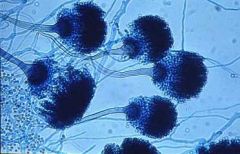
who am i
|

aspergillus niger
|
|
|
who am I?
|
aspergillus niger
|
|
|
who am i
|
aspergillus niger
|
|

who am i?
|

aspergillus niger
|
|

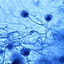
aspergillus flavus
|

aspergillus flavus
|
|

who am i?
|
aspergillus flavus
|
|

who am i?
|

aspergillus flavus
Aspergillus flavus; note the phialides arise circumferentially from the globose vesicle. |
|

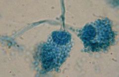
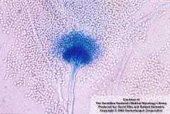
who am i?
|

Aspergillus niger in slide culture. Note how black conidia extend circumferentially from and obscure the vesicle.
|
|
|
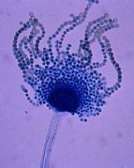
who am i
|
Aspergillus fumigatus in slide culture. Note how the phialides and conidia extend from the top half of the vesicle
|
|

who am i?
|

Aspergillus fumigatus in slide culture. Note how the phialides and conidia extend from the top half of the vesicle
|
|

who am i?
|

Aspergillus terreus.Note biseriate, "swept-forward" appearance
|
|

who am i?
|
aspergillus terrus
|
|
|
Biosafety Levels for Infectious agents:
What is level one? |
basic: Standard microbiological practices, containment by standard practices on open benches
|
|
|
Biosafety Levels for Infectious agents:
What is level two? |
Basic +1
level 1 plus stricter decon, more stringent labeling, and partial containment equip (safety cabinets) used for tissue culture and routine virology |
|
|
Biosafety Levels for Infectious agents:
What is level three? |
Containment: Level 2+
Level 2+ special laboratory clothing, controlled access, caontainment equip, (neg pressure hoods and neg pressure vent) used for TB and mycology |
|
|
Biosafety Levels for Infectious agents:
What is level four? |
maximum containment
Level 3+ maximal containment equipment and practices (full body pos pressure suits) pathogens sug as viral hemorrhagic fever |