Reading...
![]()
Play button
![]()
Play button
![]()
Use LEFT and RIGHT arrow keys to navigate between flashcards;
Use UP and DOWN arrow keys to flip the card;
H to show hint;
A reads text to speech;
44 Cards in this Set
- Front
- Back
|
Patient presents with muscle pain and atrophy of the shoulder muscles. The patient also has purple-red eyelid discoloration. What are the two variances of this disease?
|
Dermatomyositis (DM) - has skin involvement. Associated with Ab-mediated damage.
Polymyositis (Pm) - no skin involvement. Associated with T cell-mediated damage. Women 40-60. Raccoon eyes. Association with lung cancer. Labs: Increased CK. |
|
|
Patient has blue sclera. What is the inheritance pattern?
|
Osteogenic imperfecta - auto dominate, locus heterogeneity. Type I collagen defect. Blue sclera - reflection of underlying choroidal veins. Deafness.
|
|
|
Patient of short stature has a normal-sized head and vertebral column. Labs: normal GH and IGF1. Diagnosis?
|
Achondroplasia - AD - impaired proliferation of cartilage at the growth plate.
|
|
|
Patient presents with pathological fractures, anemia and visual and hearing defects. What cells are disfunctional?
|
Osteoclasts, which leads to overgrowth of sclerotic cortical bone. Osteopetrosis, aka marble bone disease. Cranial nerve compression leads to the visual/hearing defects. Anemia is due to narrowing of bone cavity.
|
|
|
Where does osteomyelitis most often occur?
|
Metaphysis.
|
|
|
What are the three most common osteomyelitis pathogens?
|
Staph aureus, Strep pyogenes, Hemophilus influenzae.
|
|
|
Define involucrum.
|
Layer of new bone growth outside existing bone seen in chronic osteomyelitis. It results from the stripping off of the periosteum by the accumulation of pus within the bone, and new bone growing from the periosteum.
|
|
|
Osteomyelitis in HbSS. What bug?
|
Salmonella paratyphi.
|
|
|
Whats the pathogenesis of senile osteoporosis?
|
Decreased ability of osteoblasts to divide and produce osteoid.
|
|
|
Young boy, 4, presents with pain in the knee and a limp. You do an MRI scan which shows increased density in the femoral head. Diagnosis?
|
Aseptic necrosis of ossification center in the femoral head - osteochondrosis.
|
|
|
What are some complications that Paget's disease patients have?
|
Pathologic fractures, risk of osteogenic sarcoma, risk for high-output heart failure (AV shunts).
Pagets - osteoclastic resoprtion (shaggy-lytic lesions) followed by osteoblastic bone formation (high ALP, weak-mosaic bone). |
|
|
Albright's syndrome.
|
Cafe-au-lait spots, precocious sexual development, polyostic (multiple bones) fibrous dysplasia (non-neoplastic).
|
|
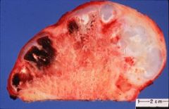
Most common benign bone tumor.
|
Osteochondroma: outgrowth of bone (exostosis) capped by benign cartilage.
|
|
|
Gardner's polyposis syndrome.
|
Autosomal dominant. Benign osteomas and desmoid tumors (fibromatosis).
|
|
|
What s the primary location of these benign tumors:
1) Osteoid osteoma 2) Osteoblastoma 3) Giant cell tumor 4) Osteoma 5) Enchondroma |
1) Cortex of proximal femur
2) Vertebra 3) Epiphysis of distal femur or proximal tibia (female) 4) Facial bones 5) Medullary location - hands/feet |
|

Male 30-60 years old. Pelvic bone. Propensity to metastasizes to lung.
|
Chondrosarcoma.
|
|
|
What are the risk factors for osteogenic sarcoma?
|
Paget's disease, familial retinoblastoma, irradiation.
|
|
|
What are the radiographic characterisitics of malignant bone disorders?
|
Sunburst appearance (spiculated pattern of calcified malignant osteoid), Codman's triangle (tumor lifting periosteum).
|
|
|
What age group does the following tumor normal affect: "onion skin" appearance around bone; fever; anemia; often affects pelvic girdle or rib.
|
Ewing's sarcoma, male 10-20.
|
|
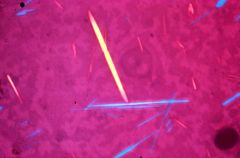
Describe the crystal analysis.
|
(1) Crystals are monoclinic (needle shaped) and could be either monosodium urate or calcium pyrophosphate, (2) if crystal is yellow when parallel to the slow ray of the compensator, it is monosodium urate and defines negative birefringence, (3) if crystal is blue when parallel to the slow ray of the compensator, it calcium pyrophosphate and defines positive birefringence
|
|
|
Patients urine turns black when oxidized. They also have sclera darkening. What musculoskeletal abnormalities will they develop?
|
Ochronosis (alkaptonuria) is AR caused by deficiency of homogentisic acid oxidase. Patients develop secondary osteoarthritis in spine, hip, and/or knee.
|
|
|
Joint findings in osteoarthritis?
|
Decreased articular cartilage, osteophytes (reactive bone formation at joint margins), subchondral cysts, dense sclerotic bone.
|
|
|
What are Heberden's nodes and Bouchard's nodes?
|
Osteophytes:
Heberden's - enlargement of DIP joint. Bouchard - enlargement of PIP |
|
|
What are the causes of neuropathic arthropathy (Charcot's joint)?
|
DM - tarsometatarsal joints
Syringomyelia - shoulder, elbow, wrist joints Tabes dorsalis - hip, knee, ankle joints |
|
|
Explain the pathogenesis of Rheumatoid arthritis.
|
B cells in joint produce RF complexes (IgM autoAbs against Fc of IgG); RF complexes activate complement and attract PMN; acute inflammation + PMNs phagocytose RF complexes producing ragocytes; chronically inflamed synovial tissue proliferates (forms pannus); pannus releases cytokines that destroys articular cartilage; joint fusion.
|
|
|
What is a Bakers cyst?
What are Rheumatoid nodules? What is Felty's syndrome? Why would an RA patient develop anemia? |
Bakers cyst - outpoutching of joint space (back of knee is common).
Nodules - fibrinoid necrosis on extensor surface of forearm. Felty's - autoimmune neutropenia/splenomegaly. Anemia - anemia of chronic disease. |
|
|
How does Juvenile RA differ from RA?
|
RF is normally absent. Common in females < 16.
Polyarticular - disabling arthritis. Pauciarticular - few joints + uveitis. |
|
|
Still's disease?
|
Juvenile rheumatoid arthritis (subtype) - fever, rash, polyarthritis, lymphadenopathy, neutrophilic leukocytosis.
|
|
|
Causes of gout?
|
Decreased renal excretion: lead poisoning, alcoholism.
Increased nucleated cell turnover: leukemia. |
|
|
Most common joint involved in gouty arthritis? (Called podagra).
|
First metatarsophalangeal joint.
|
|
|
What is tophus?
|
Monosodium urate crystals (MSU) depositing in soft tissue around joints. Granulomatous reaction with multinucleated giant cells are present.
|
|
|
What is chondrocalcinosis?
|
Pseudogout - degenerative joint disease. Calcium pyrophosphate (positive birefringences) crystals.
|
|
|
Young male presents with morning stiffness in his lower back (sacroiliitis). What are some complications this man may experience in the future? What is the HLA associated?
|
Ankylosing spondylitis. HLA-B27. Complications: fusion of vertebrae, aortitis with aortic regurgitation, uveitis (blurry vision). Association with ulcerative colitis.
|
|
|
Whats the triad of Reiter's syndrome?
|
Chlamydia trachomatis - urethritis, arthritis (achilles tendon periostitis), conjunctivitis.
|
|
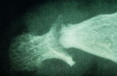
Pencil-in-cup deformity.
|
Psoriatic arthritis. Sausage shaped DIP joints. Nail pitting is also present.
|
|
|
Most common cause of septic arthritis?
|
Neisseria gonorrhoeae. Predisposed: C6-C9 (MAC) deficiency.
Staph aureus - most common nongonococcal cause. |
|
|
Patient present with arthritis in both knees and bilateral Bell's palsy. What hematological abnormality may be present in this patient?
|
Lyme's disease. Borrelia burgdorferi, spirochete, from ixode. Myocarditis and pericarditis may be present. Babesiosis (protozoa) is also carried by ixode - may cause hemolytic anemia.
|
|
|
Septic arthritis and tendinitis due to cat bite.
|
Pasteurella multocida.
|
|
|
Child, 4, has very large calf muscles. He also uses his arms to help himself stand. The child also has a waddling gait. What is the inheritance of this disease and what are the two type?
|
Duchenne's muscular dystrophy (XLR) - deficiency in dystrophin (anchors actin to membrane glycoprotein). Becker's type - defective dystrophin. Path: progressive degeneration of type I and II fibers, fibrosis and infiltration of muscle tissue by fatty tissue (pseudohypertrophy of calf muscles). Death by age 20. Labs: increased CK at birth.
|
|
|
This autosomal dominant disorder results in selective atrophy of type I muscle fibers.
|
Muscular dystrophy - (type I = slow-twitch red fibers rich in mitochondria) trinucleotide repeat sequence. Facial weakness, myotonia (inability to relax muscles), frontal balding, cataracts, testicular atrophy, cardiac involvement. Increased serum CK.
|
|
|
Adult male is unable to release his hand shake when he greats you. Diagnosis?
|
Muscular dystrophy. This is known as myotonia.
|
|
|
28 female presents with ptosis. She says her eyes aren't droopy in the mornings. What cancer is this patient at risk of developing?
|
Myasthenia Gravis - type II hypersensitivity against ACh-R. Ab's are synthesized in the thymus (thymic hyperplasia with germinal follicles - 85%). Eventually patient will develop dysphagia for solids and liquids.Tensilon test (endrophonium) - reverses muscle weakness.
|
|
|
What is the
1) Most common benign soft tissue tumor? 2) Most common adult sarcoma? 3) Most common sarcoma in children? 4) Most common tumor of nerve trunks? |
1) Lipoma
2) Liposarcoma 3) Embryonal rhabdomyosarcoma 4) Neurofibrosarcoma |
|
|
What is the most common cause of avascular necrosis?
|
Corticosteroids
|