Reading...
![]()
Play button
![]()
Play button
![]()
Use LEFT and RIGHT arrow keys to navigate between flashcards;
Use UP and DOWN arrow keys to flip the card;
H to show hint;
A reads text to speech;
173 Cards in this Set
- Front
- Back
- 3rd side (hint)
|
Bones and skeletal muscles work together to produce body movement where?
|
Joints (where two bones meet)
|
|
|
|
What is the most common joint type?
|
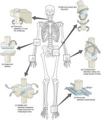
Synovial joints
|
|
|
|
what does Diarthroses mean
|
Freely movable joints
|
|
|
|
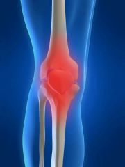
Why are certain joints classified as synovial
|
Because the joint cavity between the articulating bones is lined by a synovial membrane
|
|
|
|
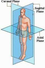
How many anatomical plains are there
|

3
|
|
|
|
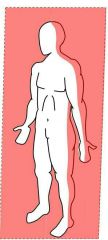
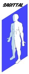
What are the three anatomical planes
|

Sagittal plane,
frontal plane, transverse plane |
|
|
|
What vertical anatomical plane divides the body or a body part into right and left portions
|
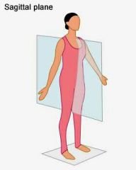
Sagittal plane
|
|
|
|
what vertical anatomical plane divides the body or body parts into anterior and posterior portions
|
Frontal plane
|
|
|
|
what anatomical plane divides the body into superior and inferior portions
|
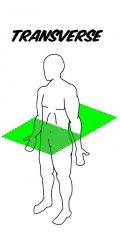
Transverse plane
|
|
|
|
How are joints classified
|
By structure and function
|
|
|
|
What are the three types of joints?
|
Fibrous.
cartilaginous. synovial |
|
|
|
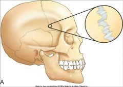
Where are suture joints found?
|

Skull
|
|
|
|
What are joints that are held by a ligament called?
|
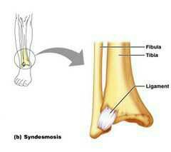
Syndesmosis
|
|
|
|
The periodontal ligament that holds teeth in their sockets are what type of joint?
|
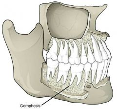
Gomphosis
|
|
|
|
what is Synarthroses?
|
Immovable joint
|
|
|
|
what is Amphiarthrosis?
|
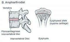
Slightly movable joints
|
|
|
|
what is Diarthrosis
|
Free movable joints
|
|
|
|
What type of joints are found predominantly in the limbs?
|

Diarthrosis
|
|
|
|
What type of joints can we both rigid and slightly movable?
|

Cartilaginous joints
|
|
|
|
What are two types of cartilaginous joints?
|

Synchondrosis.
Symphysis. |
|
|
|
What is the joint called where a bar or plate of hyaline cartilage unites the bone
|
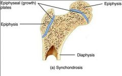
Synchondrosis
|
|
|
|
What is the joint called where fibrocartilage unites bone
|
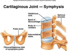
Symphysis
|
|
|
|
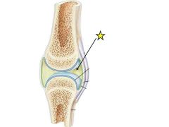
What type of joint has a particular articulating bone separated by a fluid containing joint cavity?
|
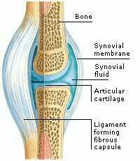
Synovial joint
|
|
|
|
What are the six distinguishing features of a synovial joint?
|

Articular cartilage.
joint cavity. articular capsule. synovial fluid. reinforcing ligaments. nervous and blood vessels. |
|
|
|
What is the unique feature of synovial joints?
|
Joint cavity
|
|
|
|
What type of fluid has an egg white consistency
|

Synovial fluid
|
|
|
|
What is the function of synovial fluid?
|

Reduces friction.
has nutrients that nourishes articulating cartilage. has phagocytes to keep joint cavity clean. |
|
|
|
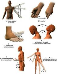
What is the combination of flexion-extension and abduction-adduction called?
|

Circumduction
|
|
|
|
what is the rotation of a bone around its own longitudinal axis called?
|
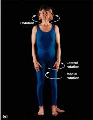
Rotation
|
|
|
|
what is it called when the forearm is rotated laterally so that the palm faces forward?
|

Supination
|
|
|
|
What is it called on forearm is rotated medially so that the palm faces backwards
|
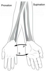
Pronation
|
|
|
|
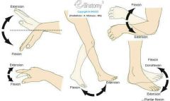
Flexing the foot is called what?
|
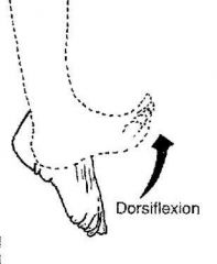
dorsiflexion
|
|
|
|
Extending the foot is called what?
|
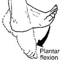
Plantar flexion
|
|
|
|
What is it called when the plantar surface (sole of foot) is moved the face medially
|
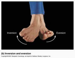
Inversion
|
|
|
|
Moving your foot so that the planter surface faces latterly is called
|
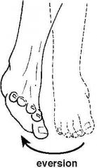
eversion
|
|
|
|
Movement of the mandible forward in the transverse plane is called
|

protraction
|
|
|
|
Movement of the mandible backwards in the transverse plane is called
|
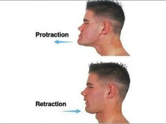
Retraction
|
|
|
|
Touching the thumb to any of the tips of the other digits is called
|
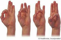
Opposition
|
|
|
|
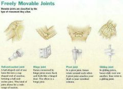
a Synovial joint ball socket allows for what type of movements
|
Flexation-extension.
abduction-adduction. circumduction. rotation. |
|
|
|
A hinge type of synovial joint allows for what type of movement
|
flexion-extension
|
|
|
|
The synovial joint ,saddle joint, allows for what type of movement
|
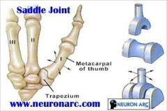
Flexion extension
abduction-adduction circumduction |
|
|
|
pivot joints allows what type of movement.
|
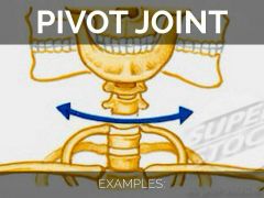
Rotation
|
|
|
|
The synovial joint "gliding plane" allows what type of movement
|
Gliding
|
|
|
|
What joints allow movement in the sagittal plane
|
Ball and socket joints.
condylar joints. hinge joints. saddle joints. |
|
|
|
What joints allow movement in the frontal plane
|
Ball and socket joints
condylar joints saddle joints |
|
|
|
What joints allow rotation?
|
Ball and socket joints
pivot joints |
|
|
|
What joints allow circumduction?
|
Ball and socket joint
condylar joint saddle joint |
|
|
|
The deep fascia is a sheet of what?
|

Dense irregular connective tissue
|
|
|
|
What is the deep fascia do?
|
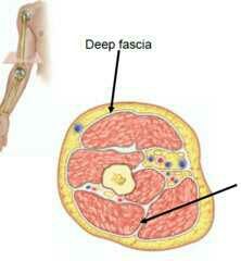
Separates the muscle compartments in the Lambs.
Separates individual muscles |
|
|
|
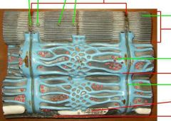
What is the Epimysium of a skeletal muscle.
|
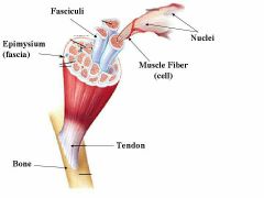
Layer of dense irregular connective tissue around the muscle
|
|
|
|
what is the Perimysium of skeletal muscle
|

A layer of less dense connective tissue surrounding each fascicle
|
|
|
|
What is the Endomysium of skeletal muscle.
|
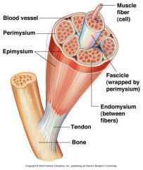
A layer of fine areolar connective tissue surrounding each muscle fiber
|
|
|
|
The connective tissues of the deep fascia, the Epimysium, Perimysium, and Endomysium continue past each end of the muscle to form. ...?
|
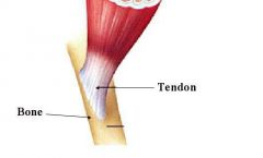
Tendon or Aponeurosis. that connects the muscle to the periosteum of a bone
|
|
|
|
What is the purpose of tendons and aponeurosis?;
|

Create extremely strong attachment between muscle and bone.
Allows the pulling force of muscle to be transferred to bone |
|
|
|
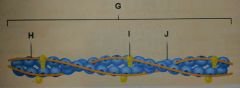
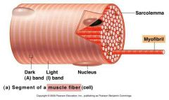
What is the structural levels of skeletal muscle
|
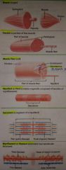
Muscle.
Fascicle Muscle Fiber Myofibril or Fibril Sarcomere Myofilament or filament |
|
|
|
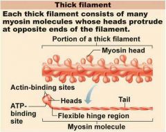
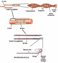
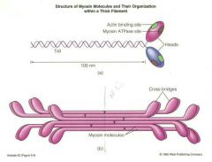
What does a thick filament consists of?
|

Myosin molecules whose heads protrude at opposite ends of the filament.
|
|
|
|
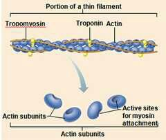
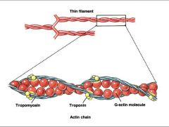
What does thin filament consists of?
|

Two strands of actin subunits twisted into a helix plus two types of regulatory proteins. (troponin and tropomyosin)
|
|
|
What is the structure
|

Myofibril
|
|
|
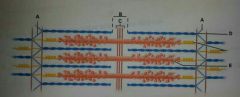
Identify the sarcomere cross section
|

I band
|
|
|

Identify the sarcomere cross section
|
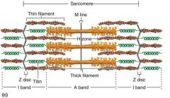
H zone
|
|
|
Identify the sarcomere cross section
|

M line
|
|
|
Identify the sarcomere cross section
|

Zone of overlap
|
|
|
Identify the molecule
|
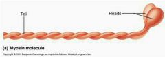
Myosin molecule
|
|
|
|
Which filament has a ATP binding site?
|

thick. ( myosin molecule)
|
|
|

Identify A
|

Z disk
|
|
|

Identify B
|

H zone
|
|
|
Identify C
|

M line
|
|
|

Identify D
|
Thin filament (actin)
|
|
|

Identify E
|
Thick filament myosin
|
|
|

Identify A
|

Actin binding sites of a Myosin molecule
|
|
|

Identified B
|

ATP binding site
|
|
|

identify C
|

Heads of Myosin molecule
|
|
|

Identified D
|

Flexible hinge region of Myosin molecule
|
|
|

Identify E
|

Tail
|
|
|
Identify F
|

myosin molecule
|
|
|

Identify G
|

thin filament
|
|
|
Identify H
|

Tropomyosin molecule
|
|
|

Identify I
|

troponin molecule
|
|
|

Identify J
|

Actin molecule
|
|
|
|
What can be described as one big continuous net that wraps around all of the myofibrils within the muscle cell
|
sarcoplasmic reticulum
|
|
|
What is the function of the sarcoplasmic reticulum (SR)
|
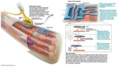
To store Ca2+
|
|
|
|
What is Terminal cisternae
|
Enlarged sarcoplasmic reticulum (SR) tubules that store more Ca2+
|
|
|
|
What does the T in T tubule stand for.
|
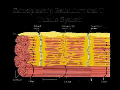
Transverse tubule
|
|
|
|
Where are the T tubules located?
|

Between each pair of terminal cisternae
|
|
|
|
How are T tubules formed
|

By an invagination of the sarcolemma and consequently continuous with it.
|
|
|
|
Each pair of terminal cisternae plus the T Tubule between them is called what?
|

Triad
|
|
|
|
What does the "Triad" of the sarcoplasmic reticulum consist of?
|
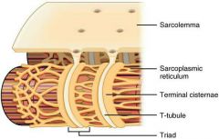
Terminal cisternae plus the T tubule between them
|
|
|
|
What part of the sarcoplasmic reticulum (SR) is continuous with the sarcolemma?
|

T tubules
|
|
|
|
Thin and thick filaments overlap only at the ends of the A band in what type of sarcomere.
|

Relaxed sarcomere
|
|
|
|
When thin filaments slide toward the M line pulling the Z discs closer together and causing the sarcomere to shorten, what kind of sarcomere is this?
|
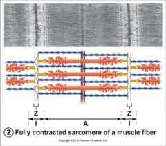
Contracted sarcomere
|
|
|
|
What is the shortening of a muscle fiber called
|
Contraction
|
|
|
|
During the contraction of a muscle fiber, what do the I bands do?
|
Shorten
|
|
|
|
During the contraction of a muscle fiber, do the thin filaments remain the same length, shorten, or disappear.
|
Remain the same like
|
|
|
|
During the contraction of a muscle fiber, do A bands remain the same length, shorten, or disappear?
|

Remain the same length
|
|
|
|
During the contraction of a muscle fiber, do the H bands remain the same length, shorten, or disappear?
|
Disappear
|
|
|
|
how many phases is muscle contraction divided into?
|
3
|
|
|
|
What are the three phases of muscle contraction?
|
Excitation,
excitation contraction coupling, contraction cross bridge cycle. |
|
|
|
What is An action potential in a nerve cell that leads to an action potential in a muscle cell
|
Excitation
|
|
|
|
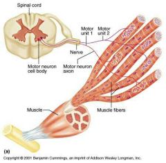
What are nerve cells that stimulate (excite) skeletal muscle cells called?
|
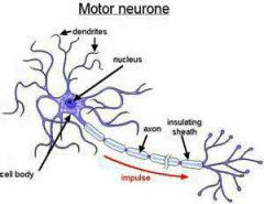
Motor neuron
|
|
|
|
What are the two types of nerve processes
|

Dendrites,
axon |
|
|
|
What is ATP's role during the cross bridge cycle
|

Moves myosin heads to their high energy position.
detaches myosin head from actin. moves Ca2+ back into the SR by primary active transport |
|
|
|
Describe the cross bridge cycle
|
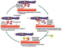
All of the thin filaments in the muscle cell or pulled towards the center of their respective sarcomeres
|
|
|
|
The brain and spinal cord are part of what nervous system
|
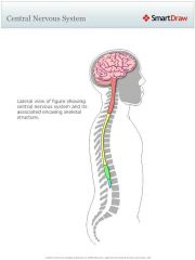
Central nervous system
|
|
|
|
The nerves and ganglia are apart of what nervous system
|
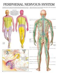
Peripheral nervous system
|
|
|
|
What is a motor unit
|

One motor neuron and all the skeletal muscle fibers it stimulates
|
|
|
|
Muscles that exert "fine control" have how many muscle fibers in each of your motor units?
|
as few as 4 muscle fibers
|
|
|
|
What kind of muscles have as many as seven hundred muscle fibers per motor unit
|
Bearing muscles
|
|
|
|
What do muscle cells store
|
ATP,
creatine phosphate, glycogen. |
|
|
|
How many thick filaments can a single muscle fiber have
|
15 billion
|
|
|
|
When the fiber is contracting how much ATP does each thick filament breakdown
|
Roughly 2500 ATP molecules per second
|
|
|
|
How much ATP do muscle cells store.
|
Enough for 6 seconds worth of contractile activity
|
|
|
|
How many ways can ATP be regenerated
|
3
|
|
|
|
What are the three ways ATP is regenerated
|
Phosphorylation of ADP by creatine phosphate.
Anaerobic regeneration. aerobic regeneration. |
|
|
|
What is good about phosphorylation of ADP by creatine phosphate?
|
Involves only one chemical reaction, so produces ATP quickly
|
|
|
|
What is bad about phosphorylation of ADP by creatine phosphate?
|
Muscle cells can store only so much creatine phosphate, so creatine phosphate is used up after about 10 seconds of rigorous contractile activity.
|
|
|
|
what is glycolysis
|
Conversion of glucose to pyruvic acid
|
|
|
|
What respiration produces the most ATP?
|

Aerobic respiration
|
|
|
|
What are the negatives to aerobic respiration?
|

Limited by 02 availability
|
|
|
|
What are the positives to aerobic respiration
|
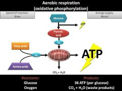
Total of 34 ATP per glucose molecule. CO2 is less toxic than lactic acid.
|
|
|
|
Why does anaerobic respiration occur
|
Because Arabic respiration is unable to supply all of the ATP for contraction
|
|
|
|
What are the positives to anaerobic respiration
|
Not limited by 02 availability
|
|
|
|
What are the negatives to anaerobic respiration
|
Total of 2 ATP per glucose molecule. Lactic acid more toxic than CO2.
|
|
|
|
during 6 seconds of intense short duration exercise where does ATP come from
|
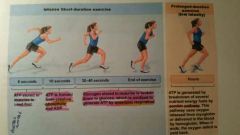
ATP stored in muscle
|
|
|
|
During 10 seconds of intense short duration exercise, where does ATP come from?
|
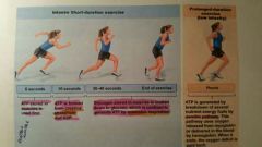
creatine phosphate and ADP
|
|
|
|
During 30 to 40 seconds of intense short-duration exercise how is ATP formed
|
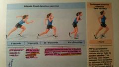
Glycogen stored in muscles is broken down into glucose, which is oxidized to generate ATP by anaerobic respiration
|
|
|
|
Which respiration provides most of the energy in sports involving intense bursts of activity lasting between approximately 15 seconds in one minute
|
Anaerobic respiration
|
|
|
|
Low intensity prolonged exercise such as marathon running uses what type of restoration
|
Aerobic respiration
|
|
|
|
Why does exercise lower blood pH?
|
anaerobic respiration produces lactic acid.
increased Arabic respiration produces more Co2. |
|
|
|
What causes the respiratory center in the brain to increase breathing during exercise
|
A drop in blood p H
|
|
|
|
Arrange the following from the molecular level to the organ level:
Actin and myosin molecules, fascicle, fiber, febrile, filament, muscle, sarcomere, |
Actin and myosin molecules,
filament, sarcomere, fibril, fiber, fascicle, muscle. |
|
|
|
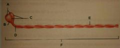
What are bundles of muscle cells called
|

Fascicles
|
|
|
|
What is the plasma membrane of a muscle fiber?
|
Sarcolemma
|
|
|
|
What is the cytoplasm of a muscle fiber
|

Sarcoplasm
|
|
|
|
What is the endoplasmic reticulum of muscle fiber
|

Sarcoplasmic reticulum (SR)
|
|
|
|
What are the sac like enlargements of sarcoplasmic reticulum for increased storage of ca2+
|

Terminal cisterns
|
|
|
|
What are tubular invaginations of sarcolemma that conduct action potentials into muscle fiber
|

Transverse tubules
|
|
|
|
What are the thread like organelles extending the length of a muscle fiber
|
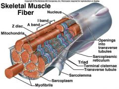
Myofibrils
|
|
|
|
Segments of a myofibril that contain the filaments
|
Sarcomere
|
|
|
|
Components of the cytoskeleton
|
myofilaments
|
|
|
|
what is o2 binding red protein pigment in fibers
|
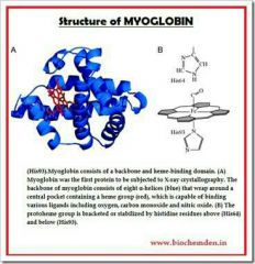
Myoglobin
|
|
|
|
What is the contractile protein composing thin filaments
|

Actin
|
|
|
|
What is the contractile protein composing thick filaments
|
Myosin
|
|
|
|
What is the regulatory protein that covers the myosin binding sites in a relaxed fiber
|
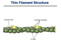
Tropomyosin
|
|
|
|
what is the regulatory protein that holds the tropomyosin of a thin filament in one place?
|

Troponin
|
|
|
|
What is the boundary between adjacent sarcomeres
|

Z disc
|
|
|
|
In the sarcomere What can contain thin, but no thick, filaments.
|

I BaNd
|
|
|
|
Contains thick but no thin filaments.
|

H zone
|
|
|
|
What is formed by supporting proteins holding the thick filaments together at the center of the H zone
|

M line
|
|
|
|
What part of the sarcomere contains both thin and thick filaments
|

Zone of overlap
|
|
|
|
Do I-bands appear darker or lighter under the microscope
|

lighter
|
|
|
|
Why do i bands appear lighter under the microscope
|
Because they are less dense
|
|
|
|
Do the zones of overlap in A bands appear lighter or darker under the microscope
|

darker
|
|
|
|
What part of the nerve cell contains a nucleus
|
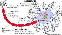
Cell body
|
|
|
|
What is the space called between the nerve cell and muscle cell
|
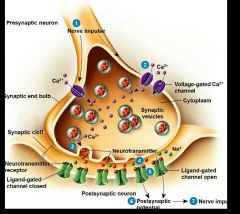
Synaptic cleft
|
|
|
|
What organelle contains ACh?
|

Synaptic vesicle
|
|
|
|
What do you call a motor neuron plus the muscle fibers it stimulates
|

Motor unit
|
|
|
|
What contains synaptic vesicles
|

axon terminal
|
|
|
|
What parts of a motor neuron are located inside the brain or spinal cord
|

Cell body and dendrites
|
|
|
|
In a sarcomere what shortens but does not disappear during contraction
|

i bands
|
|
|
|
In a sarcomere what remains the same length during contraction
|

A bands.
|
|
|
|
What does troponin tropomyosin complex do when a muscle fiber is relaxed
|

Change shape
|
|
|
|
What is responsible for the opening of the calcium channels in the SR membrane
|

Transmission of action potential down T tubules in the triads
|
|
|
|
What causes the troponin tropomyosin complex is to change shape and move out of the way of the binding sites?
|
The binding of Ca+ to troponin
|
|
|
|
Andrew sprinted the hundred meter dash in 10 seconds where does the ATP come from?
|
Stored ATP and CP
|
|
|
|
Mary lifts 200 pounds in competition, it takes 5 seconds to get the barbell over her head. where did the ATP come from?
|
Stored ATP
|
|
|
|
It takes Tom 35 seconds to swim 100 meters as fast as he can. Wear did the ATP come from?
|
Anaerobic respiration
|
|
|
|
Susan jogs 5 miles 4 times a week where does the ATP come from
|
Aerobic respiration
|
|
|
|
Glycolysis begins with what compound
|
Glucose
|
|
|
|
Besides ATP what is the other product of glycolysis
|
Pyruvic acid
|
|
|
|
When there isn't enough oxygen available to the cells pyruvic acid is broken down to what compound
|
Lactic acid
|
|
|
|
When enough oxygen is available, to what compounds is pyruvic acid broken down?
|
Carbon dioxide and water
|
|
|
|
The chemical reaction catalyzed by creatine phosphate yields ATP and what other compound?
|
Creatine
|
|
|
|
Production of too much ....... causes a drop in cytosol pH disabling enzymes and resulting in muscle fatigue
|
Lactic acid
|
|